J. Morphol. Sci., 2013, vol. 30, no. 2, p. 103-106 103
Original
article
Anatomical study of the retroarticular process in dry human skulls
Holleben, P.
1*, Louzada, FF.
2and Faig-Leite, H.
31Department of Restorative Dentistry, Instituto de Ciência e Tecnologia, Universidade Estadual Paulista – UNESP, CEP 12245-000, São José dos Campos, SP, Brasil
2Department of Dental Materials and Prosthodontics, Instituto de Ciência e Tecnologia, Universidade Estadual Paulista – UNESP, CEP 12245-000, São José dos Campos, SP, Brasil
3Department of Biosciences and Oral Diagnosis, Instituto de Ciência e Tecnologia, Universidade Estadual Paulista – UNESP, CEP 12245-000, São José dos Campos, SP, Brasil
*E-mail: epria@uol.com.br
Abstract
Introduction: The retroarticular process is a bony prominence formed by the thickening of the lateral border of the mandibular fossa, forming the posterior wall of the temporomandibular joint. Since little is known and discussed about the retroarticular process, our aim was to study its presence, shape and size, relating these findings to the shape of the skulls according to the horizontal cephalic index. Materials and Methods:We used 400 dry human skulls of the Institute of Science and Technology – UNESP Anatomy Laboratory. Each skull was classified in brachycranics, mesocranics or dolichocranics, and then positioned on a craneostat to measure the height of the retroarticular process from its lower extremity to the auriculo-orbital plane. The width was obtained by measuring the base of the process on its longer lateral axis. Results:The retroarticular process was found bilaterally in 397 skulls (99.25%). All the processes were classified into the following shapes: pyramidal (35.55%), tubercular (31.78%), mammilar (20.73%), crest-like (9.05%) and molar shape (2.89%); 254 skulls (63.50%) showed the same type of process at the right and left sides (Kappa=0.496, moderate agreement). The average height and width were 5.28 mm and 12.81 mm, respectively. Conclusion: The retroarticular process was found in almost all the skulls examined. There are no significant evidences about the relationship among the presence, shape and size of the retroarticular process and the shape of the skulls according to the horizontal cephalic index. However, our findings led us to infer that there would be a functional relationship between the process and the temporomandibular joint.
Keywords: retroarticular process, temporomandibular joint, mandibular fossa, horizontal cephalic index, auricular-orbital plane.
1 Introduction
The retroarticular process (RP) – formerly known as postglenoid tubercle – is a bony prominence formed by a thickening of the lateral border of the mandibular fossa. It is located in front of the external acoustic meatus in the tympanic part of the temporal bone, forming the posterior wall of the temporomandibular joint (TMJ). (CRUZ-RIZZOLO and MADEIRA, 2009; KATSAVRIAS and DIBBETS, 2002; McKAY, YEMM and CADDEN, 1992). It has been found in just about all the human skulls with the same frequency for both genders (OLIVEIRA, 1979).
Bony processes are the points of attachment where a muscle or ligament connects to the bone (CRUZ-RIZZOLO and MADEIRA, 2009). However, accurate information about the insertions on the RP surface is unclear (KATSAVRIAS and DIBBETS, 2002). According to Sicher and DuBrul (1970) the articular capsule of the TMJ attaches not only to the anterior surface of the RP, but also to its extremity. In accordance to Grossmann and Munerato (1996), the superior retrodiscal lamina attaches on the RP and the squamotympanic fissure. Other authors assert that the elastic fibers of the retrodiscal pad of the TMJ attach the posterior border of the articular disc to the RP (CRUZ-RIZZOLO and MADEIRA, 2009).
In a histological study of the morphology and development of the articular disk in 7 human fetuses, from the 10th to the
32nd week of pregnancy, it was observed that at the 20th week
a great amount of elastic fibers from the posterior region of the prenatal articular disk were longitudinally oriented along with the insertion on RP (VALENZA, FARINA and CARINI, 1993).
Some authors have suggested that RP hinders the direct impact of the mandibular condyles onto the tympanic wall. However, the functional meaning of the process is still unclear. It is believed that its presence contributes for the normal physiology of TMJ, consequently its absence could be considered as a predisposing factor to temporomandibular disorders (TMDs) (KATSAVRIAS and DIBBETS, 2002; DUBRUL, 1988).
In human TMJ, the mandibular condyle on the working side initially rotates to mesial and lateral. This movement could be limited by the horizontal component of the temporomandibular ligament and by the insertion of the superior retrodiscal lamina on the RP (KATSAVRIAS and DOUKOUDAKIS, 2001).
Holleben, P., Louzada, FF. and Faig-Leite, H.
J. Morphol. Sci., 2013, vol. 30, no. 2, p. 103-106 104
the apex, RP was classified as crests-like (Figure 4). If the 2 extremities were round-shaped and the base size was similar to the extremity size, RP was classified as molar shape (Figure 5).
Katsavrias and Dibbets (2002) analyzed 90 skulls between 2 and 21 years of age, aiming to quantify RP growth, and observed that 71 skulls (78.88%) presented the process. These authors made an impression of the mandibular fossa of the skulls with silicon material and digitized these impressions to measure the RP height from its lower extremity to the deepest point of the mandibular fossa. The mean height was 5.31 mm. Also, it was verified that RP growth stopped around 13 years-old, showing a uniform mean annual growth (0.27 mm/year).
The RP exhibits anatomical variation regarding its shape, size, symmetry, orientation and presence, with high-percentage prevalence in skulls of the modern man (OLIVEIRA, 1979; KATSAVRIAS and DIBBETS, 2002).
Oliveira (1979) studied the presence and shape of RP in 811 dry adult human skulls (1622 sides), identified by gender and race group. According to this author, RP can be found in 5 different shapes: mammilar (most frequent), pyramidal, tubercular, crest-like and molar (least frequent). RP presence was reported in 98.40% of the examined skulls, without significant differences in the frequencies between genders and among races. Additionally, right and left RPs were the same type in 713 (87.91%) skulls.
Because of the lack of studies on the RP bony morphology, and of the high prevalence of this anatomical structure in the skulls of the modern man, a more detailed study on RP was considered of great importance. The aim of this study was to assess the presence, shape and size of RP in dry human skulls relating these findings to the shape of the skulls according to the horizontal cephalic index (HCI).
2 Materials and Methods
To conduct this study, 400 dry human skulls (800 sides) were selected, not identified regarding gender, age or race, belonging to the Anatomy Laboratory of the School of Dentistry of São José dos Campos – UNESP. This study was submitted and approved by the Ethical Committee in Human Research under protocol number 068/2008-PH/ CEP.
Each skull was measured with the aid of a thickness caliper and classified according to HCI, correlating the maximum width and length of the skull: dolichocranics (HCI ≤ 74.9), mesocranics (75 ≤ HCI ≤ 79.9) or brachycranics (HCI ≥ 80) (CRUZ-RIZZOLO and MADEIRA, 2009).
After the HCI classification, the RP presence was quantified at each side of the skulls. If present, they were classified into 5 different types (shapes): pyramidal, mammilar, tubercular, crests-like and molar shape (OLIVEIRA, 1979).
In order to establish accurate criteria for the identification of each RP type, the skulls were observed at lateral norm. It was adopted that pyramidal RP (Figure 1) shows a pointed extremity, widening towards the base. The tubercular (Figure 2) and mammilar (Figure 3) types have a round-shape extremity, but mammilar RP (at lateral norm) shows the base width similar to its extremity while tubercular RP exhibits a base wider than its extremity. According to Oliveira (1979), it resembles the lateral aspect of the articular eminence of temporal bone.
The crests-like and molar RPs seem to present 2 extremities. At lateral norm, if these 2 extremities either round- or point-shaped, but it had their base wider than
Figure 1. Pyramidal retroarticular process (RP).
Figure 2. Tubercular retroarticular process (RP).
Anatomical study of the retroarticular process
J. Morphol. Sci., 2013, vol. 30, no. 2, p. 103-106 105
The RP classification according to its shape at lateral norm was performed based on the methodology of Oliveira (1979). The results are presented in Table 1. The Left-side RP was equal to right-Left-side RP in 254 skulls (63.50%). Aiming to assess the agreement and reproducibility of the matches observed, Kappa statistical test was applied, showing a moderate agreement degree (K=0.496).
All skulls presenting RP were placed into a craneostat to locate their auriculo-orbital plane (AOP; Frankfort horizontal plane), accurately. Then, the RP height was measured from its lower extremity to the AOP (Figure 6). Following, the skull was removed from the craneostat, and placed at basilar norm, and the greatest lateral-to-lateral width of the RP base was measured (Figure 7). Each measure (height and width) was performed 3 times with a caliper and transferred to an electronic caliper (Mitutoyo model 500-683, Santo Amaro, SP, Brazil), at 200 mm range/ 0.01 mm resolution.
To evaluate the agreement and reproducibility of the data found, Kappa statistical test was applied.
3 Results
Each one of the 400 skulls was classified according to HCI into: 176 (44%) brachycranics, 158 (39.50%) mesocranics and 66 (16.50%) dolichocranics.
The RP was found bilaterally in 397 (99.25%) skulls, unilaterally (right side) in 2 (0.50%) and absent in 1 (0.25%) skull. In the analysis of the 800 sides, 796 RPs were seen: 399 (50.13%) at the right side and 397 (49.87%) at the left side.
Figure 5. Molar retroarticular process (RP).
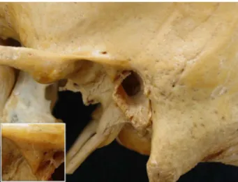
Figure 6. Skull at lateral norm showing the obtainment of the RP height. AOP: auriculo-orbital plane; po: porion point.
Figure 7. Skull at basilar norm showing the obtainment of the RP width.
Table 1. Types of RPs.
Retroarticular Process Shape
Number (%) of Retroarticular Processes Total Left Side Right Side Pyramidal 283 (35.55) 130 (32.74) 153 (38.35) Tubercular 253 (31.78) 133 (33.50) 120 (30.08) Mammilar 165 (20.73) 84 (21.16) 81 (20.30) Crests 72 (9.05) 36 (9.07) 36 (9.02) Molar 23 (2.89) 14 (3.53) 9 (2.25) Total 796 (100) 397 (100) 399 (100)
Holleben, P., Louzada, FF. and Faig-Leite, H.
J. Morphol. Sci., 2013, vol. 30, no. 2, p. 103-106 106
(13.20 mm) mean width is 2% (0.32 mm). Therefore, there were no significant differences for the mean height and width values among dolichocranics, mesocranics and brachycranics.
4 Conclusion
The RP was found in almost all the examined skulls. The pyramidal shape was the most frequent, possibly justified by the tension direction of TMJ structures which are inserted into the process, since we did not find significant evidences about the relationship between the shape and size of RP to the shape of the skulls according to the HCI. Similarly to Katsavrias and Dibbets (2002), we believe that RP absence could be a factor related to DTMs. Nevertheless, taking into consideration the analysis of the RP region of our sample (800 sides), we assume that not only the absence, but also the shape, size and inclination of the RP may contribute to DTMs when associated with other factors.
Acknowledgements: Scientific initiation scholarship FAPESP
08/57444-0. Assistant Professor Ivan Balducci (Department of Social Dentistry and Pediatric Clinic, School of Dentistry of São José dos Campos, UNESP – Univ. Estadual Paulista): statistics.
References
CRUZ-RIZZOLO, RJ. and MADEIRA, MC. Anatomia Facial com fundamentos de anatomia sistêmica geral. São Paulo: Sarvier, 2009. 355 p.
DUBRUL, EL. Sicher and DuBrul’s Oral Anatomy. Saint Louis: Ishiyaku EuroAmerica, 1988. 356 p.
GROSSMANN, E. and MUNERATO, MC. Aspectos anátomo-fisiológicos da articulação temporomandibular. Revista da Faculdade de Odontologia da Universidade de Passo Fundo, 1996, vol. 1, n. 2, p. 11-20.
KATSAVRIAS, EG. and DIBBETS, JMH. The postglenoid tubercle: prevalence and growth. Annals of Anatomy, 2002, vol. 184, n. 2, p. 185-188. http://dx.doi.org/10.1016/S0940-9602(02)80017-6
KATSAVRIAS, EG. and DOUKOUDAKIS, A. The normal temporomandibular joint [in Greek]. Athens: M Bonnissel, 2001.
McKAY, GS., YEMM, R. and CADDEN, SW. The structure and function of the temporomandibular joint. British Dental Journal, 1992, vol. 173, n. 4, p. 127-132. PMid:1389598. http:// dx.doi.org/10.1038/sj.bdj.4807966
OLIVEIRA, Y. Le processus rétro-articulaire ou tubercule rétro-mandibulaire du squelette crânien chez l’homme. Acta Anatomica, 1979, vol. 104, n. 2, p. 211-219. http://dx.doi. org/10.1159/000145069
SICHER, H. and DUBRUL, EL. Oral Anatomy. Saint Louis: The C. V. Mosby Company, 1970. 502 p.
VALENZA, V., FARINA, E. and CARINI, F. The prenatal morphology of the articular disk of the human temporomandibular joint. Italian Journal of Anatomy and Embryology, 1993, vol. 98, n. 4, p. 221-230.
WRIGHT, DM. and MOFFETT, BC. The postnatal development of the human temporomandibular joint. American Journal of Anatomy, 1974, vol. 141, n. 2, p. 235-249. PMid:4416417. http://dx.doi.org/10.1002/aja.1001410206
Received February 2, 2013 Accepted May 22, 2013
Concerning to RP height, the greatest measures obtained were 10.71 mm at the right side and 10.69 mm at the left side. The smallest heights were 1.63 mm at the right side and 1.90 mm at the left side. The mean height was 5.42 mm (5.55 mm at the right side and 5.30 mm at the left side).
The largest width obtained was 19.12 mm at the left side and 17.88 mm at the right side. The narrowest measures registered were 7.06 mm at the right side and 8.09 mm at the left side. The mean RP width was 13.31 mm (13.31 mm at the right side and 13.31 mm at the left side).
Following the classification of the skulls in dolichocranics, mesocranics and brachycranics, these data were crossed with the shape and size (height and width) of the RPs, to verify a possible correlation between the skull and the RP shape (Table 2). It was noticed that pyramidal RP was the most frequent in brachycranics. There were no significant differences between pyramidal and tubercular types in mesocranics. In dolichocranics, tubercular RP was the most found, without significant differences in tubercular and pyramidal types at the right side. Also, the pyramidal and mammilar shapes exhibited the same frequency at the left side.
Figure 8 shows the averages of RP height and width, for each side, in dolichocranics, mesocranics and brachycranics. The bars on each column represent the standard deviation (positive square root of variance). As noted, the difference between the greatest (5.94 mm) and the smallest (5.19 mm) mean height is of about 12.60% (0.75 mm). The difference between the largest (13.52 mm) and the narrowest
Table 2. Distribution (%) of RPs.
Retroarticular Process Shape
Brachycranic Mesocranic Dolichocranic
LS RS LS RS LS RS
Pyramidal 36.36 43.75 31.65 34.81 24.24 31.82 Tubercular 31.82 26.71 31.01 32.28 42.43 33.33 Mammilar 17.61 17.04 23.42 22.78 24.24 22.73 Crests 10.80 10.23 9.49 7.60 3.03 9.09 Molar 2.27 1.70 3.80 2.53 6.06 3.03 Absent 1.14 0.57 0.63 0 0 0
LS: left side; RS: right side.