Increased
μ
-Calpain Activity in Blasts of
Common B-Precursor Childhood Acute
Lymphoblastic Leukemia Correlates with
Their Lower Susceptibility to Apoptosis
Anna Mikosik1, Izabella Henc2, Katarzyna Ruckemann-Dziurdzińska2, Joanna
E. Frąckowiak1, Anna Płoszyńska3, Anna Balcerska3, Ewa Bryl2, Jacek M. Witkowski1*
1Department of Pathophysiology, Medical University of Gdańsk, Gdańsk, Poland,2Department of Pathology and Experimental Rheumatology, Medical University of Gdańsk, Gdańsk, Poland,3Clinic of Pediatrics, Hematology and Oncology, Medical University of Gdańsk, Gdańsk, Poland
Abstract
Childhood acute lymphoblastic leukemia (ALL) blasts are characterized by inhibited apopto-sis promoting fast disease progress. It is known that in chronic lymphocytic and acute mye-loid leukemias the reduced apoptosis is strongly related with the activity of calpain-calpastatin system (CCS) composed of cytoplasmic proteases—calpains—performing the modulatory proteolysis of key proteins involved in cell proliferation and apoptosis, and of their endogenous inhibitor—calpastatin. Here, the CCS protein abundance and activity was for the first time studied in childhood ALL blasts and in control bone marrow CD19+B cells by semi-quantitative flow cytometry and western blotting of calpastatin fragments resulting from endogenous calpain activity. Significantly higherμ-calpain (CAPN1) gene
transcrip-tion, protein amounts and activity (but not those of m-calpain), with calpastatin amount and transcription of its gene (CAST) greatly varying were observed in CD19+ALL blasts com-pared to control cells. Significant inverse relation between the amount/activity of calpain and spontaneous apoptosis was noted. Patients older than 10 years (considered at higher risk) displayed increased amounts and activities of blast calpain. Finally, treatment of blasts with the tripeptide calpain inhibitors II and IV significantly and in dose-dependent fashion increased the percentage of blasts entering apoptosis. Together, these findings make the CCS a potential new predictive tool and therapeutic target in childhood ALL.
Introduction
Acute lymphoblastic leukemia (ALL) is the most common type of pediatric leukemia, account-ing for over 70% of all cancer cases diagnosed in patients under 15 years of age[1,2]. In addition to an enhanced cell proliferation in ALL, leukemic blasts are also characterized by reduced sus-ceptibility to programmed cell death (apoptosis), which results in an inhibition of normal hematopoiesis and leads to the development of the disease[3–5].
OPEN ACCESS
Citation:Mikosik A, Henc I, Ruckemann-Dziurdzińska K, Frąckowiak JE, Płoszyńska A, Balcerska A, et al. (2015) Increasedμ-Calpain Activity in Blasts of Common B-Precursor Childhood Acute Lymphoblastic Leukemia Correlates with Their Lower Susceptibility to Apoptosis. PLoS ONE 10(8): e0136615. doi:10.1371/journal.pone.0136615
Editor:Pierre Bobé, INSERM-Université Paris-Sud, FRANCE
Received:December 5, 2013
Accepted:July 23, 2015
Published:August 28, 2015
Copyright:© 2015 Mikosik et al. This is an open access article distributed under the terms of the
Creative Commons Attribution License, which permits unrestricted use, distribution, and reproduction in any medium, provided the original author and source are credited.
Funding:This work was supported by Polish Ministry of Science and Higher Education grant No 2 P05E 103 30 (to AP) and by a statutory (ST-58) grant from Polish Ministry of Science and Higher Education 02-0058/07/262 (to JMW). The funders had no role in study design, data collection and analysis, decision to publish, or preparation of the manuscript.
Competing Interests:The authors have declared
Intensive development of chemotherapy together with improvements of the supportive care have contributed to a significant increase in the survival rates, which now reach more than 80% in ALL patients[2,3,6–9]. Still there are cases of refractory ALL and there are patients who relapse. Thus it remains important to search for new prognostic factors or markers of disease progression with the potential for diagnostic purposes.
We have shown before[10] that significantly reduced ability of chronic B-cell leukemia (B-CLL) cells to enter spontaneous apoptosis is associated with grossly increased total amount and activity of the proteolytic enzyme,μ-calpain, member of the so-called calpain-calpastatin system (CCS) consisting of a group of proteases–calpains—and their endogenous inhibitor– calpastatin47. Calpains are the cytoplasmic cysteine proteases, activation of which requires suf-ficiently high concentration of Ca2+[11,12]. Calpain activity was first described and isolated from the rat brain in 1964[13]; it has since become the object of an intense research in the con-text of its (patho)physiological significance.
The main part of the CCS are so called‘ubiquitous’calpains:μ-calpain (calpain I) and m-calpain (m-calpain II), named after the concentrations of Ca2+required for their full proteolytic activity in vitro:μ-calpain requires 1–100μM Ca2+and m-calpain 0.1–1 mM Ca2+respectively [11,12,14]. The two-subunit structure of these classical calpains is highly evolutionarily con-served among all vertebrates, indicating the paramount importance of these proteins for the cellular function [11,14,15].
Over a hundred of important proteins have been already identified as intracellular calpain substrates [11,12,14,16–18]. Although calpains can completely hydrolyze some of their sub-strates, for the majority of them they do not operate like protein-degrading proteases, but mod-ulate their activity and function by limited hydrolysis [11,12,19,20]. This adjustable
biomodulation of substrates can, on one hand, lead to the transmission of signals participating in the cell cycle and cell proliferation control, but on the other hand, their role in the regulation of apoptosis (indirectly or through a direct effect on its substrates, including caspases or Bcl-2 protein family members, such as Bcl-2, Bcl-xL, Bid, Bax) is postulated [11,16,21–23].
Due to the role of calpains in abovementioned cell functions, their intracellular activation and inhibition must be precisely regulated. Uncontrolled proteolysis of calpain substrates is prevented by the only known, specific endogenous calpain inhibitor–calpastatin, exerting no influence on other enzymes, which is itself a calpain substrate [12,18,24,25]. Calpastatin is first activated through limited, Ca2+-dependent proteolysis by calpain. Only after such activation does calpastatin gain the ability to competitively inhibit calpain[19,21]; thus, the system oper-ates in a feedback loop. The balance must be strictly regulated here, since the excess of one of the components of the system can lead to cell functional pathology and progression of resulting disease[21,26]. Calpains themselves can also be their own substrates in a self-limiting activa-tion/degradation cycle[20,21].
Based on the facts summarized above, in this work we investigated the role of calpains in the pathomechanism of apoptosis avoidance of ALL blasts.
Materials and Methods
Ethics statement
The study was approved by the Local Independent Committee for Ethics in Scientific Research at the Medical University of Gdansk. The patients’parents (legal guardians) gave written informed consent for participation in the study. The procedures were in accord with the Hel-sinki Declaration of 1975, as revised in 2008.
Subjects
Altogether, thirty nine ALL patients diagnosed in the Department of Pediatrics, Hematology, Oncology and Endocrinology, Medical University of Gdansk, were enrolled into this study. However, due to paucity of biological material available (BM samples remaining after diagnos-tic BM aspiration), not all experiments could be performed on all samples. The numbers of samples included in any specific experiment are given in the figure legends. The only inclusion criteria were the diagnosis of common acute lymphoblastic leukemia and material availability. There were 19 girls and 20 boys, median age 5.68 years (from 2.5 months to 17.6 years); with no significant age differences between the genders. The bone marrows were routinely assessed by the hematological diagnostic laboratory and the leukemic blasts were classified according to their morphology and immunophenotype. All patients were diagnosed as common ALL and received treatment according to the same protocol. Detailed clinical characteristics of the patient source cohort including the karyotype analysis and treatment protocol is given else-where[33].
Twenty-one children in the control group were age- and gender-matched patients of the same Department, in whom the diagnostic bone marrow aspiration was performed and the malignancy was ruled out. In choice of the control group, apart from the obvious availability matter, we considered the reports indicating that B lineage progenitor (CD19+CD34+) cells form a relatively substantial proportion of the B lineage pool in the bone marrow of children younger than 15 years[34]. The only inclusion criteria were the availability of the post-diagnos-tic bone marrow surplus aspirate and no malignancy diagnosed. There were 10 girls and 11 boys, median age 6.5 years (from 3.5 to 17.9 years). The bone marrows were routinely assessed by the hematological diagnostic laboratory and diagnosed as: thrombocytopenia (6 patients), leukopenia (3), cyclic neutropenia (3), spherocytosis (2), deficiency anemia (2) hemolytic ane-mia (2), mononucleosis (1), cytomegaly (1), and collagenosis (1). In the available literature there are no reports on significant deviation of the contents and/or activity of calpains in the BM of children suffering from any of the above mentioned diseases from these seen in healthy BM.
The event-free survival was assessed two years after the initial ex vivo analysis–an event was defined as relapse (6/39 patients); of these 6 patients, one-third obtained bone marrow trans-plant (3/39 patients) and one-third died within the follow-up period (3/39 patients).
Sample preparation
(BMMC) were isolated by density gradient centrifugation on Histopaque 1077 (Sigma– Aldrich). Interphase cells were collected and washed twice in RPMI medium (Sigma–Aldrich). Aliquots of 0.3 × 10⁶cells were used for further staining.
Immunophenotyping
Staining to identify malignant B cells by simultaneous expression of CD19 and CD34 was cho-sen based on the routine bone marrow immunophenotyping performed for diagnostic pur-poses. The percentages of blasts in all the BMs from ALL patients were 90–100%, hence there was no need for further purification of cells for the molecular tests.
CCS staining
All three anti-CCS antibodies (anti-μ-calpain, anti-m-calpain and anti-calpastatin) were mouse monoclonal non-conjugated antibodies (Abcam, UK, clone numbers 15C10, 107–82 and CSL5-10 respectively); rat monoclonal antibody (Becton Dickinson, USA, clone number A85-1) against mouse immunoglobulins, coupled with R-Phycoerythrin was used for the detection of these CCS antibodies in a two-step protocol. The intracellular labeling of CCS pro-teins was performed after staining the cells with FITC-anti-CD19/PE-Cy5-anti-CD34 mix (both from Becton Dickinson, USA, clones HIB19 I 581 respectively), fixation and permeabili-sation with 2% paraformaldehyde and 0.25% saponin (Sigma Aldrich,USA) in PBS[35]. Matched isotype controls were used throughtout (Becton Dickinson, USA).
Apoptosis assessment
Spontaneous apoptosis in ALL blasts, as well as in non-malignant BM CD19+cells was mea-sured by PE-Annexin V binding to the cell surface with simultaneous 7-aminoactinomycin D (7-AAD) staining to exclude necrotic cells (protocol by the manufacturer, Becton Dickinson, USA). Measurement of the mitochondrial membrane potential loss characteristic for earlier stages of apoptosis was performed with the JC1 probe (5,5’,6,6’-tetrachloro-1,1’,3,3’ tetraethyl-benzimidazolylcarbocyanine iodide, Molecular Probes) in ALL blasts ex vivo and in 24-hours cell cultures in vitro, according to[36–38]. Separate samples of ALL blasts and non-malignant CD19+lymphocytes were treated with 5μg/ml chelerythrine (a pan-kinase C inhibitor with potent pro-apoptotic and anti-tumor activity[39]) prior to assessment of their apoptosis by JC1 staining. These experiments uniformly yielded around 98% of cells with strongly depolarized mitochondria (apoptotic) and thus served as positive control for the test.
There are reports (including ours) suggesting that human B cell lineage might contain bothμ- and m-calpain[10]. Expression of both proteases in childhood ALL is put to test in this
work. Currently (November 2014) a PUBMED search for“calpain AND acute lymphoblastic
leukemia”or“calpain inhibition AND acute lymphoblastic leukemia”returns precisely one paper[29] describing the pro-apoptotic effect of calpain inhibitor II on established human ALL cell lines. In this work, in order to assess the effectiveness of calpain inhibition in induction of apoptosis of the ALL blasts, samples of the BMMC (2 × 10⁶cells/ml) were cultured for 24 hours with/ or without calpain inhibitor IV (CI IV, a synthetic tripeptide aldehyde LLY: Z-Leu-Leu-Tyr-CH2F (Z = benzyloxycarbonyl), Calbiochem, UK) at concentrations ranging from 1μM to 8μM [40] or with 20μg/mL calpain inhibitor II (CI II, N-Acetyl-Leu-Leu-Met-al; Cal-biochem)[10] and tested for apoptosis induction.
Flow cytometry analysis
All measurements requiring flow cytometry, i.e., phenotyping and analysis of expression of the CCS member proteins, were performed using the FACScan cytometer (Becton Dickinson, USA). At least 2x10⁴cells were acquired from each sample using the CellQuest software of the instrument and then analyzed using the Cyflogic v. 1.2.1 software. Mean fluorescence intensity (MFI) minus MFI of relevant isotype control was used as a semi-quantitative measure of anti-gen expression. In order to assure lack of influence of the possible FACS MFI readout instabil-ity, the instrument was calibrated at each session with the calibration beads (CaliBRITE 3 for three-color flow cytometer setup, Becton Dickinson), so as both the beads’MFI and CV were the same throughout the project.
Quantitative real time-PCR estimation of CCS genes
’
expression
Total RNA was isolated from flash-frozen cells of ALL patients and the control group (samples @ 2 x10⁶cells) using the miRNeasy Mini Kit (Qiagen, Netherlands) and DNA digestion was carried out using the RNase-Free DNase Set (Qiagen, Netherlands). The quality of the obtained total RNA (RNA Integrity Number) was measured by the Agilent 2100 Bioanalyzer instrument using the Agilent RNA 6000 Nano Kit and the resulting values indicated high RNA quality (>8). The reverse transcription reaction was performed using the ImProm-II Reverse Tran-scription System (Promega, USA). The resulting cDNA served as a template for Real-Time PCR reactions performed with the use of the LightCycler FastStart DNA Master SYBR Green I (Roche) and run on the LightCycler 2.0 instrument supplied with the LightCycler Software 4.05 (Roche Diagnostics, Germany). The sequence of primers (BLIRT, Poland) employed in
the Real-Time PCR reactions were as follows: 5’-ATTTCGTTTGCTGCCTGGTG-3’and
5’-ATGGTCAGCTGCAACCACTTA-3’forμ-calpain, 5’-GCATACGCCAAGATCAACGG-3’
and 5’-GGAGGGGGCTTCTTCAACTC-3’for m-calpain, 5’-CCCAAGCCTCGGAGTGAA
TC-3’and 5’-AGCGGCCTTAGATTCTTCTGT -3’for calpastatin, 5’-CAGTCAGCCGCA
TCTTCTTT-3’and 5’-GACCAAATCCGTTGACTCCG-3’for GAPDH as the reference gene.
The reaction consisted of a pre-incubation step (95°C, 10 minutes), a quantification step which included 40 cycles (95°C, 10 seconds; 62°C, 10 seconds; 72°C, 5 seconds), a melting step (65°C, 15 seconds) and a cooling step (40°C, 30 seconds). The obtained Ct values ofμ-calpain, m-cal-pain, calpastatin and the reference gene GAPDH[41] were analyzed using theΔΔCt method and expressed as fold change over the expression of GAPDH.
Measurement of the endogenous
μ
-calpain activity ex vivo
nonspecific binding was blocked in 3% solution of skimmed milk powder in TBS (Tris-buffered saline) with 0.05% Tween-20 for 1 hour, then twin membranes were carefully washed and incu-bated with either 1:1000 anti-calpastatin mouse monoclonal antibody or 1:2000 anti-actin mouse monoclonal antibody (both Abcam, Great Britain) overnight, at 4°C. Then, washed mem-branes were incubated with 1:2000 HRP-conjugated rabbit polyclonal antibody against mouse IgG for 2 hours. Bound antibodies were detected by chemiluminescence (SuperSignal West Pico Chemiluminescent Substrate, Thermo Scientific, USA) recorded on the x-ray film. The devel-oped and fixed films were digitalized using the GDS-8000 System and dedicated acquisition soft-ware Labworks Image Acquisition and Analysis Softsoft-ware Version 4.0 (UVP Bioimaging System, UK). Relative ex vivo calpain activity was assessed by the detection and densitometric quantifica-tion of the protein bands containing the degraded fragments and native calpastatin. Densitomet-ric analysis was performed using the Scion Image Beta (version 4.0.2) program.
Statistical analysis
Statistical analyses were performed with STATISTICA 8 (StatSoft, Poland) software using the non-parametric Mann-Whitney U-test to compare two independent groups (relative to the groups or to the variable) after confirming the lack of normality of data distribution. All values are shown in the graphs as‘box and whisker’plots corresponding to median, 25thand 75th quartiles and data range.
Results
1. Comparison of the proportions of ALL blasts and non-malignant
CD19
+B cells containing detectable
μ
-, m-calpain and calpastatin and of
amounts of these proteins in these cells
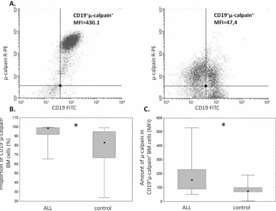
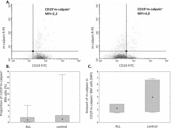
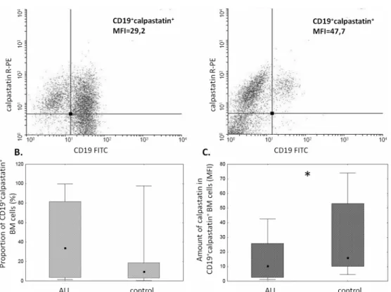
The proportion ofμ-calpain+cells was significantly higher among CD19+ALL blasts compared to B lymphocytes in the control group (Fig 1B). In the light of published data, assuming gener-ally thatμ-calpain is present in all leucocytes[10,42] the only explanation of these percentages being lower than 100% is that in some B lymphocytes the levels of this enzyme were below the detection threshold of the method used. Also the assessment ofμ-calpain amounts measured semi-quantitatively as MFI in ALL blasts and non-malignant CD19+cells showed significantly higher amount of this enzyme in the blasts (Fig 1C). Unlike that ofμ-calpain, the percentage of m-calpain+cells as well as the m-calpain MFI did not differ between CD19+ALL blasts and healthy CD19+cells (Fig 2). It is worth noting here that the proportion of m-calpain-expressing cells was extremely low, as was the amount of the enzyme in the cells, regardless their benign or malignant character. Finally, although the cytometrically assessed proportions of calpasta-tin-positive cells did not differ between the BM CD19+ALL blasts and nonmalignant B cells, the actual levels of calpastatin expressed as corrected MFI were significantly lower in the blasts (Fig 3). As ALL blasts of the patients were confirmed to be CD19+CD34+, in some experiments the processed BM cells from control individuals were gated for CD34 positivity, then the levels of expression of calpains were additionally assessed in the non-malignant CD19+CD34+. We did not see any difference between the detected amounts of all three CCS proteins when they were compared in the general BM CD19+ and in the CD19+CD34+subpopulation of non-malignant BMs (not shown).
2. Comparison of the CCS genes
’
expression
results analysis we could demonstrate that the expression ofμ-calpain gene (CAPN1,Fig 4A) was significantly higher the ALL blasts compared to the B cells from the control group (mean fold change ALL vs control = 2.3), while the expression of both m-calpain (CAPN2,Fig 4B) and calpastatin (CAST,Fig 4C) genes did not differ between the compared groups (fold changes 0.93 and 0.895 respectively). Interestingly, expression of all three CCS genes in the control cells was very uniform and, in case ofCAPN1andCAST, practically at the same level, while that ofCAPN1and especiallyCASTgene in the blasts was widely dispersed among the samples.
3. Assessment of the endogenous calpain activity in ALL blasts ex vivo
Our own method based on the detection of calpain-specific degradation of cellular calpastatin was used; seeMaterials and Methodsfor details[35]. All 37 samples of ALL blast lysates tested contained specifically degraded calpastatin indicating endogenous calpain activity, with the majority (27/37) showing variable activities ranging from limited to almost total degradation of available calpastatin (not shown). No calpain activity could be detected in nonmalignant BM B lymphocytes using the same technique. A representative result of one such experiment, where lysates from non-malignant BM B cells, ALL blasts from a 12 year-old patient and the same
Fig 1. Proportions ofμ-calpain-positive cells and relative amounts ofμ-calpain are elevated among ALL blasts.A. Representative two-parameter plots (dot plots) resulting from simultaneous staining of BM samples (left panel–ALL, right panel–control) with anti-CD19, anti-CD34 and anti-μ-calpain antibodies. Actual corrected MFI values for calpain signal in CD19+cells are shown. B. Significant difference between the
proportion ofμ-calpain-positive cells among ALL blasts and nonmalignant BM B cells. Box-and-whisker plots depict the medians, 25thand 75thpercentile and range respectively. Asterisk signifies p = 0.02; N(ALL) = 20,
N(control) = 9. C. Amount ofμ-calpain is significantly higher in ALL blasts than in nonmalignant B lymphocytes. Comparison of relative intensities (MFI) ofμ-calpain–bound antibody in CD19+ALL blasts
(ALL) and non-malignant B cells (control). Asterisk denotes p = 0.03; N(ALL) = 16, N(control) = 9). For the details seeMaterials and Methods.
blasts treated with 4μM CI IV for 18 hours are tested for calpastatin expression by Western blot is shown in theFig 5A. Detection of some native calpastatin in the calpain inhibitor-treated blasts clearly shows that calpain is indeed responsible for calpastatin cleavage in the blasts and that the method can be used for the determination of endogenous calpain activity. As patient age is a risk factor, with children older than ten years being at higher risk associated with poorer response after relapse (revieved by Bhojwani and Pui[43]) and decreasing event-free survival (reviewed by Hochberg et al., [44]), we have examined whether this endogenous calpain activity in the ALL blasts is different if our patients were subdivided according to age. The older patients (age>10 years) had on average demonstrated high to very high calpain activity (in no sample from that group had native calpastatin been detected) (Fig 5B).
4. Analysis of the in vivo and in vitro relation between ALL blast
apoptosis and CCS system
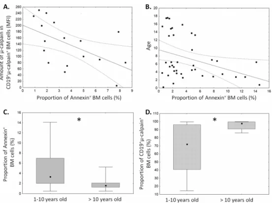
Spontaneous apoptosis of ALL blasts measured after 18 hours in vitro ranged from 1 to 14% (Fig 6). This variation allowed us to seek possible relation between the level of ALL blast apo-ptosis ex vivo and the amounts ofμ-calpain and proportions of calpain-positive blasts. Signifi-cantinverserelation between the amount ofμ-calpain [MFI] and spontaneous apoptosis was noted in ALL blasts (Fig 6A). Similarly significant negative correlation between the percentage of apoptotic cells and the age of the ALL patients was found—the older the ALL patients, the
Fig 2. Similar proportions of m-calpain-positive cells and amounts of m-calpain in ALL blasts and control cells.A. Representative two-parameter plots (dot plots) resulting from simultaneous staining of BM samples (left panel–ALL, right panel–control) with anti-CD19 and anti-m-calpain antibodies. Actual corrected MFI values for calpain signal in CD19+cells are shown. Details in Materials and Methods. B, C. No significant
difference between the proportion of m-calpain-positive cells among ALL blasts and nonmalignant BM B cells (B) and between amount (MFI) (C). Box-and-whisker plots depict the medians, 25thand 75thpercentile and
range respectively. N(ALL) = 6, N(control) = 6. For the details seeMaterials and Methods.
stronger inhibition of apoptosis in ALL blasts (Fig 6B). Also, when patients were subdivided into age groups (below and above 10 years of age), the older subgroup exhibited significantly lower proportion of spontaneously apoptotic ALL blasts simultaneously with significantly higher proportion ofμ-calpain-positive blasts (Fig 6C and 6D). Finally, despite relatively short
Fig 3. The amounts of calpastatin differ between CD19+ALL blasts and non-malignant B cells.A.
Representative two-parameter plots (dot plots) resulting from simultaneous staining of BM samples (left panel–ALL, right panel–control) with anti-CD19 and anti-calpastatin antibodies. Actual corrected MFI values for calpastatin signal in CD19+cells are shown. B. No significant difference between the proportions of calpastatin-positive cells among ALL blasts and nonmalignant BM B cells. C. Significantly lower calpastatin amount (MFI) in the blasts (C). Box-and-whisker plots depict the medians, 25thand 75thpercentile and range
respectively. N(ALL) = 30, N(control) = 17. For the details seeMaterials and Methods.
doi:10.1371/journal.pone.0136615.g003
Fig 4. Expression ofμ-calpain but not other CCS genes is different in ALL blasts and nonmalignant BM B cells.A. Significantly higher expression ofCAPN1(μ-calpain) gene in ALL blasts compared to control
B cells. B,C. No differences between expression ofCAPN2(m-calpain) andCAST(calpastatin) genes in ALL
blasts vs non-malignant B cells. Please mark huge variability of expression of both theCAPN1and especially
CASTgenes. CCS gene expression is shown as proportion of the expression ofGAPDHhousekeeping gene considered 1. Box-and-whisker plots depict the means, SEM and SD respectively. P values (Kruskall-Wallis test) are given in the graphs; N(ALL) = 6, N(control) = 6. For the details seeMaterials and Methods.
Fig 5. Endogenous calpain activity is present in ALL blasts. A. Representative result of western blot determinantion of calpastatin and its immunoreactive fragments resulting from calpain activity in non-malignant BM CD19+cells (lane 1), ALL blasts from a 12-year old patient (lane 2) and blasts from the same
patient as in lane 2, but incubated in vitro for 24 hours with 4μM calpain inhibitor IV (lane 3). Actin was used as a reference protein. For further details seeMaterials and Methods. B. Endogenous calpain activity in ALL blasts measured by degree of calpastatin degradation (loss of the native form) is significantly higher in the children more than 10 years old. Box-and-whisker plots depict the medians, 25thand 75thpercentile and
range respectively. Asterisk signifies p = 0.01; N (1–10 years old ALL patients) = 27, N(>10 years old ALL patients) = 10.
doi:10.1371/journal.pone.0136615.g005
Fig 6. Levels of spontaneous apoptosis of ALL blasts depend on patient age andμ–calpain amount.
Apoptosis was determined as the proportion of AnnexinV+ blasts and plotted against the amount ofμ–calpain in the blasts (A, N = 17, r = -0.54, p<0.05) and against patients’age (B, N = 39, r = -0.31, p<0.05). See
Materials and Methodsfor details. When the patients were subdivided into below and above 10 years of age subgroups, the latter were characterized by significantly lower apoptosis (C, asterisk signifies p = 0.01) and significantly higher proportion of theμ–calpain positive blasts (D, asterisk signifies p = 0.04). N (1–10 years old ALL patients) = 29, N(>10 years old ALL patients) = 10.
time of follow-up, we had already observed a significantly lower proportion (p = 0.03) of sponta-neously apoptotic cells with higherμ-calpain amount and activity in our 6 patients who, at the end of follow-up period, were recorded to exhibit adverse events (relapse followed by bone mar-row transplant in 3/6 or by death in another 3/6) versus those 33 exhibiting event-free survival.
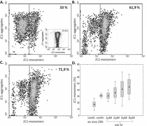
As the ex vivo studies presented above strongly suggested the relation between the amount and activity ofμ-calpain and spontaneous apoptosis of the ALL blasts, we decided to check if we can induce blast apoptosis by inhibiting the enzyme activity. A significant, calpain inhibitor dose-dependent increase in the percentage of cells containing monomeric form of JC1, corre-sponding to a severe mitochondrial depolarization (i.e., increase of early apoptosis), was shown in blasts treated with calpain inhibitor IV for 24 hours. The strongest induction of blast apopto-sis have been noticed when the cells were treated with 2–8μM calpain inhibitor IV, while 1μM had no effect. (Fig 7). Pretreatment of blasts with 20μg/mL calpain inhibitor II for 24 hours produced the effect similar to that of 4μM calpain inhibitor IV, proving that the effect is not related to direct cytotoxicity of the single inhibitor (not shown).
Discussion
The ubiquitous calpain-calpastatin system (CCS) activities are long recognized and well docu-mented to participate in the cellular proliferation and apoptosis, both in nonmalignant and
Fig 7. Inhibition of calpain activity in vitro induces ALL blast apoptosis in a dose-dependent manner.
Representative cytometric result of the changes in the mitochondrial potential (relative increase of JC1 monomer fluorescence) in ALL blasts incubated over 24 hours without (A) and with calpain inhibitor IV at concentrations of 2 and 4μM (B, C). An insert in A shows the result of a positive control experiment, where BM blasts were treated with 5μg/ml chelerythrine. D—The effect of calpain inhibitor IV concentration on apoptosis of ALL blasts. Box-and-whisker plots depict the medians, 25thand 75thpercentile and range
respectively; N = 6. SeeMaterials and Methodsfor details.
cancer cells; reviewed inŁopatniuk and Witkowski[21] and Storr et al[45]. We have shown before that excessive amount and activity ofμ-calpain is associated with reduced ability of chronic B-cell leukemia (B-CLL) cells to undergo apoptosis[10]. The only other report pub-lished so far on the activity of calpains in hematological malignancies is that by Niapour et al, demonstrating that in acute myelogenous leukemia the calpain activity was also greatly ele-vated and inversely correlated with calpastatin levels; also, the calpain activity correlated with patients response to treatment[30].
Similarly to the B-CLL cells, the ALL blasts are known to escape both spontaneous and induced apoptosis, the feature which may not only quicken the development of the disease, but also lay basis to relatively frequent therapeutic failure, including no or delayed remission, early relapse and death[1,46,47]. We show here that not only there is an increased amount of the μ-calpain in the ALL blasts, but the enzyme is actually (potentially permanently) active (unlike in non-malignant, resting lymphocytes). This in vivo activity leads to degradation of the natural inhibitor of calpains–calpastatin–coexisting with the protease in the cytoplasm, and thus shifts the stoichiometry of the CCS towards an uncontrolled proteolysis of the relevant substrates. At the functional level, we show here that not only the cellular levels ofμ-calpain strongly corre-late with spontaneous apoptosis of the blasts (Fig 6), but also it is possible to significantly (on average more than twofold) increase the rate of ALL blast apoptosis in vitro by treatment with membrane-penetrating calpain inhibitor (Fig 7).
One of the well known risk factors in childhood ALL is patient age above 10 years, associ-ated with poorer response after relapse (reviewed in[43]); and decreasing event-free survival (reviewed in[44]). Despite a relatively small group of patients under study, we were able to show that both the proportion of ALL blasts with detectable levels ofμ-calpain, as well as the endogenous activity of the enzyme was significantly higher in the patients older than 10 years, correlating with lower proportion of the blasts entering spontaneous apoptosis (Fig 6).
Interestingly, BM B cells and ALL blasts seem not to have significant amounts of m-calpain, very unlike normal peripheral blood T and B cells, and even the B-CLL cells[10,48]. At this moment it is difficult to speculate if lack of the m-calpain is of any consequence for the ALL blast biology; thus, further studies would be indicated.
What could be the molecular mechanism of the observed increased amount and endoge-nous activity ofμ-calpain in ALL blasts? Our analysis of transcription levels of all three CCS genes (CAPN1,CAPN2andCAST) had demonstrated that only the activity ofCAPN1 (μ-cal-pain) gene was significantly, on average more than twice, higher in the blasts than in the con-trol B cells; we have demonstrated earlier that this was the case for B-CLL cells[10].CAPN1
endogenous calpain activity we observed in the ALL blasts was at least in some cases strong enough to degrade the available calpastatin beyond the possibility of immunological detection by FACS, assisting to lower transcription of its gene. There is an interesting possibility here, related to the fact thatCAST(calpastatin) gene resides in the 5q15 region of the fifth chromo-some, which was reported to be partially or altogether deleted at least in some cases of child-hood ALL[49–52]. However, karyotype analysis of our patient samples yielded just one (1/39) case of the deletion in 5thchromosome and a single case of translocation between the 5thand 10thchromosome, which speaks against the involvement of such mutation in the phenomenon observed. On the other hand, reported common childhood ALL karyotype changes (chromo-somal mutations) do not involve the chromosome 11, being the site ofCAPN1gene (reviewed by Lo Nigro[53]), which seems to corroborate with us being unable to demonstrate significant differences in the overallCAPN1transcription levels between ALL and non-ALL samples. In fact, we had recorded a translocation involving chromosome 11–t(4,11)q21,q23–in a single case among our ALL patients. Of course one cannot exclude the possibility of some other, pos-sibly more common, ALL-associated mutation resulting in the observed downregulation of the
CASTgene in ALL blasts, perhaps via the change in the pattern of expressed miRNAs. How-ever, at the moment, there are no known/reported miRNAs associated with the expression of large subunit ofCAPN1. Still, at this time it would be a pure speculation to say what mecha-nisms (genetic? epigenetic? other) lie behind the increased transcription ofCAPN1and vari-ability ofCASTgene transcription observed in the blasts and the issue requires further investigation.
Another precondition forμ-calpain activity is Ca2+concentration in the cytoplasm exceed-ing the typical restexceed-ing level of 100 nM and, at least locally, approachexceed-ing micromolar concentra-tions. Is such a precondition fulfilled in the ALL? Interestingly, ALL is frequently complicated by hypercalcemia, a condition which is relatively frequent and more typical for older children [54], and sometimes considered a harbinger of the disease[55]. Thus, extracellular levels of Ca2+may be elevated in ALL patients, facilitating its entry into the cytoplasm. On the other hand, there are (unfortunately scarce) reports demonstrating elevated intracellular Ca2+in ALL blasts, for instance in those cases of ALL where BCR-Abl kinase was present and active [56],. Elevation of cytoplasmic Ca2+concentration in the pre-B ALL cells may be induced by CXCL12 chemokine stimulation[57]. CXCL12 levels were reported to be elevated in ALL[58], making feasible such a mechanism of cytoplasmic Ca2+increase. Finally, also the cytokine bFGF is strongly inducing intracellular Ca2+in ALL blasts, for which it is a pro-survival factor [59]. Increase in circulating bFGF in ALL has been reported[60]; we had also observed rela-tively increased bFGF concentrations in the sera of our ALL patients (Ruckemann-Dziurd-zińska et al, in preparation). Summarizing, ALL blasts seem prone to elevated intracellular Ca2+which may be responsible for observed endogenous activation ofμ-calpain in these cells.
Concluding, the assessment of the levels and activities of CCS proteins and our successful attempt to modulate their activity leading to the induction of blast apoptosis may help to understand the pathomechanism of ALL better; it may also contribute to the development of new prognostic markers and possibly therapeutic strategies, whereμ-calpain, and/or perhaps calpastatin may become potential targets for new (supplementary) anti-ALL therapy. As shown by our data presented here, increasedCAPN1amount and activity is more likely to occur in the patients older than 10 years, who also do have lower levels of spontaneous blast apoptosis (Fig 6). They would be one of the two primary targets for adjuvant anti-calpain ther-apy, the other being children with detected 5q chromosome deletion.
Author Contributions
Conceived and designed the experiments: JMW. Performed the experiments: AM IH KRD JEF. Analyzed the data: AM IH KRD JEF AP AB EB JMW. Contributed reagents/materials/analysis tools: JMW AP AB. Wrote the paper: AM EB JEF JMW.
References
1. Pui CH. (2010) Recent research advances in childhood acute lymphoblastic leukemia. J Formos Med Assoc 109: 777–787. doi:10.1016/S0929-6646(10)60123-4PMID:21126650
2. Pui CH, Jeha S. (2007) New therapeutic strategies for the treatment of acute lymphoblastic leukaemia. Nat Rev Drug Discov 6: 149–165. PMID:17268486
3. Volm M, Zintl F, Sauerbrey A, Koomagi R. (1999) Proliferation and apoptosis in newly diagnosed and relapsed childhood acute lymphoblastic leukemia. Anticancer Res 19: 4327–4331. PMID:10628395
4. Nakagawa Y, Yamaguchi S, Hasegawa M, Nemoto T, Inoue M, Suzuki K, et al. (2004) Differential expression of survivin in bone marrow cells from patients with acute lymphocytic leukemia and chronic lymphocytic leukemia. Leuk Res 28: 487–494. PMID:15068902
5. Fulda S. (2009) Therapeutic opportunities for counteracting apoptosis resistance in childhood leukae-mia. Br J Haematol 145: 441–454. doi:10.1111/j.1365-2141.2009.07603.xPMID:19298593
6. Panzer-Grumayer ER, Schneider M, Panzer S, Fasching K, Gadner H. (2000) Rapid molecular response during early induction chemotherapy predicts a good outcome in childhood acute lympho-blastic leukemia. Blood 95: 790–794. PMID:10648387
7. Yeoh EJ, Ross ME, Shurtleff SA, Williams WK, Patel D, Mahfouz R, et al. (2002) Classification, subtype discovery, and prediction of outcome in pediatric acute lymphoblastic leukemia by gene expression pro-filing. Cancer Cell 1: 133–143. PMID:12086872
8. Pui CH, Robison LL, Look AT. (2008) Acute lymphoblastic leukaemia. Lancet 371: 1030–1043. doi:10. 1016/S0140-6736(08)60457-2PMID:18358930
9. Hunger SP, Raetz EA, Loh ML, Mullighan CG. (2011) Improving outcomes for high-risk ALL: translating new discoveries into clinical care. Pediatr Blood Cancer 56: 984–993. doi:10.1002/pbc.22996PMID:
21370430
10. Witkowski JM, Zmuda-Trzebiatowska E, Swiercz JM, Cichorek M, Ciepluch H, Lewandowski K, et al. (2002) Modulation of the activity of calcium-activated neutral proteases (calpains) in chronic lympho-cytic leukemia (B-CLL) cells. Blood 100: 1802–1809. PMID:12176903
11. Sorimachi H, Ishiura S, Suzuki K. (1997) Structure and physiological function of calpains. Biochem J 328 (Pt 3): 721–732.
12. Sorimachi H, Hata S, Ono Y. (2011) Calpain chronicle—an enzyme family under multidisciplinary char-acterization. Proc Jpn Acad Ser B Phys Biol Sci 87: 287–327. PMID:21670566
13. Guroff G. (1964) A neutral, calcium-activated proteinase from the soluble fraction of rat brain. J Biol Chem 239: 149–155. PMID:14114836
14. Goll DE, Thompson VF, Li H, Wei W, Cong J. (2003) The calpain system. Physiol Rev 83: 731–801. PMID:12843408
15. Smith TP, Simmen FA, Zhao G, Vallet JL. (2001) Rapid communication: Nucleotide sequences of two isoforms of porcine micromolar calcium-activated neutral protease 1 cDNA. J Anim Sci 79: 552–553. PMID:11219468
17. Reverter D, Sorimachi H, Bode W. (2001) The structure of calcium-free human m-calpain: implications for calcium activation and function. Trends Cardiovasc Med 11: 222–229. PMID:11673052
18. Tompa P, Mucsi Z, Orosz G, Friedrich P. (2002) Calpastatin subdomains A and C are activators of cal-pain. J Biol Chem 277: 9022–9026. PMID:11809743
19. Franco SJ, Huttenlocher A. (2005) Regulating cell migration: calpains make the cut. J Cell Sci 118: 3829–3838. PMID:16129881
20. Wang KK. (2000) Calpain and caspase: can you tell the difference? Trends Neurosci 23: 20–26. PMID:
10631785
21. Lopatniuk P, Witkowski JM. (2011) Conventional calpains and programmed cell death. Acta Biochim Pol 58: 287–296. PMID:21887410
22. Lu T, Xu Y, Mericle MT, Mellgren RL. (2002) Participation of the conventional calpains in apoptosis. Bio-chim Biophys Acta 1590: 16–26. PMID:12063165
23. Momeni HR. (2011) Role of calpain in apoptosis. Cell J 13: 65–72. PMID:23507938
24. Porn-Ares MI, Samali A, Orrenius S. (1998) Cleavage of the calpain inhibitor, calpastatin, during apo-ptosis. Cell Death Differ 5: 1028–1033. PMID:9894609
25. Chua BT, Guo K, Li P. (2000) Direct cleavage by the calcium-activated protease calpain can lead to inactivation of caspases. J Biol Chem 275: 5131–5135. PMID:10671558
26. Hanna RA, Campbell RL, Davies PL. (2008) Calcium-bound structure of calpain and its mechanism of inhibition by calpastatin. Nature 456: 409–412. doi:10.1038/nature07451PMID:19020623
27. Leloup L, Wells A. (2011) Calpains as potential anti-cancer targets. Expert Opin Ther Targets 15: 309– 323. doi:10.1517/14728222.2011.553611PMID:21244345
28. Cheng G, Shan J, Xu G, Huang J, Ma J, Ying S, et al. (2003) Apoptosis induced by simvastatin in rat vascular smooth muscle cell through Ca2+-calpain and caspase-3 dependent pathway. Pharmacol Res 48: 571–578. PMID:14527821
29. Zhu DM, Uckun FM. (2000) Calpain inhibitor II induces caspase-dependent apoptosis in human acute lymphoblastic leukemia and non-Hodgkin's lymphoma cells as well as some solid tumor cells. Clin Can-cer Res 6: 2456–2463. PMID:10873099
30. Niapour M, Farr C, Minden M, Berger SA. (2012) Elevated calpain activity in acute myelogenous leuke-mia correlates with decreased calpastatin expression. Blood Cancer J 2: e51. doi:10.1038/bcj.2011. 50PMID:22829235
31. Lin CW, Manshouri T, Jilani I, Neuberg D, Patel K, Kantarjian H, et al. (2002) Proliferation and apoptosis in acute and chronic leukemias and myelodysplastic syndrome. Leuk Res 26: 551–559. PMID:
12007503
32. Choi J, Hwang YK, Sung KW, Lee SH, Yoo KH, Jung HL, et al. (2007) Expression of Livin, an antiapop-totic protein, is an independent favorable prognostic factor in childhood acute lymphoblastic leukemia. Blood 109: 471–477. PMID:16990595
33. Ploszynska A, Ruckemann-Dziurdzinska K, Jozwik A, Mikosik A, Lisowska K, Balcerska A, et al. (2012) Cytometric evaluation of transferrin receptor 1 (CD71) in childhood acute lymphoblastic leuke-mia. Folia Histochem Cytobiol 50: 304–311. PMID:22763969
34. Lucio P, Parreira A, van den Beemd MW, van Lochem EG, van Wering ER, Baars E, et al. (1999) Flow cytometric analysis of normal B cell differentiation: a frame of reference for the detection of minimal residual disease in precursor-B-ALL. Leukemia 13: 419–427. PMID:10086733
35. Mikosik A, Zaremba A, Puchalska Z, Daca A, Smolenska Z, Lopatniuk P, et al. (2007) Ex vivo measure-ment of calpain activation in human peripheral blood lymphocytes by detection of immunoreactive prod-ucts of calpastatin degradation. Folia Histochem Cytobiol 45: 343–347. PMID:18165173
36. Kroemer G, Reed JC. (2000) Mitochondrial control of cell death. Nat Med 6: 513–519. PMID:
10802706
37. Bedner E, Li X, Gorczyca W, Melamed MR, Darzynkiewicz Z. (1999) Analysis of apoptosis by laser scanning cytometry. Cytometry 35: 181–195. PMID:10082299
38. Smiley ST, Reers M, Mottola-Hartshorn C, Lin M, Chen A, Smith TW, et al. (1991) Intracellular hetero-geneity in mitochondrial membrane potentials revealed by a J-aggregate-forming lipophilic cation JC-1. Proc Natl Acad Sci U S A 88: 3671–3675. PMID:2023917
39. Kaminskyy V, Kulachkovskyy O, Stoika R. (2008) A decisive role of mitochondria in defining rate and intensity of apoptosis induction by different alkaloids. Toxicol Lett 177: 168–181. doi:10.1016/j.toxlet. 2008.01.009PMID:18325696
41. Melo RC, Longhini AL, Bigarella CL, Baratti MO, Traina F, Favaro P, et al. (2014) CXCR7 is highly expressed in acute lymphoblastic leukemia and potentiates CXCR4 response to CXCL12. PLoS One 9: e85926. doi:10.1371/journal.pone.0085926PMID:24497931
42. Schwarz-Benmeir N, Glaser T, Barnoy S, Kosower NS. (1994) Calpastatin in erythrocytes of young and old individuals. Biochem J 304 (Pt 2): 365–370.
43. Bhojwani D, Pui CH. (2013) Relapsed childhood acute lymphoblastic leukaemia. Lancet Oncol 14: e205–e217. doi:10.1016/S1470-2045(12)70580-6PMID:23639321
44. Hochberg J, Khaled S, Forman SJ, Cairo MS. (2013) Criteria for and outcomes of allogeneic haemato-poietic stem cell transplant in children, adolescents and young adults with acute lymphoblastic leukae-mia in first complete remission. Br J Haematol 161: 27–42. doi:10.1111/bjh.12239PMID:23384118
45. Storr SJ, Carragher NO, Frame MC, Parr T, Martin SG. (2011) The calpain system and cancer. Nat Rev Cancer 11: 364–374. doi:10.1038/nrc3050PMID:21508973
46. Pui CH, Campana D, Pei D, Bowman WP, Sandlund JT, Kaste SC, et al. (2009) Treating childhood acute lymphoblastic leukemia without cranial irradiation. N Engl J Med 360: 2730–2741. doi:10.1056/ NEJMoa0900386PMID:19553647
47. Morris-Jones PH, Craft AW. (1990) Childhood cancer: cure at what cost? Arch Dis Child 65: 638–640. PMID:2116116
48. Mikosik A, Foerster J, Jasiulewicz A, Frackowiak J, Colonna-Romano G, Bulati M, et al. (2013) Expres-sion of calpain-calpastatin system (CCS) member proteins in human lymphocytes of young and elderly individuals; pilot baseline data for the CALPACENT project. Immun Ageing 10: 27. doi: 10.1186/1742-4933-10-27PMID:23835405
49. Berger R, Le CM, Derre J. (1992) 5q- anomaly in acute lymphoblastic leukemia. Cancer Genet Cyto-genet 61: 201–203. PMID:1638504
50. South ST, Frazer JK, Brothman AR, Chen Z. (2006) Unexpected cytogenetic finding in acute lympho-blastic leukemia: a case of del(5q) with a cryptic t(12;21). Cancer Genet Cytogenet 168: 177–178. PMID:16843112
51. Johansson B, Mertens F, Mitelman F. (1994) Secondary chromosomal abnormalities in acute leuke-mias. Leukemia 8: 953–962. PMID:8207990
52. Loncarevic IF, Roitzheim B, Ritterbach J, Viehmann S, Borkhardt A, Lampert F, et al. (1999) Trisomy 21 is a recurrent secondary aberration in childhood acute lymphoblastic leukemia with TEL/AML1 gene fusion. Genes Chromosomes Cancer 24: 272–277. PMID:10451708
53. Lo NL. (2013) Biology of childhood acute lymphoblastic leukemia. J Pediatr Hematol Oncol 35: 245– 252. doi:10.1097/MPH.0b013e31828f8746PMID:23612374
54. Trehan A, Cheetham T, Bailey S. (2009) Hypercalcemia in acute lymphoblastic leukemia: an overview. J Pediatr Hematol Oncol 31: 424–427. doi:10.1097/MPH.0b013e3181a1c12bPMID:19648791
55. Mittal MK. (2007) Severe hypercalcemia as a harbinger of acute lymphoblastic leukemia. Pediatr Emerg Care 23: 397–400. PMID:17572525
56. Feldhahn N, Klein F, Mooster JL, Hadweh P, Sprangers M, Wartenberg M, et al. (2005) Mimicry of a constitutively active pre-B cell receptor in acute lymphoblastic leukemia cells. J Exp Med 201: 1837– 1852. PMID:15939795
57. Catusse J, Wollner S, Leick M, Schrottner P, Schraufstatter I, Burger M. (2010) Attenuation of CXCR4 responses by CCL18 in acute lymphocytic leukemia B cells. J Cell Physiol 225: 792–800. doi:10.1002/ jcp.22284PMID:20568229
58. Mowafi F, Cagigi A, Matskova L, Bjork O, Chiodi F, Nilsson A. (2008) Chemokine CXCL12 enhances proliferation in pre-B-ALL via STAT5 activation. Pediatr Blood Cancer 50: 812–817. PMID:17914737
59. Pegahi R, Poyer F, Legrand E, Cazin L, Vannier JP, Lamacz M. (2005) Spontaneous and cytokine-evoked production of matrix metalloproteinases by bone marrow and peripheral blood pre-B cells in childhood acute lymphoblastic leukaemia. Eur Cytokine Netw 16: 223–232. PMID:16266864
60. Aguayo A, Kantarjian H, Manshouri T, Gidel C, Estey E, Thomas D, et al. (2000) Angiogenesis in acute and chronic leukemias and myelodysplastic syndromes. Blood 96: 2240–2245. PMID:10979972
61. Malempati S, Tibbitts D, Cunningham M, Akkari Y, Olson S, Fan G, et al. (2006) Aberrant stabilization of c-Myc protein in some lymphoblastic leukemias. Leukemia 20: 1572–1581. PMID:16855632
62. Li H, Nepal RM, Martin A, Berger SA. (2012) Induction of apoptosis in Emu-myc lymphoma cells in vitro and in vivo through calpain inhibition. Exp Hematol 40: 548–563. doi:10.1016/j.exphem.2012.02.002
PMID:22366408