between Open Biopsy and Minimally Invasive
Ultrasound-Guided Core Biopsy
Shan-Chi Yu2, Chun-Nan Chen1,3, Hsin-I Huang2, Tseng-Cheng Chen1, Cheng-Ping Wang1, Pei-Jen Lou1, Jenq-Yuh Ko1, Tzu-Yu Hsiao1, Tsung-Lin Yang1,4*
1Department of Otolaryngology, National Taiwan University Hospital and National Taiwan University College of Medicine, Taipei, Taiwan,2Department of Pathology, National Taiwan University Hospital, Taiwan,3Graduate Institute of Clinical Medicine, National Taiwan University College of Medicine, Taipei, Taiwan,4Research Center for Developmental Biology and Regenerative Medicine, National Taiwan University, Taipei, Taiwan
Abstract
Kikuchi-Fujimoto disease (KFD) is a self-limited disease without any need of surgical treatments. Sampling of tissue is the only invasive procedure during the clinical course. However, the standard sampling procedure with accuracy, minimal invasiveness, and esthetic maintenance has not been established yet. In this study, a retrospective review of clinical utility and pathological presentations of the ultrasound-guided core biopsy (USCB) and the open biopsy (OB) in consecutive KFD patients. From 2010 to 2012, 34 consecutive patients were enrolled. USCB was performed in 11 patients, and OB was done in 26 patients. KFD was confirmed in 82% cases by USCB. Similar pathological presentations were found both in the specimens of USCB and OB. In the three patients who had received both USCB and OB, KFD was confirmed by USCB in one case, while two by OB. Sampling errors were found both in USCB and OB. For diagnosing KFD, USCB can serve as the first-line diagnostic tool. OB can be applied only in the failed cases of USCB.
Citation:Yu S-C, Chen C-N, Huang H-I, Chen T-C, Wang C-P, et al. (2014) Diagnosis of Kikuchi-Fujimoto Disease: A Comparison between Open Biopsy and Minimally Invasive Ultrasound-Guided Core Biopsy. PLoS ONE 9(5): e95886. doi:10.1371/journal.pone.0095886
Editor:Syed A. Aziz, Health Canada and University of Ottawa, Canada
ReceivedOctober 25, 2013;AcceptedApril 1, 2014;PublishedMay 2, 2014
Copyright:ß2014 Yu et al. This is an open-access article distributed under the terms of the Creative Commons Attribution License, which permits unrestricted use, distribution, and reproduction in any medium, provided the original author and source are credited.
Funding:The authors have no support or funding to report.
Competing Interests:The authors have declared that no competing interests exist.
* E-mail: yangtl@ntu.edu.tw
Introduction
Kikuchi-Fujimoto disease (KFD), also known as histiocytic necrotizing lymphadenitis, is a reactive disease. Although the pathogenesis remains unknown, the clinical and pathological features of KFD had been described in several series [1–3]. Cervical lymphadenopathy and fever are the most common clinical presentations [4,5]. Skin rash, arthritis, hepatosplenomeg-aly, leukopenia, elevated erythrocyte sediment rate, and anemia are also noted [6].
KFD has a self-limited clinical course. Most KFD patients recover without any treatments. Therefore, a biopsy for tissue proof is the most invasive procedure during clinical courses. The open biopsy (OB) is routinely used as a standard method to harvest specimens. However, OB is invasive, and sometimes needs general anesthesia and hospital care [7]. Scars are left on the neck, which deteriorate cosmetic outcomes. Because of these clinical draw-backs, to develop an alternative sampling method with minimal invasiveness is required. Surgical interventions are not necessary if the specimens are sufficient to confirm the diagnosis.
Some methods with minimal invasiveness have been clinically used for tissue sampling of cervical diseases. Fine needle aspiration (FNA) is a well-established procedure. However, FNA is limited in some cervical diseases because inadequate specimens and false-negative cytological readings are frequently encountered [7,8]. Ultrasound-guided core biopsy (USCB) is another sampling method of cervical diseases. In contrast to cytological findings
provided by FNA only, USCB can harvest specimens for a standard examination of histopathology and immunohistochem-istry. Recently, a growing number of studies demonstrate clinical utility of USCB in diagnosing cervical diseases [9–11]. It is possible for USCB to achieve a higher specificity in detecting malignancy, and a higher sensitivity in confirming lymphoma [12]. Accord-ingly, USCB is a promising alternative to OB for cervical diseases [13,14].
Although USCB has been used for cervical lymphadenopathy, its role of diagnosing KFD has seldom been addressed. In this study, we aimed to evaluate the clinical utility and pathological findings of USCB and OB in diagnosing KFD. The report provides useful information of selecting the appropriate sampling approach of KFD.
Materials and Methods
Patients
Procedure of USCB
The patient was placed in a supine position with the neck hyper-extended. Each patient signed informed consents. The neck skin was disinfected and draped in the standard manner of antiseptic procedure. An ultrasound examination was performed with a 12 MHz linear probe (Toshiba Aplio SSA790 diagnostic ultra-sound system, Tochigi-ken, Japan). For USCB, sonographic features and the location of targeted lymph nodes were evaluated and recorded. A color-duplex model was used to avoid vascular injury during USCB. After identifying the safest path to the targeted lesions, local anesthesia was applied. An 18-gauge biopsy needle (Temno Evolution Biopsy Devices, Cardinal Health Inc., Dublin, USA) was guided by ultrasound for sampling. After tissue harvesting, the specimen was removed from the needle notch, checked for quality and quantity, and fixed in formalin. One tissue sample was first collected. If the tissue quality and quantity was not good enough in the gross examination, the second shot was performed. Specimens harvested from cervical lesions were sent for pathological diagnosis. After the procedure, oozing from the puncture wound was controlled with pressure for 5 minutes. The patient was observed for 30 minutes. If there were no signs of complications, the patient was then discharged (Fig 1).
The procedure of open biopsy
An incision was made just above the palpable mass along the skin crease after local anesthesia. Dissection was performed in a subplastymal manner to carefully separate the surrounding soft tissue, which facilitate to expose the lymph nodes. Either excisional or incisional biopsies were performed for harvesting the tissue samples of lymph nodes. The specimens were fixed and sent for pathological examinations. After hemostasis, the wound was closed and cared in a standard manner. If general anesthesia was required for the procedure of OB, especially for the deep-seated lymph nodes, endotracheal intubation was used. After the procedure, the patient was admitted and stayed in hospital for post-operative care.
Histopathological examination
The slides of all patients retrieved from the archive of Department of Pathology, were evaluated in the standard pathological examinations, including hematoxylin-eosin stain, histochemistry and immunohistochemistry. The findings of KFD were reviewed independently by two independent pathologists who were blind to clinical information. The pathological features were scored semi-quantitatively. The amount of plasmacytoid dendritic cell was evaluated by immunohistochemical stain for CD123 (IL-3RA, 1:50, BD Biosciences, USA).
Statistical analysis
For statistical analysis, continuous data were compared by Student’s t-test, paired t-test, and ANOVA, and categorical data were compared by Fisher’s exact test and Chi-square test. Statistical significance was set atp,.05. Statistical analysis was performed using GraphPad Prism5.
Results
Patients
Thirty-four consecutive patients diagnosed as KFD were enrolled. The diagnoses of all patients were confirmed patholog-ically by the features compatible with that of Kikuchi’s disease. The mean age of all patients was 27 years old. There were 15 males and 19 females. Cervical lymphadenopathy was the most common clinical presentation. Twenty-nine cases had unilateral cervical lymphadenopathy, either solitary or multiple, and the other five patients had bilateral neck lymphadenopathy. Fever was found in 10 patients. Abnormal hemograms were noted in 12 patients with a wide variety of abnormality including anemia, leukocytosis, leukopenia, and mild thrombocytopenia. Elevated serum lactate dehydrogenase, increased C-reactive protein, and positive anti-nuclear antibody were found. The patients were only followed up without any treatment. During the average 12 months follow-ups, no patients had developed systemic lupus erythema-tosus (SLE) or malignant lymphoma (Table 1).
Methods of sampling
Among all enrolled patients, 11 patients received USCB, and 26 patients received OB. USCB specimens were obtained from neck lymph nodes under ultrasound guidance without any notable complications. Seven patients needed general anesthesia for OB. The average time of OB was 32 minutes while that of USCB was 10 minutes (Table 2).
Of the 11 patients receiving USCB, KFD was diagnosed in 9 patients (82%). Atypical lymphoid infiltrates were found in the other 2 patients and subsequent OB was arranged. Of the 9 patients who had a diagnosis of KFD by USCB, one patient also received OB. In summary, three patients received both USCB and OB. Overall, there were 11 pathological specimens from USCB and 26 from OB.
Pathological presentation of specimens harvested from open biopsy
Twenty-six specimens harvested from OB specimens were analyzed. Necrosis was present in 22 cases (84%), 11 (42%) with large geographic necrosis and 11 (42%) with smaller necrotic foci. The necrosis often has karyorrhexis, histiocytes (Fig 2a) and sometimes immunoblasts. Karyorrhexis was conspicuous in all cases (Fig 2b). Fourteen (54%) had extensive karyorrhexis while 12 (46%) had focal lesions. Histiocyte infiltration was found in all, and foamy histiocyte infiltration was marked in 6 (23%) cases (Fig 2c).
Figure 1. Sonographic presentation and USCB of KFD. a.
Multiple lymph nodes with heterogeneous echogenicity and well-defined margins. b. Color duplex mode showed central lymphatic hilus in the enlarged nodes. c. A core needle (arrow) was inserted into the lymph nodes for tissue harvest. d. The inner needle (arrow) was pushed forward to expose the notch for harvesting tissue specimens within the lymph nodes.
Clustered plasmacytoid dendritic cell, which was highlighted by CD123 staining, was marked in 24 (92%) cases (Fig 2d). Small amount of plasma cells were noted in 3 cases (12%). Fibrovascular organization was seen in 2 cases (8%). Pericapsular inflammation was noted in 8 cases (31%). Because of extensive necrosis and histiocyte infiltration, the morphology of follicles varied (Table 3).
The heterogeneous presentations of specific KFD pathological characterization were noted either within the same lymph nodes or among different lymph nodes. These typical pathological finding of KFD was either widespread (9 cases, 35%), or limited in focal area (17 cases, 65%) (Fig 3). Among 11 patients with multiple
Table 1.Demographic and clinical features of patients.
Parameters
Patient number 34
Mean age (years old), 27 (8–45)
Sex (Male :Female) 15: 19 (1: 1.27)
Associated disease
Autoimmune disease 3 (9%)
Malignancy 2 (6%)
Chief complaint
Neck mass 31 (91%)
Fever 3 (9%)
Mean duration (days) 25 (1–150) days
Extent of lymphadenopathy
Unilateral, solitary 15 (44%)
Unilateral, multiple 14 (41%)
Bilateral 5 (15%)
Symptoms and signs
Fever 10 (29%)
Night sweating 1 (3%)
Body weight loss 1 (3%)
URI symptoms 2 (6%)
Laboratory data
Abnormal complete blood count 12/26 (46%)
Elevated lactate dehydrogenase 18/21 (86%)
Elevated C-reactive protein 2/8 (25%)
Positive anti-nuclear antibody 4/8 (50%)
Positive tissue culture 0/10 (0%)
Follow-up (months) 12 (4–37)
doi:10.1371/journal.pone.0095886.t001
Table 2.Comparison of the specimens harvested by USCB or Open Biopsy.
USCB OB p-value
Number of specimens 11 26
Age (years) 27.669.6 (18–46) 27.1611.7 (9–49) NS
Gender NS
Male 2 13
Female 9 13
Anesthesia ,0.05
General 0 10
Local 11 16
Procedure time (cm) 10.063.2(5–16) 32.4610.6 (19–65) ,0.05
Hospital stays (days) 0 (0–0) 3.3 (0–5) ,0.05
NS: non-significant.
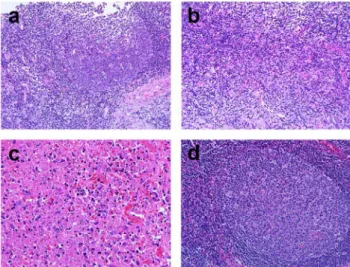
Figure 2. Representative photographs of an open biopsy
specimen (400x).a. Necrosis, usually with blurred cell shadow, was
mixed with karyorrhexis and histiocytes. b. Numerous histiocytes engulfed karyorrhectic debris. c. Foamy histiocytes were identified. d. Immunohistochemical stain for CD123 highlighted plasmacytoid dendritic cells. Hematoxylin counterstain revealed abundant karyor-rhexis around the plasmacytoid dendritic cells.
doi:10.1371/journal.pone.0095886.g002
Table 3.Pathological presentation in the specimens harvested from OB.
Histological features Case number Percentage
Karyorrhexis
Focal 12 46%
Extensive 14 54%
Necrosis
Absent 4 15%
Small foci 11 42%
Large geographic 11 42%
Histiocyte aggregation
Mild 17 65%
Marked 9 35%
Crescentic histiocyte 26 100%
Foamy histiocyte aggregation
Absent 15 58%
Mild 5 19%
Marked 6 23%
Clustered PDC
Mild 2 8%
Marked 24 92%
Presence of plasma cells 3 12%
Fibrovascular organization 2 8%
Pericapsular inflammation 8 31%
Lymphoid follicle
Hyperplastic 8 31%
Normal 6 23%
Atrophic 1 4%
Indiscernible 11 42%
PDC: plasmacytoid dendritic cells. doi:10.1371/journal.pone.0095886.t003
Figure 3. Variable pathological presentations of KFD were
shown within one lymph node.The left panel is the scanning view
of a bisected lymph node. (Scale bar: 1 mm). The high power view of areas b, c, and d were shown in the right panels (Scale bar: 200mm). Area b showed nonspecific reactive lymphoid hyperplasia. Area c showed necrosis with karyorrhexis. Area d showed. histiocytic infiltration with karyorrhexis. It demonstrated the possibility of sampling bias if distinct sites of the lesion were harvested.
lymph nodes, the morphological variation among lymph nodes was evident in 9 cases (82%) (Fig 4).
Pathological presentations in the specimens harvested from USCB
In addition to the specimens harvested from OB, the pathological findings of those from USCB were reviewed. The pathological presentation of individual USCB specimen was listed in table 4. Diagnosis was made by USCB alone in case 1 to 9. The diagnosis of case 10 and 11 was confirmed by subsequent OB. Necrosis and karyorrhexis were distinctive features of KFD in USCB specimens. Necrosis was mixed with karyorrhexis and histiocytes in 5 cases (45%) (Fig 5a). Karyorrhexis was present in 9 cases (82%), either in necrotic or viable area (Fig 5b). Crescentic histiocytes were noted in 9 cases (82%). Clustered plasmacytoid dendritic cells was present in 7 cases (64%) (Fig 5c). Histiocyte infiltration was found in 11 cases (100%) (Fig 5d). No foamy
Figure 4. Variable pathological presentations of KFD among
different lymph nodes obtained from a surgical biopsy.a. One
lymph node showed reactive follicular hyperplasia without evidence of KFD. b. The other lymph node showed specific features of KFD with paracortical necrosis and histiocytic infiltration. (Scale bar: 1 mm). doi:10.1371/journal.pone.0095886.g004
Figure 5. Representative photographs of a USCB specimen.a.
Necrosis was mixed with karyorrhexis and histiocytes (H&E, 400x). b. Conspicuous karyorrhexis accompanied by histiocyte aggregation (H&E, 400x). c. Clustered plasmacytoid dendritic cells highlighted by CD123 (200x). d. Increased histiocytes were observed (H&E, 400x).
histiocytes, multinucleate giant cells or granuloma formation was noted. Plasma cells were present in one case (9%).
Comparison of pathological findings of the specimens harvested from open biopsies and USCB
In our series, three patients had received both USCB and OB. One case was diagnosed as KFD by USCB, and another two cases were confirmed by OB. In the case diagnosed by USCB, the specimen showed specific features of KFD including evident necrosis, numerous karyorrhexis, and increased immunoblasts (Fig 6a, 6b). Samples taken from OB merely showed nonspecific reactive lymphoid hyperplasia and fibrovascular organization (Fig 6c). There was only one small focus with coagulative necrosis, karyorrhexis, accompanied by plasma cell infiltration (Fig 6d). This case indicated that diagnosis of KFD is sometimes easier to be made by USCB than OB. On the other two cases, the histopathological pictures of USCB specimens showed benign lymphoid tissue with a few lymphoid follicles (Fig 7a). Mild histiocyte infiltration was present, but no karyorrhectic debris was seen (Fig 7b). These pictures favored the diagnosis of reactive lymphoid hyperplasia. In the specimens harvested from OB, there was prominent geographic necrosis and karyorrhexis at paracortex accompanied by histiocytic infiltration. (Fig 7c, d).
Discussion
In this study, we demonstrated that KFD could be diagnosed either by USCB or OB. The sampling errors may happen both in USCB and OB. When the diagnosis is confirmed by USCB, further OB is unnecessary and the invasive surgical intervention can be avoided.
KFD is a self-limited disease which resolves spontaneously. Confirmation of diagnosis based on tissue sampling is critical. Incisional or excisional biopsies are usually regarded as diagnostic tools of KFD. However, OB is an invasive surgical intervention. It is time-consuming and leaves a visible scar on the neck. General anesthesia, wound care, and hospital stay may be required for OB.
For a self-limited disease like KFD, invasive diagnostic procedures are unnecessary if an alternative method is available. To this end, USCB and FNA are alternative choices. A few series of KFD using FNA concluded that FNA is not useful [15,16]. Most cases suspicious of KFD can be diagnosed by USCB in our series. USCB is an office-based procedure that has the characteristics of minimal invasiveness and preciseness. Most importantly, the pathological information including immunohistochemical staining can be provided by USCB. Using USCB, patient suffering, wound care, and medical cost can be reduced.
The major concern of USCB in diagnosing KFD is accuracy, which is largely dependent on the pathological features presented in the harvested specimens. KFD usually shows paracortical foci of necrosis with abundant karyorrhectic debris and numerous histiocytes [1,2]. Similar diagnostic features of KFD are also found in the specimens harvested by USCB. Necrosis, usually mixed with karyorrhexis and histiocytes, is observed. The morphology of histiocytes may vary, including crescentic histio-cytes and foamy histiohistio-cytes, but multinucleate giant cells or epithelioid histiocytes are not seen in KFD. Immunohistochemical stain for CD123 can highlight clustered plasmacytoid dendritic cells, which is beneficial to confirm the diagnosis of KFD in USCB specimens.
It is important to make the differential diagnosis of KFD. Therefore, the advantages and disadvantages of different sampling techniques should be verified based on pathological evidences. KFD may mimic malignant lymphoma by showing abundant immunoblasts. The pictures of extensive karyorrhexis of KFD which are found both in USCB and OB can be used for differentiation. The immunohistochemical results from either USCB or OB are also helpful to distinguish two disease entities. SLE lymphadenopathy has nearly identical histopathological features of KFD [17]. Furthermore, the histopathological features of several infectious diseases such as Yersinia enterolitica and typhoid lymphadenopathy may resemble those of KFD [18,19]. Bartonella henselae, Epstein Barr virus, cytomegalovirus,
Toxo-Figure 7. A case of KFD diagnosed by the specimens harvested
from OB rather than USCB. a, b. Specimens of USCB. Reactive
lymphoid follicles with tingible bodies (a, 200x). Mildly increased histiocytes were noted between follicles, but no karyorrhexis was seen (b, 200x). c, d. Specimens of OB. Distinctive histology with necrosis and karyorrhexis was prominent (c, 400x). Reactive follicular hyperplasia was observed in the viable parts (d, 200x).
doi:10.1371/journal.pone.0095886.g007
Figure 6. A case of KFD diagnosed by the specimens harvested
from USCB instead of OB. a, b. Specimens of USCB. A focus of
necrosis mixed with karyorrhexis, histiocytes and immunoblasts (H&E. a, 100x b, 400x). c, d. Specimens of OB. Nonspecific reactive lymphoid hyperplasia with occasional fibrovascular organization (c, 40x). There was only one small focus of aggregated histiocytes accompanied by karyorrhexis (d, x400).
plasma gondii, and syphilis are also associated with necrotizing lymphadenitis [4]. However, their clinical presentations are different from KFD. Tuberculosis is one of the infectious diseases that should be carefully distinguished form KFD because the clinical presentations and radiological findings are similar in both diseases [20–23]. Tuberculosis mimics KFD when the specimen is composed of abundant caseous necrosis with few or absent epithelioid cells. In contrast to tuberculosis, KFD has no epithelioid histiocytes, granuloma or multinucleate giant cells. An acid fast stain is suggested for the cases suspicious of tuberculosis. In our study, none of the 9 KFD patients diagnosed by USCB had evidence of lymphoma, SLE or specific infectious diseases. All of them had a benign clinical course during follow-up. Because KFD exhibits marked variation within a lymph node, or among different lymph nodes, sampling error is inevitable. Three cases of our series demonstrated the possibility of sampling bias, no matter in USCB or OB. In some cases, the typical morphology of KFD is restricted locally, which may not be easily sampled by USCB, leading to inadequate sampling and inaccurate diagnosis (Fig 3). Similar effect is also observed even with OB. Because KFD may exhibit different features among lymph nodes, only few lymph nodes taken form OB may also result in a diagnostic bias (Fig 4). Certainly, more lymph nodes taken for evaluation indeed significantly increase the diagnostic rate of KFD. However, the invasiveness might correspondingly increase, which is not favored for a self-limited, benign disease like KFD. With meticulous histopathological examinations by experienced physicians and pathologists, many KFD cases can be successfully diagnosed by USCB. Therefore, USCB may serve as an alternative sampling method of OB for KFD diagnosis. For routine practices, USCB can provide specimens for pathological examinations and has the advantages of minimal invasiveness, convenience, and low cost. Therefore, USCB can serve as the
first-line diagnostic tool of KFD. Multiple shots of USCB are suggested to increase the diagnostic accuracy because of the heterogeneous pathological nature of KFD. An OB can be a backup sampling tool when USCB is futile.
For suspicious KFD patients, USCB can serve as the first-line diagnostic tool. USCB is a sampling procedure with the characteristics of minimal invasiveness and preciseness, and can provide pathological information for diagnosis. Using USCB, the possibility of scar formation, wound care, and medical cost can be reduced.
Supporting Information
Checklist S1 A STARD checklist for reporting of diag-nostic accuracy in this study.
(DOC)
Flow Chart S1 A STARD flow chart shows the process of recruiting patients with suspicious KFD for diagnostic procedures.
(PDF)
Acknowledgments
The authors thank National Science Council of the Republic of China and National Taiwan University Hospital for their support. The authors also thank the staff of the Eighth Core Lab, Department of Medical Research, National Taiwan University Hospital for technical support.
Author Contributions
Conceived and designed the experiments: SCY TLY. Performed the experiments: SCY CNC HIH TLY. Analyzed the data: SCY CNC HIH TLY. Contributed reagents/materials/analysis tools: CNC TCC CPW PJL JYK TYH TLY. Wrote the paper: SCY CNC TLY.
References
1. Kuo TT (1995) Kikuchi’s disease (histiocytic necrotizing lymphadenitis). A clinicopathologic study of 79 cases with an analysis of histologic subtypes, immunohistology, and DNA ploidy. Am J Surg Pathol 19:798–809. 2. Tsang WY, Chan JK, Ng CS (1994) Kikuchi’s lymphadenitis. A morphologic
analysis of 75 cases with special reference to unusual features. Am J Surg Pathol 18:219–231.
3. Turner RR, Martin J, Dorfman RF (1983) Necrotizing lymphadenitis. A study of 30 cases. Am J Surg Pathol 7:115–123.
4. Hutchinson CB, Wang E (2010) Kikuchi-Fujimoto disease. Arch Pathol Lab Med 134:289–293.
5. Onciu M, Medeiros LJ (2003) Kikuchi-Fujimoto lymphadenitis. Adv Anat Pathol 10:204–211.
6. Kucukardali Y, Solmazgul E, Kunter E, Oncul O, Yildirim S, et al. (2007) Kikuchi-Fujimoto Disease: analysis of 244 cases. Clin Rheumatol 26:50–54. 7. Pfeiffer J, Kayser G, Ridder GJ (2009) Sonography-assisted cutting needle biopsy
in the head and neck for the diagnosis of lymphoma: can it replace lymph node extirpation? Laryngoscope 119:689–695.
8. Hehn ST, Grogan TM, Miller TP (2004) Utility of fine-needle aspiration as a diagnostic technique in lymphoma. J Clin Oncol 22:3046–3052.
9. Kim BM, Kim EK, Kim MJ, Yang WI, Park CS, et al. (2007) Sonographically guided core needle biopsy of cervical lymphadenopathy in patients without known malignancy. J Ultrasound Med 26:585–591.
10. Screaton NJ, Berman LH, Grant JW (2002) Head and neck lymphadenopathy: evaluation with US-guided cutting-needle biopsy. Radiology 224:75–81. 11. Chen CN, Hsiao TY, Ko JY, Lou PJ, Wang CP, et al. (2014) Ultrasound-guided
core biopsy for hypopharyngeal cancer with difficult endoscopic approaches: Our experience in eleven patients. Clin Otolaryngol DOI: 10.1111/coa.12196. 12. Novoa E, Gurtler N, Arnoux A, Kraft M (2012) Role of ultrasound-guided core-needle biopsy in the assessment of head and neck lesions: a meta-analysis and systematic review of the literature. Head Neck 34:1497–1503.
13. Carbone A, Ferlito A, Devaney KO, Rinaldo A (2008) Ultrasound-guided core-needle biopsy: is it effective in the diagnosis of suspected lymphomas presenting in the head and neck? J Surg Oncol 98:4–5.
14. Chen CN, Yang TL (2012) Ultrasound-guided core needle biopsy of advanced hypopharyngeal cancer. J Med Ultrasound 20:240–243.
15. Viguer JM, Jimenez-Heffernan JA, Perez P, Lopez-Ferrer P, Gonzalez-Peramato P, et al. (2001) Fine-needle aspiration cytology of Kikuchi’s lymphadenitis: a report of ten cases. Diagn Cytopathol 25:220–224. 16. Hsueh EJ, Ko WS, Hwang WS, Yam LT (1993) Fine-needle aspiration of
histiocytic necrotizing lymphadenitis (Kikuchi’s disease). Diagn Cytopathol 9:448–452.
17. Hu S, Kuo TT, Hong HS (2003) Lupus lymphadenitis simulating Kikuchi’s lymphadenitis in patients with systemic lupus erythematosus: a clinicopatholog-ical analysis of six cases and review of the literature. Pathol Int 53:221–226. 18. Feller AC, Lennert K, Stein H, Bruhn HD, Wuthe HH (1983) Immunohistology
and aetiology of histiocytic necrotizing lymphadenitis. Report of three instructive cases. Histopathology 7:825–839.
19. Gatter KC, Delso G, Warnke RA, Pezella F (2012) The diagnosis of lymphoproliferative diseases, 2nd ed. Oxford, UK: Wiley-Blackwell. 20. Song JY, Cheong HJ, Kee SY, Lee J, Sohn JW, et al. (2007) Disease spectrum of
cervical lymphadenitis: analysis based on ultrasound-guided core-needle gun biopsy. J Infect 55:310–316.
21. Rammohan A, Cherukuri SD, Manimaran AB, Manohar RR, Naidu RM (2012) Kikuchi-Fujimoto disease: a sheep in wolf’s clothing. J Otolaryngol Head Neck Surg 41:222–226.
22. Lee S, Yoo JH, Lee SW (2012) Kikuchi disease: differentiation from tuberculous lymphadenitis based on patterns of nodal necrosis on CT. AJNR Am J Neuroradiol 33:135–140.