259
Chojniak R et al. CT-guided biopsy of soft tissue tumors
Radiol Bras. 2012 Set/Out;45(5):259–262
Percutaneous computed tomography-guided core needle
biopsy of soft tissue tumors: results and correlation with
surgical specimen analysis
*
Biópsia percutânea por agulha grossa de tumores de partes moles guiada por tomografia computadorizada: resultados e correlação com análise da peça cirúrgica
Rubens Chojniak1, Henrique Ramos Grigio2, Almir Galvão Vieira Bitencourt3, Paula Nicole Vieira Pinto3, Chiang J. Tyng3, Isabela Werneck da Cunha4, Samuel Aguiar Junior5, Ademar Lopes6
Objective: To evaluate the efficacy of percutaneous computed tomography (CT)-guided core needle biopsy of soft-tissue tumors in obtaining appropriate samples for histological analysis, and compare its diagnosis with the results of the surgical pathology as available. Materials and Methods: The authors reviewed medical records, imaging and histological reports of 262 patients with soft-tissue tumors submitted to CT-guided core needle biopsy in an oncologic reference center between 2003 and 2009. Results: Appropriate samples were obtained in 215 (82.1%) out of the 262 patients. The most prevalent tumors were sarcomas (38.6%), metastatic carcinomas (28.8%), benign mesenchymal tumors (20.5%) and lymphomas (9.3%). Histological grading was feasible in 92.8% of sarcoma patients, with the majority of them (77.9%) being classified as high grade tumors. Out of the total sample, 116 patients (44.3%) underwent surgical excision and diagnosis confirmation. Core biopsy demonstrated 94.6% accuracy in the identification of sarcomas, with 96.4% sensitivity and 89.5% specificity. A significant intermethod agreement about histological grading was observed between core biopsy and surgical resection (p < 0.001; kappa = 0.75). Conclusion: CT-guided core needle biopsy demonstrated a high diagnostic accuracy in the evaluation of soft tissue tumors as well as in the histological grading of sarcomas, allowing an appropriate therapeutic planning.
Keywords: Sarcoma; Needle biopsy; Computed tomography.
Objetivo: Avaliar a eficácia da biópsia percutânea por agulha grossa (BPAG) de tumores de partes moles guiada por tomografia computadorizada (TC), em relação ao sucesso na obtenção de amostra para análise, e comparar o diag-nóstico da BPAG com o resultado anatomopatológico da peça cirúrgica, quando disponível. Materiais e Métodos:
Foram revisados os prontuários e laudos diagnósticos de 262 pacientes com tumores de partes moles submetidos a BPAG guiada por TC em um centro de referência oncológico entre 2003 e 2009. Resultados: Das 262 biópsias rea-lizadas, foi possível a obtenção de amostra adequada em 215 (82,1%). Os tumores mais prevalentes foram os sarco-mas (38,6%), carcinosarco-mas metastáticos (28,8%), tumores mesenquimais benignos (20,5%) e linfosarco-mas (9,3%). Foi possível realizar graduação histológica em 92,8% dos pacientes com sarcoma, sendo a maioria (77,9%) classificada como alto grau. Do total de pacientes, 116 (44,3%) realizaram cirurgia para exérese e confirmação diagnóstica. A BPAG mostrou acurácia de 94,6% na identificação de sarcomas, com sensibilidade de 96,4% e especificidade de 89,5%. A graduação histológica teve concordância significativa entre a BPAG e a peça cirúrgica (p < 0,001; kappa = 0,75).
Conclusão: A BPAG guiada por TC demonstrou elevada acurácia diagnóstica na avaliação de tumores de partes moles e na graduação histológica dos sarcomas, permitindo um adequado planejamento terapêutico.
Unitermos: Sarcoma; Biópsia por agulha; Tomografia computadorizada.
Abstract
Resumo
* Study developed at Hospital A. C. Camargo, São Paulo, SP, Brazil.
1. PhD, Titular and Director, Department of Imaging, Hospital A. C. Camargo, Professor at Faculdade de Medicina da Univer-sidade Nove de Julho, São Paulo, SP, Brazil.
2. Graduate Student of Medicine, Faculdade de Medicina da Universidade Nove de Julho, Scholar, Programa Institucional de Bolsas de Iniciação Científica (PIBIC), Hospital A. C. Camargo, São Paulo, SP, Brazil.
3. Fellow PhD degree of Oncology, Titular, Department of Im-aging, Hospital A. C. Camargo, São Paulo, SP, Brazil.
Chojniak R, Grigio HR, Bitencourt AGV, Pinto PNV, Tyng CJ, Cunha IW, Aguiar Junior S, Lopes A. Percutaneous computed tomography-guided core needle biopsy of soft tissue tumors: results and correlation with surgical specimen analysis. Radiol Bras. 2012 Set/ Out;45(5):259–262.
0100-3984 © Colégio Brasileiro de Radiologia e Diagnóstico por Imagem ORIGINAL ARTICLE
INTRODUCTION
Soft tissue tumors comprise a great number of neoplasms, among them, mes-enchymal tumors (the benign ones and sar-comas) and other tumors such as lympho-mas, melanomas and metastatic carcino-mas(1). Malignant mesenchymal tumors, 4. PhD, Titular, Department of Anatomic Pathology, Hospital
A. C. Camargo, São Paulo, SP, Brazil.
5. PhD, Titular, Department of Pelvic Surgery, Hospital A. C. Camargo, São Paulo, SP, Brazil.
6. PhD, Titular and Director, Department of Pelvic Surgery, Hospital A. C. Camargo, São Paulo, SP, Brazil.
Mailing Address: Dr. Rubens Chojniak. Hospital A. C. Camargo – Departamento de Imagem. Rua Professor Antônio Prudente, 211, Liberdade. São Paulo, SP, Brazil, 01509-010. E-mail: [email protected]
260
Chojniak R et al. CT-guided biopsy of soft tissue tumors
Radiol Bras. 2012 Set/Out;45(5):259–262
called sarcomas, are characterized for be-ing solid, with considerable heterogeneity in their anatomy, histological subtypes and degree of biological aggressiveness. The approach to be adopted is significantly in-fluenced by the location and staging of the tumor(2).
Anatomopathological analysis allows the identification of tumor type, so it is essential for the therapeutic planning. The preoperative determination of grading and histological type of the sarcomas is critical to identify those patients at higher risk for metastasis, influencing the decision mak-ing on the the need for neoadjuvant treat-ment and surgery extent(3,4). The current
parameters most utilized by pathologists for grading soft tissue sarcomas are the following: cellular differentiation, cellular-ity, amount of necrosis and number of mi-toses(5).
There are several techniques for obtain-ing soft tissue tumors specimens for histo-logical analysis. Open surgical biopsy al-lows direct access to the tumor and gener-ally the collection of larger amounts of material than in percutaneous biopsies, which tends to favor a correct diagnosis, increasing the capability of differentiating benign from malignant tissues. However, its main disadvantages are the high cost and morbidity, as it is the case in any open sur-gical procedure(6). For such reasons,
percu-taneous core needle biopsy (CNB) is many
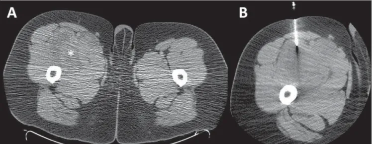
times utilized as a diagnostic method pro-viding tissue fragments for histological analysis, with a low rate of complications. Percutaneous biopsies are usually guided by imaging methods such as ultrasonogra-phy (US) or computed tomograultrasonogra-phy (CT), particularly in cases of deeply located tu-mors(7,8) (Figure 1). However, percutaneous
biopsy results may underestimated tumor aggressiveness estimate, as only a small part of the tumor is analyzed(9).
The main objectives of the present study were to evaluate the effectiveness of CT-guided CNB of soft tissue tumors with re-spect to success in obtaining specimens for analysis, and comparing the diagnosis ob-tained by means of CNB with the anatomo-pathological result of the surgical speci-men, when available.
MATERIALS AND METHODS
The present retrospective study re-viewed records and diagnostic reports from patients with soft tissue tumors submitted to CT-guided CNB in a reference oncology center, in the period from April 22, 2003 to June 30, 2009. The present study was ap-proved by the Committee for Ethics in Research of the institution before the com-mencement of data collection.
A standardized data form was retrospec-tively filled in for each patient included in the study. The collected information
com-prised epidemiological data, clinical data, imaging findings, CNB results, informa-tion on the surgery and respective anato-mopathological report. For histological grading of the sarcomas, the World Health Organization (WHO) criteria based on a two-level reference classification (“low grade” versus “high grade”) were utilized.
Whenever the utilized grading system was different, the conversion of such grading into the two-level system was performed, as recommended(10).
All the data were stored in a databank for statistical analysis by means of the SPSS software for Windows, version 17.0 (SPSS Inc.; Chicago, IL, USA). The de-scriptive analysis comprised the calculation of simple and relative frequencies of the studied variables. Sensitivity, specificity, predictive values and CNB accuracy were calculated for the differentiation between benign and malignant mesenchymal tu-mors. For the analysis of agreement be-tween data regarding CNB results and sur-gical specimens analysis, the kappa test was utilized, with p-value ranging between
0 and 1, indicating higher agreement as closer to 1.
RESULTS
The present study included 262 patients, with a mean age of 51 years (ranging from 1 to 93 years), 132 (50.4%) men. Table 1
261
Chojniak R et al. CT-guided biopsy of soft tissue tumors
Radiol Bras. 2012 Set/Out;45(5):259–262
presents the age distribution of patients submitted to CNB. Most of the tumors were located in the trunk (n = 127; 48.5%),
followed by extremities (n = 78; 29.8%),
retroperitoneum (n = 55; 21.0%) and head
& neck (n = 2; 0.8%).
Out of the 262 biopsies, appropriate specimens could be obtained in 215 cases (82,1%). Out of the 47 (18%) cases where the specimens were considered as being inappropriate, the histological result was absence of tumor in 33 cases (70%) fol-lowed by extensive necrosis in 8 cases (17%); collection artifacts in 3 cases (6%); inflammatory process in 2 cases (4%) and cellular debris intermingled with hemosid-erin and red blood cells in one case (2%). The diagnoses of 23 of such patients was later obtained by tumor excision in 19 (83%) cases, open biopsy in 3 (13%) cases and additional CNB in 1 (4%) case. For the remaining 24 cases, no follow-up data was available.
Table 2 presents the quantitative and percentage distribution of the histological results obtained by CNB.
Among the 44 benign mesenchymal neoplasms, it was possible to define the histological subtype by means of CNB in 34 cases (77.3%), with lipoma being the
most frequent type (n = 17; 50%). As
re-gards sarcomas, the histological sub-type was defined in 74 of the 83 cases (89.2%), with such cases being described on Table 3. Histological grading could be performed in 77 (92.8%) of the 83 patients with sar-comas, 60 (77.9%) of them being classified as high grade and 17 (22.1%) being classi-fied as low grade.
Out of the 215 patients with tumors di-agnosed by CNB, 116 (44.3%) underwent surgery for tumor excision and diagnosis confirmation. Among these cases, 74 had been classified as mesenchymal tumors at CNB – 19 benign and 55 malignant. Among the 19 tumors classified as benign at CNB, 17 were confirmed by the surgi-cal specimens analysis and two results were discordant – one liposarcoma in a lesion classified as lipoma at CNB, and one syn-ovial sarcoma in a lesion previously diag-nosed as leiomyoma. Out of the 55 tumors classified as malignant by CNB, 2 (3.6%) presented discordant results in the surgical specimen analysis – one seminoma and one synovial cyst in two lesions classified as undifferentiated sarcomas at CNB.
Thus, CNB demonstrated 94.6% accu-racy in the identification of sarcomas among the mesenchymal tumors, with sen-sitivity and positive predictive value of 96.4% and specificity and negative predic-tive value of 89.5%.
Out of the 77 patients with sarcomas whose histological grade could be deter-mined by CNB, 49 were submitted to sur-gery, and only 4 (8.2%) of them presented difference in grading between CNB and surgical specimen analysis. All of such cases were classified as low grade at CNB and reclassified as high grade at the surgi-cal specimen analysis. There was a signifi-cant agreement between CNB and surgical specimen analysis in the determination of the histological grade (high and low grade) (p < 0.001; kappa = 0.75; CI 95%: 0.48–
1.00).
DISCUSSION
Soft tissue sarcomas are rarely found and the clinical/radiological differentiation between benign tumors and sarcomas is sometimes impossible. Furthermore, sarco-mas may not be preoperatively suspected, with the diagnosis being confirmed only after the lesion resection. Enucleation (excisional biopsy) is acceptable in cases of benign lesions, but it is not appropriate for sarcomas, as the surgeon misses the opportunity to perform a more effective treatment, with better local management of the disease. Also, incisional biopsy is not routinely indicated for sarcomas because of the high rate of wound complications, which may compromise the local
treat-Table 1 Age distribution of 262 patients with soft tissue tumors submitted to computed tomography-guided core needle biopsy.
Age (years)
0–20
21–40
41–60
61–80
> 80
n
24
45
103
70
20
%
9.2%
17.2%
39.3%
26.7%
7.6%
Table 2 Histological diagnoses obtained with percutaneous computed tomography-guided core needle biopsy in 215 patients with soft tissue tu-mors.
Histological diagnosis
Mesenchymal tumors
Malignant
Benign
Benign/malignant
Metastatic carcinomas
Lymphomas
Melanoma
n
130
83
44
3
62
20
3
%
60.5%
38.6%
20.5%
1.4%
28.8%
9.3%
1.4%
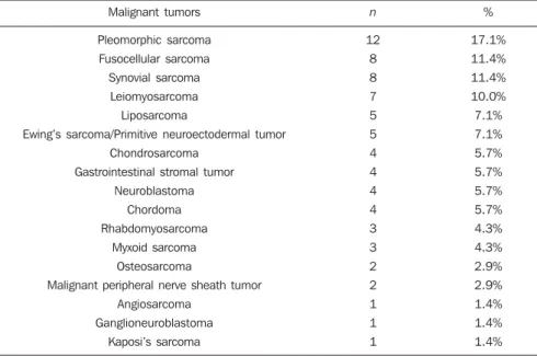
Table 3 Distribution of histological sub-types of sarcomas diagnosed by computed tomography-guided core needle biopsy in 83 patients.
Malignant tumors
Pleomorphic sarcoma Fusocellular sarcoma Synovial sarcoma
Leiomyosarcoma
Liposarcoma
Ewing’s sarcoma/Primitive neuroectodermal tumor Chondrosarcoma
Gastrointestinal stromal tumor
Neuroblastoma Chordoma Rhabdomyosarcoma
Myxoid sarcoma
Osteosarcoma
Malignant peripheral nerve sheath tumor Angiosarcoma
Ganglioneuroblastoma
Kaposi’s sarcoma
n
12 8 8 7
5 5 4 4
4 4 3 3
2 2 1 1
1
%
17.1% 11.4% 11.4% 10.0%
7.1% 7.1% 5.7% 5.7%
5.7% 5.7% 4.3% 4.3%
2.9% 2.9% 1.4% 1.4%
262
Chojniak R et al. CT-guided biopsy of soft tissue tumors
Radiol Bras. 2012 Set/Out;45(5):259–262
ment(11). For this reason, CNB is the
method of choice for preoperative investi-gation in patients with suspected soft tis-sue tumors(12). Fine needle aspiration
bi-opsy is not recommended as a first-line diagnostic modality, however it can be uti-lized in selected cases, as in the suspicion of recurrence(12).
Imaging-guided CNB provides addi-tional advantage for being capable of evaluating, at the collection moment, the best site for fragment collection, avoiding, for example, areas of necrosis and/or fibro-sis. Narvani et al.have demonstrated that imaging-guided procedures improve the accuracy of CNB in 78% to 95%, specially in cases of small and deeply located le-sions(13). Such procedure must be
per-formed in a reference center, with special-ized radiologists and pathologists, and the case must always be previously discussed with the surgeon for a better plan-ning(12,14,15). During the procedure, the
bi-opsy pathway must be planned in such a way that it can be resected at the moment of the definitive surgery, and several frag-ments from different areas should be col-lected for appropriate sampling of the tu-mor. According to Wu et al., at least four fragments must be collected in soft tissue tumors CNB in order to allow an appropri-ate histological diagnosis(16).
CT and US have been utilized to guide percutaneous biopsy of soft tissue tumors, with good results being reported in the lit-erature(17–19). No study was found
suggest-ing the superiority of either of the methods. One should select the method in which the lesion is best characterized and the one in which the radiologist is more experiented. In general, US is preferred for superficial lesions, while CT is utilized for deeper le-sions.
In the present study, CNB provided ap-propriate specimens for analysis in 82% of the cases, allowing the differentiation be-tween benign and malignant lesions, iden-tification of the histological type of the tumors, and the histological grading in most of the cases. Such results confirm the accuracy of the method reported in litera-ture, demonstrating that the sub-type and
grading of the tumors can be determined in 80% to 95% of the CNBs(20). In the present
study, sarcomas were the most prevalent soft tissue tumors, followed by metastatic carcinomas, benign mesenchymal tumors and lymphomas.
In the present casuistry, the diagnostic accuracy of CNB in the identification of sarcomas was high, with significant agree-ment in relation to the histological grading of the tumor by biopsy and by surgical specimen analysis. Such results were simi-lar to those from other studies published in the literature. Ray-Coquard et al.have dem-onstrated 95% accuracy of CNB in the di-agnosis of soft tissue sarcomas(21). Strauss
et al.have evaluated 530 patients with soft tissue tumors submitted to CNB, reporting 426 mesenchymal tumors (225 malignant and 201 benign), and found 97.6% accu-racy in the differentiation between malig-nancy and benignity, and 86.3% accuracy in the differentiation between high and low histological grade tumors(22).
Woon et al. have evaluated 94 patients submitted to surgery for soft tissue tumors and demonstrated that 95% of the patients preoperatively submitted to preoperative CNB had definitive one-stage surgery, as compared with 45% of those patients who were not submitted to biopsy(20).
CONCLUSION
CT-guided CNB demonstrated to be an effective diagnostic method in the evalua-tion of soft tissue tumors and in the histo-logical grading of sarcomas, allowing for appropriate therapeutic planning.
REFERENCES
1. Weiss S, Goldblum J. Enzinger and Weiss’s soft tissue tumors. 4th ed. St Louis, MO: Mosby; 2001. 2. Clark MA, Fisher C, Judson I, et al. Soft-tissue sarcomas in adults. N Engl J Med. 2005;353:701– 11.
3. Mankin HJ, Hornicek FJ. Diagnosis, classifica-tion, and management of soft tissue sarcomas. Cancer Control. 2005;12:5–21.
4. Lopes A, Ferreira FO, Aguiar Junior S, et al. Sar-comas de partes moles no adulto. In: Kowalski LP, Guimarães GC, Salvajoli JV, et al., organizado-res. Manual de condutas diagnósticas e terapêu-ticas em oncologia. 3ª ed. São Paulo, SP: Âmbito Editores; 2006. p. 681–90.
5. Nascimento A, Oliveira A. Patologia geral. In: Lopes A, organizador. Sarcomas de partes moles. Rio de Janeiro, RJ: Medsi; 1999. p. 41–68. 6. Cormier JN, Pollock RE. Soft tissue sarcomas. CA
Cancer J Clin. 2004;54:94–109.
7. Chojniak R, Isberner RK, Viana LM, et al. Com-puted tomography guided needle biopsy: experi-ence from1,300 procedures. Sao Paulo Med J. 2006;124:10–4.
8. Heslin MJ, Lewis JJ, Woodruff JM, et al. Core needle biopsy for diagnosis of extremity soft tis-sue sarcoma. Ann Surg Oncol. 1997;4:425–31. 9. Hoeber I, Spillane A, Fisher C, et al. Accuracy of
biopsy techniques for limb and limb girdle soft tissue tumors. Ann Surg Oncol. 2001;8:80–7. 10. Sobin L, Wittekind C. Tumores ósseos e de
par-tes moles. In: TNM Classificação de tumores malignos. 6ª ed. Rio de Janeiro, RJ: INCA; 2004. p. 115–24.
11. Serpell JW, Pitcher ME. Pre-operative core biopsy of soft-tissue tumours facilitates their surgical management. Aust N Z J Surg. 1998;68:345–9. 12. Grimer R, Judson I, Peake D, et al. Guidelines for the management of soft tissue sarcomas. Sar-coma. 2010;2010:506182.
13. Narvani AA, Tsiridis E, Saifuddin A, et al. Does image guidance improve accuracy of core needle biopsy in diagnosis of soft tissue tumours? Acta Orthop Belg. 2009;75:239–44.
14. Thway K, Fisher C. Histopathological diagnos-tic discrepancies in soft tissue tumours referred to a specialist centre. Sarcoma. 2009;2009:741975. 15. Rydholm A. Improving the management of soft tissue sarcoma. Diagnosis and treatment should be given in specialist centres. BMJ. 1998;317: 93–4.
16. Wu JS, Goldsmith JD, Horwich PJ, et al. Bone and soft-tissue lesions: what factors affect diag-nostic yield of image-guided core-needle biopsy? Radiology. 2008;248:962–70.
17. Zornoza J, Bernardino ME, Ordonez NG, et al. Percutaneous needle biopsy of soft tissue tumors guided by ultrasound and computed tomography. Skeletal Radiol. 1982;9:33–6.
18. Soudack M, Nachtigal A, Vladovski E, et al. Sonographically guided percutaneous needle bi-opsy of soft tissue masses with histopathologic correlation. J Ultrasound Med. 2006;25:1271–7. 19. Issakov J, Flusser G, Kollender Y, et al. Computed tomography-guided core needle biopsy for bone and soft tissue tumors. Isr Med Assoc J. 2003;5: 28–30.
20. Woon DT, Serpell JW. Preoperative core biopsy of soft tissue tumours facilitates their surgical management: a 10-year update. ANZ J Surg. 2008;78:977–81.
21. Ray-Coquard I, Ranchère-Vince D, Thiesse P, et al. Evaluation of core needle biopsy as a substi-tute to open biopsy in the diagnosis of soft-tissue masses. Eur J Cancer. 2003;39:2021–5. 22. Strauss DC, Qureshi YA, Hayes AJ, et al. The role