Polymorphism and Mucosal Expression of Dectin-1 in
Inflammatory Bowel Disease
Hilbert S. de Vries1, Theo S. Plantinga2,3, J. Han van Krieken4, Rinke Stienstra2,3, Ad A. van Bodegraven5, Eleonora A. M. Festen6, Rinse K. Weersma6, J. Bart A. Crusius7, Ronald K. Linskens8, Leo A. B. Joosten2,3, Mihai G. Netea2,3, Dirk J. de Jong1*
1Department of Gastroenterology and Hepatology, Radboud University Nijmegen Medical Centre, Nijmegen, The Netherlands,2Department of Medicine, Radboud University Nijmegen Medical Centre, Nijmegen, The Netherlands,3Nijmegen Institute for Infection, Inflammation and Immunity (N4i), Nijmegen, The Netherlands, 4Department of Pathology, Radboud University Nijmegen Medical Centre, Nijmegen, The Netherlands,5Department of Gastroenterology and Hepatology, VU University Medical Centre, Amsterdam, The Netherlands,6Department of Gastroenterology and Hepatology, University Medical Centre Groningen, Groningen, The Netherlands, 7Department of Pathology, Laboratory of Immunogenetics, VU University Medical Centre, Amsterdam, The Netherlands,8Department of Gastroenterology, Saint Anna Hospital, Geldrop, The Netherlands
Abstract
Background:Dectin-1 is a pattern recognition receptor (PRR) expressed by myeloid cells that specifically recognizesb-1,3 glucan, a polysaccharide and major component of the fungal cell wall. Upon activation, dectin-1 signaling converges, similar to NOD2, on the adaptor molecule CARD9 which is associated with inflammatory bowel disease (IBD). An early stop codon polymorphism (c.714T.G) inDECTIN-1results in a loss-of-function (p.Y238X) and impaired cytokine responses, including TNF-a, interleukin (IL)-1band IL-17 uponin vitrostimulation withCandida albicansorb-glucan. The aim of the present study was to test the hypothesis that the DECTIN-1 c.714T.G (p.Y238X) polymorphism is associated with lower disease susceptibility or severity in IBD and to investigate the level of dectin-1 expression in inflamed and non-inflamed colon tissue of IBD patients.
Methodology: Paraffin embedded tissue samples from non-inflamed and inflamed colon of IBD patients and from diverticulitis patients were immunohistochemically stained for dectin-1 and related to CD68 macrophage staining. Genomic DNA of IBD patients (778 patients with Crohn’s disease and 759 patients with ulcerative colitis) and healthy controls (n = 772) was genotyped for the c.714T.G polymorphism and genotype-phenotype interactions were investigated.
Principal Findings:Increased expression of dectin-1 was observed in actively inflamed colon tissue, as compared to non-inflamed tissue of the same patients. Also an increase in dectin-1 expression was apparent in diverticulitis tissue. No statistically significant difference inDECTIN-1c.714T.G allele frequencies was observed between IBD patients and healthy controls. Furthermore, no differences in clinical characteristics could be observed related toDECTIN-1genotype, neither alone, nor stratified forNOD2genotype.
Conclusions:Our data demonstrate that dectin-1 expression is elevated on macrophages, neutrophils, and other immune cells involved in the inflammatory reaction in IBD. The DECTIN-1 c.714T.G polymorphism however, is not a major
susceptibility factor for developing IBD.
Citation:de Vries HS, Plantinga TS, van Krieken JH, Stienstra R, van Bodegraven AA, et al. (2009) Genetic Association Analysis of the Functional c.714T.G Polymorphism and Mucosal Expression of Dectin-1 in Inflammatory Bowel Disease. PLoS ONE 4(11): e7818. doi:10.1371/journal.pone.0007818
Editor:Stefan Bereswill, Charite´-Universita¨tsmedizin Berlin, Germany
ReceivedJune 9, 2009;AcceptedOctober 19, 2009;PublishedNovember 12, 2009
Copyright:ß2009 de Vries et al. This is an open-access article distributed under the terms of the Creative Commons Attribution License, which permits unrestricted use, distribution, and reproduction in any medium, provided the original author and source are credited.
Funding:M.G.N. was supported by a Vidi grant of the Netherlands Organization for Scientific Research. T.S.P. was supported by TI-Pharma Institute. The funders had no role in study design, data collection and analysis, decision to publish, or preparation of the manuscript.
Competing Interests:The authors have declared that no competing interests exist. * E-mail: D.deJong@mdl.umcn.nl
Introduction
Inflammatory bowel disease (IBD), is an idiopathic, chronic, relapsing inflammatory disorder of the gastrointestinal tract. It is commonly accepted that IBD is caused by an exaggerated cell mediated immune response to intestinal microbiota in genetically susceptible individuals[1,2]. IBD mainly involves two distinct diseases, which show some overlap: Crohn’s disease (CD) and ulcerative colitis (UC). Genetic susceptibility is more pronounced
in CD compared to UC[3]. Several susceptibility loci for developing CD have been identified in the past decades including the NOD2 gene within the IBD1 locus[4]. The established association ofNOD2 (CARD15)with CD emphasizes the important role of the intestinal microbiota in the pathogenesis of CD, since NOD2 acts as an intracellular pattern recognition receptor (PRR) recognizing bacterial peptidoglycans[5,6].
polysaccharide and component of the fungal cell wall. As a result, dectin-1 is involved in recognition of fungi such asCandida albicans and Aspergillus fumigatus. Upon activation, dectin-1 recruits spleen tyrosine kinase (Syk) which in turn activates NF-kB, requiring the adaptor molecule Caspase Activating Recruitment Domain 9 (CARD9), a key adaptor for non-Toll Like Receptor (TLR) signal transduction[7]. Although not the exclusive pathway, CARD9 also has a critical function in NOD2-mediated activation of the kinases p38 and Jnk, required for the production of pro-inflammatory cytokines in innate immune responses to intracellular patho-gens[8]. LeibundGut-Landmann et al. showed that dectin-1-Syk-CARD9 signaling induces dendritic cell (DC) maturation and secretion of pro-inflammatory cytokines like interleukin (IL)-6, TNF-a, IL-17 and IL-23[9]. Furthermore, Zhernakova et al. identifiedCARD9as a susceptibility locus for IBD[10]. Recently, theDECTIN-1polymorphism c.714T.G on chromosome 12p13 has been described, with a transition from a tyrosine to an early stop codon on amino acid position 238 (p.Y238X)[11]. The functional consequence of this polymorphism is a complete loss-of-function, and immune cells expressing this truncated protein produce significantly less cytokines, including TNF-a, IL-1band IL-17, upon in vitro stimulation with b-glucan or Candida albicans[12].
Th17 responses are considered to be involved in the pathogenesis of auto-immune diseases. This T cell subset appears to play a role in the etiology of CD since IL-17 is up-regulated in the intestine of IBD patients[13]. Interestingly, both NOD2 and dectin-1 are shown to be capable of inducing Th17 responses after activation[9,14]. In this respect, the DECTIN-1 c.714T.G polymorphism could influence the Th17 response towards fungi such as Candida albicans, a commensal microorganism of the gastrointestinal tract.Candida albicans isalso one of the immuno-gens for developing antibodies against Saccharomyces cerevisiae (ASCA), which are regularly observed in patients with CD[15,16]. Taking together the data from various studies, a dysregulation of the immune response to the commensalCandida albicansthrough dectin-1 and IL-17 release might play a role in the pathogenesis of CD.
As stated above, activation of NOD2 and dectin-1 leads to signaling through a shared pathway (CARD9). The importance of this pathway in CD is demonstrated by the fact that mutations in NOD2 and CARD9 and the presence of circulating ASCA, are associated with CD. Since the c.714T.G polymorphism within DECTIN-1results in a loss of function, we hypothesized that this polymorphism could be potentially protective against developing IBD. Therefore, we aimed to elucidate the role of theDECTIN-1 c.714T.G (p.Tyr238X) polymorphism in patients with IBD, focusing on the occurrence and the clinical severity of IBD.
Methods Patients
Patients with a diagnosis of IBD were recruited from the outpatient clinics of three university hospitals in the Netherlands: Radboud University Nijmegen Medical Centre (CD: n = 161, UC: n = 212), VU University Medical Centre Amsterdam (CD: n = 177, UC: n = 148) and the University Medical Centre Groningen (CD: n = 308, UC: n = 214), and one regional hospital: St. Anna Hospital, Geldrop (CD: n = 132, UC: n = 185). Healthy controls were recruited at the Radboud University Nijmegen Medical Centre and at the University Medical Centre Groningen (n = 772).
Diagnosis of IBD was based on accepted clinical, endoscopic, radiological and histological findings[17]. Clinical data on patients
were retrieved by retrospective collection from patients’ clinical charts. Clinical data on patients from the VU University Medical Centre were collected prospectively. The following data were obtained from patients with CD: age, age at diagnosis, gender, familial or sporadic IBD, disease localization and behavior of disease (according to the Vienna classification[18]), extra-intestinal manifestations, peri-anal disease, and surgery for CD. For patients with UC, the following data were obtained: age, age at diagnosis, disease location (according to the Montreal classification[19]), familial or sporadic IBD, extra intestinal manifestations, surgery for UC, and occurrence of colorectal cancer.
The ethical committee of region Nijmegen and Arnhem reviewed and approved the protocol under number CWOM-nr 9804-0100. Verbal informed consent was obtained from each patient before study participation in agreement with the approval and all samples were anonymized. Given the fact that all research data were anonymously collected, at least verbal informed consent was needed according to national regulations. Therefore, since written informed consent was not required, no written informed consent procedure was introduced at time of data collection.
Genotyping of c.714T.G polymorphism in DECTIN-1 Genomic DNA was isolated from peripheral venous blood using standard techniques and stored at 4uC. Genotyping of the c.714T.G (p.Y238X) polymorphism in exon 6 of theDECTIN-1 gene in the patient and healthy control groups from Nijmegen, Amsterdam and Geldrop was performed by applying the prede-signed TaqMan SNP assay C_33748481_10 (rs 16910526) on the 7300 ABI Real-Time PCR system (both from Applied Biosystems, Foster City, CA, USA) using 96-well plates. Genotyping of the IBD cohort and healthy controls from Groningen, was performed at the Department of Genetics, UMC Groningen, the Netherlands, applying the same predesigned TaqMan SNP assay, using the 7900 ABI Real-Time PCR system (Applied Biosystems, Foster City, CA, USA). The patient and control DNA samples from Groningen were processed in 384-well plates and each plate also contained 16 genotyping controls (4 duplicates of the Centre d’Etudes du Polymorphisme Humain (CEPH) DNA samples 123002,102405,090203 and 081505). For all polymor-phisms we obtained .99.8% concordance between our CEPH genotype data and the CEU (European ancestry) data available from HapMap.
Genotyping of NOD2 variants
Data on the three common NOD2variants were available for CD patients from Groningen and Nijmegen (p.Arg702Trp; n = 437, p.Gly908Arg; n = 446, p.Leu1007ProfsX2; n = 436). Genotyping of these three NOD2 variants (c.2104C.T (p.Arg702Trp), c.2722G.C (p.Gly908Arg), c.3019_3020insC (p.Leu1007ProfsX2)) has been described before[20].
Immunohistochemical staining
Dectin-1 and CD68 protein expression were evaluated by immunohistochemical staining in paraffin embedded normal and inflamed colon tissue of five IBD patients and 4 patients with diverticulitis, all homozygous for theDECTIN-1 wild-type allele (T/T). The applied primary antibodies were a monoclonal mouse-anti-human dectin-1 antibody (MAB 1859, purchased from R&D Biosystems, Minneapolis, MN, USA), used in a concentration of 5mg/ml and a monoclonal mouse-anti-human antibody against
1 hour with a secondary antibody after washing with PBS. Subsequently the staining was visualized by applying ABC complex and DAB solution. Sections were counterstained with haematoxylin.
Statistics
Statistical analysis was performed by using SPSS statistical software, version 16.0 (SPSS Inc., Chicago, IL). Controls and IBD patients were tested for Hardy Weinberg equilibrium. Allele frequencies were compared between patients and controls using the x2
test. P-values were obtained by comparing individuals carrying at least oneDECTIN-1G allele (G/G genotype and T/G genotype) with wild-type individuals (T/T genotype). Continuous variables were compared using Student t-tests. Strength of association between genotype and phenotype is given as odds ratio with 95% confidence interval (CI). Statistical interaction between NOD2 variants and the c.714T.G polymorphism regarding clinical characteristics, was investigated by comparing patients carrying aNOD2susceptibility allele in combination with carrying one or two copies of theDECTIN-1G allele, to patients not bearing any of theseNOD2orDECTIN-1minor alleles. This combined analysis for c.714T.G and NOD2was performed for each of the threeNOD2susceptibility alleles. AP-value,0.05 was considered significant.
Results
Protein expression of dectin-1 in intestinal tissue Dectin-1 and CD68 staining was performed on matched intestinal tissue samples from five IBD patients, either inflamed or non-inflamed, as depicted in Figure 1 and 2. Dectin-1
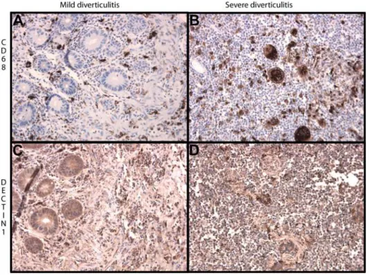
expression is mainly present on macrophages as showed by staining for CD68 (Figure 1). Furthermore, dectin-1 also appears to be weakly expressed on neutrophils, the membrane of endothelial and epithelial cells and in the submucosal neuronal plexus of Meissner (not shown). Dectin-1 appeared to be up-regulated within inflamed colon tissue due to increased expression of dectin-1 on inflammatory cells and increased influx of inflammatory cells (Figure 1). In order to test whether this increased expression of dectin-1 is IBD specific, additional staining was performed on matched intestinal tissue samples from 4 patients with diverticulitis. As shown in Figure 2, increased expression of dectin-1 was also observed in patients with severe diverticulitis compared to mild diverticulitis. As is true for patients with IBD, expression of dectin-1 is mainly present on macrophages as shown by CD68 staining.
Distribution of the DECTIN-1 c.714T.G polymorphism in IBD patients and healthy controls
Characteristics of the study population and healthy controls are depicted in Table 1. Genotype frequencies of healthy controls were in Hardy Weinberg equilibrium. Carriage of at least one copy of the G allele of theDECTIN-1polymorphism was 14% in the CD, 14% in the UC and 17% in the healthy control group. The frequency of the G allele was 9.8% in the healthy controls, 8.1% in CD patients and 7.7% in UC patients. Overall, no significant differences were observed between patients with IBD and healthy controls regarding allele frequencies of theDECTIN-1 polymorphism. However, a slight trend towards association of homozygosity for theDECTIN-1G allele with IBD was observed (G allele frequency of 1.0% in healthy controls vs. 0.5–0.6% in CD and UC, Table 1).
Figure 1. Representative immunohistochemical staining of DECTIN-1 and CD-68 in inflamed and non-inflamed intestine of the same specimen in Crohn’s disease (2506magnified).Macrophages are present in non-inflamed intestinal tissue but are present in increased numbers in inflamed tissue (pictures A and C). The expression of DECTIN-1 is increased in inflamed intestinal tissue compared to non-inflamed intestinal tissue (pictures B and D).
Correlation of the DECTIN-1 c.714T.G polymorphism with clinical characteristics of IBD patients
Patients with CD carrying one or two copies of theDECTIN-1 G allele were compared to patients with the wild-type genotype T/ T with regard to age at diagnosis, gender, family history of IBD, localization of disease and disease behavior, extra intestinal and peri-anal disease and surgery related to CD (Table 2). Patients with UC were likewise compared according to the DECTIN-1 genotypes regarding age at diagnosis, gender, locali-zation of disease, extra-intestinal disease, development of malig-nancies, surgery related to UC and a positive family history for IBD (Table 3). No statistical significant associations were ob-served between the c.714T.G polymorphism and specific phenotypes.
The DECTIN-1 c.714T.G polymorphism stratified by NOD2 status and clinical characteristics of IBD patients
CD patients carrying one or two copies of the G allele of the DECTIN-1 gene were stratified by NOD2 status. A NOD2 risk
genotype was defined as carrying at least one of the three common NOD2 disease susceptibility alleles (c.2104C.T (p.Arg702Trp), c.2722G.C (p.Gly908Arg), c.3019_3020insC (p.Leu1007ProfsX2)). Combinations of NOD2 risk carriers and DECTIN-1 c.714T.G carriers were compared to patients not bearing any of theseNOD2or DECTIN-1 minor alleles, regarding clinical characteristics. No statistical significant interaction between DECTIN-1 c.714T.G and one of theNOD2variants was observed (data not shown).
Discussion
Signaling through dectin-1, known for its recognition of the fungal componentb-glucan, has been described to be involved in several immunological pathways. Dectin-1 amplifies pro-inflam-matory cytokine production induced by TLR2 and TLR4, and primes Th1, Th17 and cytotoxic T cell responses induced by dendritic cells[9,21]. The DECTIN-1 c.714T.G polymorphism results in a loss-of-function of dectin-1, and we hypothesized that this polymorphism could be potentially protective in either the susceptibility to or the disease severity of IBD.
Figure 2. Representative immunohistochemical staining of dectin-1 and CD68 in mild and severe diverticulitis (2506magnified). Macrophages are present in intestinal tissue of mild diverticulitis but are present in increased numbers in severe diverticulitis (pictures A and B). Also, the expression of dectin-1 is increased in severe diverticulitis compared to mild diverticulitis (pictures C and D).
doi:10.1371/journal.pone.0007818.g002
Table 1.Distribution of genotypes of wild-type, heterozygous and homozygous individuals for the c.714T.G polymorphism.
DECTIN-1status Controls Crohn’s disease P-value* Ulcerative colitis P-value*
Total cohort, number 772 (100%) 778 (100%) 759 (100%)
T/T 642 (83.2%) 667 (85.7%) 0.16 655 (86.3%) 0.09
T/G 122 (15.8%) 106 (13.6%) 100 (13.2%)
G/G 8 (1.0%) 5 (0.6%) 4 (0.5%)
Values are presented as absolute numbers (percentages).
As shown by immunohistochemical staining of intestinal tissue, dectin-1 is mainly present on macrophages, but also weakly on epithelial and endothelial layers of the intestine. Similar findings in mice have been demonstrated by Wong and co-workers, who demonstrated that dectin-1 is mainly expressed on populations of myeloid cells (monocyte/macrophage and neutrophil lineag-es)[22]. In addition, they demonstrated that dectin-1 is also expressed in the Peyer’s patches and along the lamina propria of the mouse intestine[23,24]. Interestingly, dectin-1 expression
appeared to be elevated in inflamed intestinal tissue compared to normal tissue, due to the increased infiltration of immune cells and increased dectin-1 expression on the cell membrane of immune cells.
However, intestinal expression of dectin-1 did not appear to be disease specific but rather dependent on influx of macrophages. In fact, expression of dectin-1 was also present in intestinal samples from patients with diverticulitis. Increased infiltration of macro-phages in severe diverticulitis showed an increased expression of
Table 2.Association betweenDECTIN-1genotypes and clinical characteristics in a subset of Crohn’s disease patients from whom
detailed phenotypic data were available (N = 778).
Characteristic Total cohort CD (%) T/T (%) T/G (%) G/G (%) Odds ratio* 95% CI*
Mean age at diagnosis, yr (SD) 29.58 (612.29) 29.37 (612.64) 30.76 (617.32) Male gender (%) 261 (33.5) 222/667 (33.3) 37/106 (34.9) 2/5 (40.0)
Familial IBD (N = 631) 131 (20.8) 107/588 (20.3) 24/101 (24.0) 0/5 (0) 1.32 0.78 2.17
Localization (Vienna Classification) (%) (n = 778)
L1: ileum 196 164/667 (24.6) 32/106 (30.2) 0/0 (0) 1.24 0.80 1.94
L2: colon 194 163 (24.2) 28 (26.4) 3 (60.0) 1.20 0.76 1.88
L3: ileocolonic 388 340 (51.0) 46 (43.4) 2 (40.0) 0.73 0.49 1.10
L4: upper disease 43 36 (5.4) 7 (6.6) 0 (0) 1.18 0.51 2.72
Disease behavior (Montreal classification) (%) (n = 776)
B1: non structuring, non penetrating (%)
291 (37.5) 250/665 (37.6) 39/106 (36.8) 2/5 (40.0) 0.97 0.64 1.47
B2: structuring 215 (27.7) 187 (28.1) 27 (25.5) 1 (20.0) 0.86 0.54 1.37
B3: penetrating 270 (34.8) 228 (34.3) 40 (37.7) 2 (40.0) 1.16 0.77 1.77
Extraintestinal disease (%) (n = 750)
151 (20.1) 125/642 (19.5) 26/103 (25.2) 0/5 (0) 1.31 0.81 2.13
Perianal disease (%) (n = 643) 177 (27.5) 149/548 (27.2) 26/90 (28.9) 2/5 (40.0) 1.12 0.69 1.81
Surgery (n = 774) 411 (53.1) 355/664 (53.5) 54/105 (51.4) 2/5 (40.0) 0.90 0.60 1.35
Values are presented as absolute numbers (percentages).
*Carriers of the mutant allele (T/G and G/G) were compared to wild-types (T/T). doi:10.1371/journal.pone.0007818.t002
Table 3.Association betweenDECTIN-1genotypes and clinical characteristics in a subset of ulcerative colitis patients from whom detailed phenotypic data were available (N = 759).
Characteristic Total cohort UC (%) T/T (%) T/G (%) G/G (%) Odds ratio* 95% CI*
Age at diagnosis (SD) 36.3 (614.4) 33.9 (612.9) 35.5 (610.4)
Male gender 401 (52.8) 346/655 (52.8) 52/100 (52.0) 3/4 (75.0)
Localization (Montreal) (n = 721)
E1 (Proctitis) 124 (17.2) 110/623 (17.7) 14/95 (14.7) 0/3 (0) 0.78 0.43 1.42
E2 (Left sided) 245 (44.0) 212 (34.0) 33 (34.7) 0 (0) 0.98 0.63 1.54
E3 (Extended/pancolitis) 352 (48.8) 301 (48.3) 48 (50.5) 3 (100) 1.16 0.76 1.79
Extraintestinal disease (n = 228)
42 (18.4) 33/183 (18.0) 8/43 (18.6) 1/2 (50) 1.14 0.50 2.59
Surgery (n = 759) 145 (19.1) 120/655 (18.3) 24/100 (24.0) 1/4 (25.0) 1.41 0.86 2.31
Malignancy (n = 384) 2 (0.5) 1/329 (0.3) 1/53 (1.9) 0/2 (0) 6.07 0.37 98.57
Family diagnosis of IBD (n = 547)
81 (14.8) 72/472 (15.3) 9/72 (12.5) 0/3 (0) 0.76 0.36 1.59
Values are presented as absolute numbers (percentages).
dectin-1, compared to mild diverticulitis which is accompanied by less infiltration of macrophages.
Cohorts of CD (n = 778) and UC (n = 759) patients were screened for the DECTIN-1c.714T.G polymorphism and compared to a group of healthy subjects (n = 772). Subsequently, these genetic data were correlated with clinical parameters reflecting disease severity. This analysis revealed no statistical significant association between the prevalence of the DECTIN-1 c.714G allele and IBD, neither in disease occurrence nor in disease severity. However, one can observe that homozygous individuals bearing theDECTIN-1polymorphism were twice as frequent in healthy controls compared to IBD patients. This may suggest that complete absence of dectin-1 function could protect against IBD. It is important to realize that the only two dectin-1 isoforms capable of binding b-glucans (isoforms A and B) are structurally equally affected by theDECTIN-1c.714T.G polymor-phism[25]. The occurrence of splicing isoforms with residual function could therefore be excluded. The potential mechanism of protection are likely to include the lower production of pro-inflammatory cytokines, including IL-17, in the individuals with defective dectin-1. In these series, statistical power to preclude any functional difference is insufficient due to the low prevalence of homozygous individuals. Additional studies in homozygous and heterozygous subpopulations are needed to confirm the reported observations.
All together, the reports of the association of mutations within NOD2 andCARD9 in patients with CD, the presence of ASCA, and the shared signaling pathway of dectin-1 and NOD2, points toward a possible link between NOD2 and dectin-1[26]. Since
mutations in NOD2 in CD patients are associated with ileal involvement and increased need for surgery and stricturing disease[27], a potential interaction between NOD2 mutations and the DECTIN-1 c.714T.G polymorphism with regard to phenotypical characteristics was investigated. However, no statistical interaction could be demonstrated (data not shown).
Our data demonstrate that dectin-1 expression is elevated on macrophages, neutrophils, and other immune cells involved in the inflammatory reaction in IBD. The DECTIN-1 c.714T.G polymorphism is not a major susceptibility factor for protection against IBD, although a trend towards a lower frequency of the polymorphism in CD and UC cohorts was observed, in particular in the number of individuals homozygous for the DECTIN-1 polymorphism. These genetic findings warrant further investiga-tion of this pathogenetic pathway.
Acknowledgments
The authors thank Prof. Cisca Wijmenga (University of Groningen) and Dr. Astrid Oude Lashof for providing DNA of healthy controls.
Author Contributions
Conceived and designed the experiments: HSdV TSP LABJ MGN DJdJ. Performed the experiments: HSdV TSP JHvK RS EAF. Analyzed the data: HSdV TSP. Contributed reagents/materials/analysis tools: AAvB RKW BC RKL. Wrote the paper: HSdV TSP LABJ MGN DJdJ.
References
1. Sartor RB (2008) Microbial influences in inflammatory bowel diseases. Gastroenterology 134: 577–594.
2. Xavier RJ, Podolsky DK (2007) Unravelling the pathogenesis of inflammatory bowel disease. Nature 448: 427–434.
3. Tysk C, Lindberg E, Jarnerot G, Floderus-Myrhed B (1988) Ulcerative colitis and Crohn’s disease in an unselected population of monozygotic and dizygotic twins. A study of heritability and the influence of smoking. Gut 29: 990–996. 4. Cho JH, Weaver CT (2007) The genetics of inflammatory bowel disease.
Gastroenterology 133: 1327–1339.
5. Girardin SE, Boneca IG, Viala J, Chamaillard M, Labigne A, et al. (2003) Nod2 is a general sensor of peptidoglycan through muramyl dipeptide (MDP) detection. J Biol Chem 278: 8869–8872.
6. Inohara N, Ogura Y, Fontalba A, Gutierrez O, Pons F, et al. (2003) Host recognition of bacterial muramyl dipeptide mediated through NOD2. Implications for Crohn’s disease. J Biol Chem 278: 5509–5512.
7. Gross O, Gewies A, Finger K, Schafer M, Sparwasser T, et al. (2006) Card9 controls a non-TLR signalling pathway for innate anti-fungal immunity. Nature 442: 651–656.
8. Hsu YM, Zhang Y, You Y, Wang D, Li H, et al. (2007) The adaptor protein CARD9 is required for innate immune responses to intracellular pathogens. Nat Immunol 8: 198–205.
9. LeibundGut-Landmann S, Gross O, Robinson MJ, Osorio F, Slack EC, et al. (2007) Syk- and CARD9-dependent coupling of innate immunity to the induction of T helper cells that produce interleukin 17. Nat Immunol 8: 630–638.
10. Zhernakova A, Festen EM, Franke L, Trynka G, van Diemen CC, et al. (2008) Genetic analysis of innate immunity in Crohn’s disease and ulcerative colitis identifies two susceptibility loci harboring CARD9 and IL18RAP. Am J Hum Genet 82: 1202–1210.
11. Ferwerda B, Ferwerda G, Plantinga TS, Willment JA, van Spriel AB, Venselaar H, et al. A family with human dectin-1 deficiency and mucocutanous fungal infections. New England Journal of Medicine, In press.
12. Plantinga TS, van der Velden WJ, Ferwerda B, van Spriel AB, Adema G, et al. (2009) Early stop polymorphism in human DECTIN-1 is associated with increased candida colonization in hematopoietic stem cell transplant recipients. Clin Infect Dis 49: 724–732.
13. Seiderer J, Elben I, Diegelmann J, Glas J, Stallhofer J, et al. (2008) Role of the novel th17 cytokine IL-17F in inflammatory bowel disease (IBD): Upregulated colonic IL-17F expression in active Crohn’s disease and analysis of the IL17F p.Hisl6lArg polymorphism in IBD. Inflammatory Bowel Diseases 14: 437–445. 14. van Beelen AJ, Zelinkova Z, Taanman-Kueter EW, Muller FJ, Hommes DW, et al. (2007) Stimulation of the intracellular bacterial sensor NOD2 programs dendritic cells to promote interleukin-17 production in human memory T cells. Immunity 27: 660–669.
15. Standaert-Vitse A, Jouault T, Vandewalle P, Mille C, Seddik M, et al. (2006) Candida albicans is an immunogen for anti-Saccharomyces cerevisiae antibody markers of Crohn’s disease. Gastroenterology 130: 1764–1775.
16. Quinton JF, Sendid B, Reumaux D, Duthilleul P, Cortot A, et al. (1998) Anti-Saccharomyces cerevisiae mannan antibodies combined with antineutrophil cytoplasmic autoantibodies in inflammatory bowel disease: prevalence and diagnostic role. Gut 42: 788–791.
17. Podolsky DK (2002) Inflammatory bowel disease. New England Journal of Medicine 347: 417–429.
18. Gasche C, Scholmerich J, Brynskov J, D’Haens G, Hanauer SB, et al. (2000) A simple classification of Crohn’s disease: Report of the Working Party for the world congresses of gastroenterology, Vienna 1998. Inflammatory Bowel Diseases 6: 8–15.
19. Silverberg MS, Satsangi J, Ahmad T, Arnott ID, Bernstein CN, et al. (2005) Toward an integrated clinical, molecular and serological classification of inflammatory bowel disease: Report of a Working Party of the 2005 Montreal World Congress of Gastroenterology. Can J Gastroenterol 19 Suppl A: 5–36. 20. Oostenbrug LE, Nolte IM, Oosterom E, van der Steege G, Meerman GJT, et al.
(2006) CARD15 in inflammatory bowel disease and Crohn’s disease phenotypes: An association study and pooled analysis. Digestive and Liver Disease 38: 834–845. 21. Ferwerda G, Meyer-Wentrup F, Kullberg BJ, Netea MG, Adema GJ (2008)
Dectin-1 synergizes with TLR2 and TLR4 for cytokine production in human primary monocytes and macrophages. Cellular Microbiology 10: 2058–2066. 22. Taylor PR, Brown GD, Reid DM, Willment JA, Martinez-Pomares L, et al.
(2002) The beta-glucan receptor, dectin-1, is predominantly expressed on the, surface of cells of the monocyte/macrophage and neutrophil lineages. Journal of Immunology 169: 3876–3882.
23. Reid DM, Montoya M, Taylor PR, Borrow P, Gordon S, et al. (2004) Expression of the beta-glucan receptor, dectin-1, on murine leukocytes in situ correlates with its function in pathogen recognition and reveals potential roles in leukocyte interactions. Journal of Leukocyte Biology 76: 86–94.
24. Taylor PR, Brown GD, Reid DM, Willment JA, Martinez-Pomares L, et al. (2002) The beta-glucan receptor, dectin-1, is predominantly expressed on the, surface of cells of the monocyte/macrophage and neutrophil lineages. Journal of Immunology 169: 3876–3882.
25. Willment JA, Gordon S, Brown GD (2001) Characterization of the human beta -glucan receptor and its alternatively spliced isoforms. J Biol Chem 276: 43818–43823.
26. Underhill D, Braun J (2008) Current understanding of fungal microflora in inflammatory bowel disease pathogenesis. Inflammatory Bowel Diseases 14: 1147–1153.