www.jcol.org.br
Journal of
Coloproctology
* Corresponding author.
E-mail: [email protected] / raquell @fcm.unicamp.br (R. F. Leal) 2237-9363/$ - see front matter. © 2013 Elsevier Editora Ltda. All rights reserved.
Original article
Autophagy and proinl ammatory cytokine expression in the
intestinal mucosa and mesenteric fat tissue of patients with
Crohn’s disease
Raquel Franco Leal
a,b,c,*, Marciane Milanski
b, Cláudio Saddy Rodrigues Coy
a,c,
Mariana Portovedo
b, Viviane Soares Rodrigues
a,b, Andressa Coope
b,
Maria de Lourdes Setsuko Ayrizono
a,c, João José Fagundes
a,c, Lício Augusto Velloso
ba Coloproctology Unit, Surgery Department, Escola de Ciências Médicas, Universidade Estadual de Campinas (UNICAMP), São Paulo, SP,
Brazil
b Laboratory of Cell Signaling, Internal Medicine Department, Escola de Ciências Médicas, UNICAMP, São Paulo, SP, Brazil c Brazilian Society of Coloproctology (TSBC), Rio de Janeiro, RJ, Brazil
a r t i c l e i n f o
Article history:
Received 1 June 2012 Accepted 10 October 2012
Keywords:
Crohn’s disease
Inl ammatory bowel diseases Autophagy
Cytokines Mesenteric fat
a b s t r a c t
Background: Recently, mesenteric fat has been proposed to play a role in the pathophysiol-ogy of Crohn’s disease (CD), as fat hypertrophy is detected close to the affected intestinal area; however, there are few studies regarding autophagy and creeping fat tissue in CD.
Objective: Evaluate autophagy-related proteins and proinl ammatory cytokines in intestinal mucosa and mesenteric fat in patients with CD and controls.
Patients and methods: Ten patients with CD, eight with non-inl ammatory disease who underwent surgery, and eight with normal ileocolonoscopy were studied. The expression of LC3-II, TNF-α and IL-23 was determined by immunoblot of protein extracts. In addition, total RNA of LC3 and Atg16-L1 were determined using RT-PCR.
Results: The expression of LC3-II was signii cantly lower in the mesenteric tissue of CD when compared to controls (p < 0.05). In contrast, the intestinal mucosa of the CD group had higher levels of LC3-II (p < 0.05). However, mRNA expression of autophagy-related pro-teins was similar when compared to mesenteric fat groups. TNF-α and IL-23 expressions were higher in intestinal mucosa of CD than in control (p < 0.05).
Conclusion: These i ndings suggest a defect in the autophagic activity of the creeping fat tissue in CD, which could be involved with the maintenance of the inl ammatory process in the intestinal mucosa.
Palavras-chave:
Doença de Crohn
Doença inl amatória intestinal Autofagia
Citocinas
Gordura mesenterial
r e s u m o
Autofagia e expressão de citocinas pró-inl amatórias na mucosa intestinal e no tecido mesenterial de pacientes com doença de Crohn
Introdução: Recentemente, tem se proposto que o tecido mesenterial possa participar da i siopatologia da DC, uma vez que é notória a hipertroi a da gordura mesenterial próxima ao segmento intestinal afetado pela doença. Entretanto, há poucos estudos relacionando autofagia e tecido mesenterial na DC.
Objetivo: Avaliar autofagia e citocinas na mucosa intestinal e no mesentério de pacientes com DC.
Pacientes e métodos: Dez pacientes com DC, oito sem doença inl amatória intestinal que foram submetidos à cirurgia, e oito com ileocolonoscopia normal, foram estudados. As ex-pressões de LC3-II, TNF-α e IL-23 foram determinadas por imunoblot de extrato protéico total. Além disso, expressão gênica de LC3 e de Atg16-L1 foi realizada por RT-PCR.
Resultados: A expressão de LC3-II foi signii cativamente menor no tecido mesenterial de pa-cientes com DC quando comparada à dos controles (p < 0,05); as amostras de tecido intes-tinal do grupo DC apresentaram maior expressão de LC3-II (p < 0,05). Entretanto, as expres-sões gênicas relacionadas à autofagia foram similares nos grupos de tecido mesenterial. Os níveis de TNF-α e de IL-23 foram maiores na mucosa intestinal do grupo CD (p < 0,05).
Conclusão: Estes achados sugerem alteração da autofagia no mesentério na DC, o que pode estar envolvido com a manutenção da inl amação na mucosa intestinal.
© 2013 Elsevier Editora Ltda. Todos os direitos reservados.
Introduction
Autophagy seems to be essential for intestinal mucosal im-munity, and recent studies show that mutations in autoph-agy-related genes are associated with CD.1–4 The autophagy process has important functions, such as regulation of cell survival and cell death. When properly activated, the pro-cess promotes survival through the degradation of cytoplas-mic organelles and content via lysosomes, thereby providing metabolic fuel for mitochondrial oxidation,5,6 as well as ac-counting for the removal of proteins generated from cellular stress. However, exacerbated autophagy may also lead to cell death.7,8 Autophagy can be induced by extracellular and intra-cellular signals, including oxidative stress, growth factors, ce-ramide, endoplasmic-reticulum (ER) stress, and glucose and amino acid serum starvation.9
Association of CD with the gene loci ATG16L1 was refer-eed and patients who had more mutant alleles, develop more severe course of disease (stenosis and i stulae), compared to those patients with inl ammatory CD phenotype only.10
Although there is phenotypic variation in surgical speci-mens from CD patients, macroscopic aspects are notori-ous; particularly regarding mesenteric fat thickening near the affected intestinal area.11 Mesenteric fat hypertrophy and bowel wrapping are characteristic of CD.12 Histological analyses revealed abnormalities in adipose tissue, including ini ltration of macrophages and T cells, perivascular i brosis and inl ammation. Moreover, there are increased numbers of adipocytes in CD creeping fat, which have been reported to be smaller in CD than in controls.13,14 Similar to macrophages and epithelial cells, adipocytes from normal individuals are able to synthesize several proinl ammatory and anti-inl am-matory cytokines, and fat hormones.15 Indeed, adipocytes
can express TLR4 for the recognition of local or systemic bacterial antigens.16–18
There have been few reports discussing the role of the mesenteric fat tissue in CD.19–24 However, there are no stud-ies evaluating autophagy in this tissue. Therefore, in order to compare the autophagic activity (called ‘macroautophagy’) and the expression of proinl ammatory cytokines in fat and intestinal tissue between CD patients and controls, we used immunoblotting to determine LC3, TNF-α, and IL-23 protein expression. In addition, we performed RT-PCR assays to de-termine the relative RNA quantii cation related to the step of membrane elongation and autophagosome formation (LC3II and Atg16L1) in mesenteric fat tissue groups.25,26
Patients and methods
Mucosal biopsies were taken from 10 patients with ileocecal CD [mean age 34.9 (range, 14–60) years; male 50%; female 50%]. The presence of disease activity was assessed by colo-noscopy before surgery, and all patients had Crohn’s disease activity index (CDAI)27 over 250 points. The control groups were composed of eight patients who underwent retosig-moidectomy for non-inl ammatory disease (megacolon), with normal distal ileum (ileum mesenteric fat tissue con-trol group) [mean age 55.6 (range, 39–70) years; male 62.5%; female 37.5%], and eight patients with normal ileocolonos-copy (intestinal tissue control group) [mean age 50.4 (range, 33–60) years; male 37.5%; female 62.5%].
Mucosal biopsies from the terminal ileum and mesenteric fat tissue near the affected intestinal area were snap-frozen in liquid nitrogen and stored at −80 °C until ready to be used.
Western blotting analysis
For total protein extract preparation, the fragments were ho-mogenized in solubilization buffer at 4 ºC [1% Triton X-100, 100 mM Tris-HCl (pH 7.4), 100 mM sodium pyrophosphate, 100 mM sodium l uoride, 10 mM EDTA, 10 mM sodium orthovana-date, 2.0 mM phenylmethylsulfonyl l uoride (PMSF), and 0.1 mg aprotinin/mL] with a Polytron PTA 20S generator (model PT 10/35; Brinkmann Instruments, Westbury, NY) operated at maximum speed for 30 sec. Insoluble material was removed by centrifugation (20 min at 110,000 rpm at 4 °C). Protein concen-tration in the supernatants were determined by the Bradford dye binding method.28 Aliquots of the resulting supernatants containing 50 μg total proteins were separated by SDS-PAGE, transferred to nitrocellulose membranes and blotted with anti-LC3, anti-TNF-α and anti-IL-23 antibodies.29
Reagents for SDS-PAGE and immunoblotting were from Bio-Rad Laboratories (Richmond, CA). Phenylmethylsulfonyl l uo-ride, aprotinin, Triton X-100, Tween 20, and glycerol were from Sigma (St. Louis, MO). Nitrocellulose paper (BA85, 0.2 μm) was from Amersham (Aylesbury, UK). The anti-LC3 (M115-3, mouse monoclonal) antibody was purchased from MBL International (MA). The anti-TNF-α (sc-1347, rabbit polyclonal) and anti-IL-23 (sc-50303, rabbit polyclonal) antibodies were purchased from Santa Cruz Biotechnology, Inc. (Santa Cruz, CA). Protein molec-ular weight was assessed by the PageRulerTM from Fermentas (Glenburnie, MD). The signal was detected by chemilumines-cent reaction (SuperSignal®West Pico Chemiluminescent Sub-strate from Pierce Biothecnology, Inc. Rockford, IL).
All numerical results are expressed as mean ± SEM of the indicated number of experiments. Blotting results are pre-sented as direct comparisons of bands in autoradiographs and quantii ed by densitometry using the Gel-Pro Analyzer 6.0 software (Exon-Intron Inc., Farrell, MD). Data were analyzed by the t-Test, comparing the mesenteric fat tissue of the CD group and its respective fat control group; and comparing, separately, the intestinal tissue of the CD group and its respective intesti-nal control group. The level of signii cance was set at p < 0.05.
RT-PCR analysis
Total RNA was extracted using Trizol (Invitrogen) according to the manufacturer instructions. RNA purity and concentration were determined by UV spectrophotometry at 260 nm. RNA was treated with RNase-free Dnase (RQ1 RNase-free Dnase, Promega), then reverse transcribed using oligo (dT) primers and reverse transcriptase (RevertAid™ Kit, Fermentas). The reaction mixture (20 μl) was incubated at 42 ºC for 60 min, then 10 min at 70 ºC, and cooled on ice. RT-PCR was performed on resulting cDNA, using the manufacturer protocol, in 25 μL reaction volume per capillary. Gene-specii c primers (Applied Biosystems™) were: Hs00261291 (MAP1LC3-LC3); Hs00250530 (ATG16-L1); NM_002046.3 (GAPDH). The reaction mixture con-tained SYBR®Premix Ex Taq™ II (2x), cDNA template, primer
pair mixture and dH2O. RT-PCR amplii cation consisted of an initial denaturation step (50 ºC for 2 min and 95 ºC for 10 min), 40 cycles of denaturation (95 ºC for 15 seconds), anneling (53 ºC for 20 seconds) and extension (72 ºC for 20 seconds), followed by a i nal incubation at 60 ºC for 1 min. All measure-ments were normalized by the expression of GAPDH gene, considered as a stable housekeeping gene. Gene expression was determined using the delta-delta Ct method: 2−▲▲CT
(▲▲CT=[Ct(target gene) – Ct(GAPDH)]patient – [Ct(target gene) – Ct(GAPDH)]control)
Real-time PCR analysis of gene expression was performed in a 7500 SDS sequence detection system (Applied Biosystems). The optimal concentration of cDNA and primers, as well as the maximum efi ciency of amplii cation, were obtained through i ve-point, two-fold dilution curve analysis for each gene. Real-time data were analyzed using the Sequence Detector System 1.7 (Applied Biosystems). The t-test was used for statistical analyses, comparing the mesenteric fat tissue of the CD group and its respective fat control group; and comparing, separately, the intestinal tissue of CD group and its respective intestinal control group. The level of signii cance was set at p < 0.05.
Results
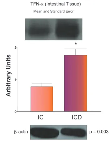
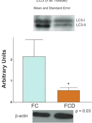
LC3-II protein expression was signii cantly lower in the mesen-teric fat tissue of CD patients (FCD group), when compared to controls (FC group) (p < 0.05). Patients with CD also had signii -cantly higher levels of LC3-II, TNF-α and IL-23 in intestinal mu-cosa (ICD group) when compared to intestinal control (IC group) (p < 0.05). No difference in cytokine levels was observed among the mesenteric groups. Results are showed in Figs. 1 to 6.
Regarding LC3 and ATG16L1 gene expressions, no statisti-cally signii cant differences were detected when the mesen-teric fat tissue groups were compared (p > 0.05). Figs. 7 and 8 illustrate these i ndings.
Discussion
Recent genome-wide association studies (GWAS) have in-creased the number of CD associated susceptibility genes. Two of these genes (ATG16L1 and IRGM) modulate autophagy, and the variant T300A of ATG16L1 results in a specii c defect in bacteria-induced autophagy.3,30,31 ATG16L1 and NOD2 risk vari-ants, in addition to Paneth cell defect, could modify intestinal epithelial cell antimicrobial responses.30 However, there is no report of autophagy-related protein levels in distinct tissues affected by CD, such as intestinal and mesenteric fat tissues.
Fig. 3 – Representative Western blot analyses and
determination of IL-23 protein expression in the fat tissue of control (FC) and Crohn’s disease (FCD) groups. For illustration purposes each band represents one patient. For the FCD Group, n = 10; for the FC Group, n = 8, *p < 0.05 vs control. Fig. 1 – Representative Western blot analyses and
determination of TNF-α protein expression in the fat tissue of control (FC) and Crohn’s disease (FCD) groups. For illustration purposes each band represents one patient. For the FCD Group, n = 10; for the FC Group, n = 8, *p < 0.05 vs control.
Fig. 2 – Representative Western blot analyses and
determination of TNF-α protein expression in the intestinal tissue of control (IC) and Crohn’s disease (ICD) groups. For illustration purposes each band represents one patient. For the ICD Group, n = 10; for the IC Group, n = 8, *p < 0.05 vs control.
Fig. 4 – Representative Western blot analyses and
determination of IL-23 protein expression in the intestinal tissue of control (IC) and Crohn’s disease (ICD) groups. For illustration purposes each band represents one patient. For the ICD Group, n = 10; for the IC Group, n = 8, *p < 0.05 vs control. TFN-α (Fat Tissue)
β-actin p = 0.61
p = 0.003 p = 0.007
p = 0.96
*
*
β-actin
β-actin β-actin
TFN-α (Intestinal Tissue)
IL - 23 (Fat Tissue)
IL - 23 (Intestinal Tissue)
Mean and Standard Error
FC
22
0.451 0.532
0.369
0.287
0.205
0.123
0.041 0 1
1
1
0
0
0
Arbitrary Units
Arbitrary Units
Arbitrary Units
Arbitrary Units
FC
IC
IC
FCD
FCD
ICD
ICD
Mean and Standard Error
Mean and Standard Error
LC3-II is localized on the cytoplasmic surface of autophago-somes and is delipidated by Atg4B to recycle LC3-I for further autophagosome formation.33,34 Indeed, Atg16L1 determines the site of LC3 conjugation, essential for autophagosome for-mation, suppressing inl ammasome activity and maintaining Paneth cells.35,36 Despite great progress since the identii cation of ATG1,37 many questions regarding the molecular regulation of autophagy remain unresolved.38
Moreover, autophagy and inl ammatory pathways are closely linked. Proinl ammatory cytokines secretion, such as IL-1β and IL-18, were enhanced in ATG16L1 or ATG7 deleted macrophages in response to lipopolysaccharide (LPS), displaying increased susceptibility to a murine model of colitis.39 Autophagy also sup-presses endotoxin-induced inl ammatory immune responses.40 Defects of autophagy activity and bacterial receptors (NOD2) have been associated with impaired antigen presentation, en-gaged intracellular removal of pathogenic bacteria and, conse-quently, higher expression of proinl ammatory cytokines.41–45
Autophagy contributes to cellular survival against nutrient starvation and the turn-over of injured organelles,46,47 explain-ing why in the present study was verii ed high expression of autophagy protein in the intestinal tissue of CD patients when compared to controls. However, this ability was not verii ed in the hypertrophied mesenteric fat tissue of CD patients, other-Fig. 6 – Representative Western blot analyses and
determination of LC3-II protein expression in the intestinal tissue of control (IC) and Crohn’s disease (ICD) groups. For illustration purposes each band represents one patient. For the ICD Group, n = 10; for the IC Group, n = 8, *p < 0.05 vs control. Fig. 5 – Representative Western blot analyses and
determination of LC3-II protein expression in the fat tissue of control (FC) and Crohn’s disease (FCD) groups. For illustration purposes each band represents one patient. For the FCD Group, n = 10; for the FC Group, n = 8, *p < 0.05 vs control.
p = 0.01 p = 0.03
LC3-I
LC3-I LC3-II
LC3-II
*
*
β-actin
β-actin
LC3 (Intestinal Tissue) LC3 (Fat Tissue)
2
0.287 0.328
0.246
0.205
0.164
0.123
0.082
0.041
0 1
0
Arbitrary Units
Arbitrary Units
Fig. 8 – Results of ATG16-L1 gene expression determined by RT-PCR. For FCD Group, n = 10; for FC Group, n = 8, *p < 0.05 vs control.
Fig. 7 – Results of LC3 gene expression determined by RT-PCR. For FCD Group, n = 10; for FC Group, n = 8, *p < 0.05 vs control.
LC3
p > 0.36 p > 0.62
1.5
4 1.0
3 0.5
2 0.0
1
0
FC
FC FCD
FCD
ATG16-L1
Gene expression
(fold control GAPDH)
Gene expression
(fold control GAPDH)
FC
IC
FCD
ICD
wise, was found low expression of LC3-II when compared to mesenteric tissue of patients without IBD. This LC3-II expres-sion could lead to an impaired clearance of bacterial species, as well as the accumulation of unprocessed unnecessary pro-teins, which activate proinl ammatory pathways involved in CD pathogenesis. The higher expression of proinl ammatory cyto-kines in intestinal mucosa, such as TNF-α and IL-23, which sig-nalize respective Th1 and Th17 lymphocyte response, could also explain the higher levels of autophagy protein seen in this study.
Although LC3-II protein expression was lower in the mesen-teric fat tissue and higher in the intestinal tissue of CD patients, no statistical differences were seen among these groups, con-sidering LC3 and ATG16L1 gene expressions. Reports on the role of microRNAs (miRNAs) in autophagy may lead to insights into these complex processes. MicroRNAs represent an important and still relatively unexplored manner of regulating protein synthesis. It does not encode for proteins, but exert catalytic, structural or regulatory activities by annealing to specii c target RNAs, and downregulating their stability and/or translation.48,49 It has been reported that miRNA-101 is a potent inhibitor of autophagy.50 Regarding CD, Brest et al., showed that a family of miRNAs, miR-196, is overexpressed in the inl ammatory intes-tinal epithelia of these individuals and there is subsequent loss of the tight regulation of IRGM expression, which compromises the control of intracellular replication of CD-associated adher-ent invasive Escherichia coli by autophagy. The association of IRGM with CD arises from a miRNA-based alteration in IRGM regulation that affects the efi cacy of autophagy.51 The results of the protein and gene expression in the present study sug-gest that miRNA alterations could be involved in the complete gene transcription, resulting in the defective production of the protein. Only one study reporting a miRNA (miR-204) regulat-ing LC3-II protein has been published, which reported an im-portant role for this mechanism in myocardial injury;52 how-ever, there are no studies of miRNA, autophagy regulation, and CD in the literature.
Recent studies, in mice, provide insights into how defective autophagy in cells, such as macrophages and Paneth cells, may contribute to CD,39,53 although none of these studies describe these alterations in adipose tissue. It is possible that the defec-tive autophagy in this tissue could be responsible for maintain-ing the local production of inl ammatory mediators and lead to intestinal involvement, especially on mesenteric longitudinal side, forming ulcers, characteristic of CD, as described by Crohn et al.54 In late stages of the disease, there is a decrease in adipo-cytes number, as these are substituted by impaired autophagic cells of the immune system, such as macrophages,55,56 possible explaining why the present study showed a defective autopha-gy in the mesenteric adipose tissue of CD patients.
The importance in knowing the autophagy pathway in creep-ing fat tissue in CD can lead us to understand more about the phenotype and molecular biology involved in CD and its patho-genesis, as well as the active role of adipose tissue in the mainte-nance of the inl ammatory process during the course of disease.
Conclusion
The present study shows a defective expression of protein-related autophagy in the mesenteric fat tissue of CD patients,
when compared to controls. Data suggest that the primary defects of cell autophagy may not occur at all tissue involved by CD, however, it may occur mainly in the adipose tissue, close to the affected intestinal area. The mesenteric fat tissue could play an important role in the maintenance of local in-l ammation in these patients. Further studies wiin-lin-l determine whether miRNA has a role in autophagy regulation in patients with CD, and whether autophagy can be used for therapeutic approaches in the treatment of CD.
Conl ict of interest
The authors declare no conl ict of interest.
Financial support
Fundação de Amparo à Pesquisa do Estado de São Paulo (FAPESP).
R E F E R E N C E S
1. Heatha RJ, Xavier RJ. Autophagy, immunity and human disease. Curr Opin Gastroenterol 2009;25:512–20.
2. Cummings JR, Cooney R, Pathan S, Anderson CA, Barrett JC, Beckly J, et al. Coni rmation of the role of ATG16L1 as a Crohn’s disease susceptibility gene. Inl amm Bowel Dis 2007;13:941–6. 3. Hampe J, Franke A, Rosenstiel P, Till A, Teuber M, Huse K,
et al. A genome-wide association scan of nonsynonymous SNPs identii es a susceptibility variant for Crohn’s disease in ATG16L1. Nat Genet 2007;39:207–11.
4. Parkes M, Barrett JC, Prescott NJ, Tremelling M, Anderson CA, Fisher SA, et al. Sequence variants in the autophagy gene IRGM and multiple other replicating loci contribute to Crohn’s disease susceptibility. Nat Genet 2007;39:830–2.
5. Eskelinen EL. Maturation of autophagic vacuoles in Mammalian cells. Autophagy 2005;1:1–10.
6. Lum JJ, Bauer DE, Kong M, Harris MH, Li C, Lindsten T, et al. Growth factor regulation of autophagy and cell survival in the absence of apoptosis. Cell 2005;120:237–48.
7. Abedin MJ, Wang D, McDonnell MA, Lehmann U, Kelekar A. Autophagy delays apoptotic death in breast cancer cells following DNA damage. Cell Death Differ 2007;14:500–10. 8. Colell A, Ricci JE, Tait S, Milasta S, Maurer U, Bouchier-Hayes L,
et al. GAPDH and autophagy preserve survival after apoptotic cytochrome c release in the absence of caspase activation. Cell 2007;129:983–97.
9. Tanida I. Autophagosome formation and molecular mechanism of autophagy. Antioxid Redox Signal 2011;14:
2201–14.
10. Weersma RK, Stokkers PCF, vanBodegraven AA, vanHogezand RA, Verspaget HW, deJong DJ, et al. Molecular prediction of disease risk and severity in a large Dutch Crohn’s disease cohort. Gut 2009;58(3):388–95.
11. Golder WA. The “creeping fat sign”-really diagnostic for Crohn’s disease? Int J Colorectal Dis 2009;24(1):1–4.
12. Bertina B, Desreumauxa P, Dubuquoy L. Obesity, visceral fat and Crohn’s disease. Curr Op Clin Nutr Metab Care 2010;13:574–80. 13. Sheehan AL, Warren BF, Gear MW, Shepherd NA. Fat-wrapping
in Crohn’s disease. pathological basis and relevance to surgical practice. Br J Surg 1992;79:955–8.
15. Vettor R, Milan G, Rossato M, Federspil G. Review article: adipocytokines and insulin resistance. Aliment Pharmacol Ther 2005;22(2):3–10.
16. Lin Y, Lee H, Berg AH, Lisanti MP, Shapiro L, Scherer PE. The lipopolysaccharide-activated Toll-like receptor (TLR)-4 induces synthesis of the closely related receptor TLR-2 in adipocytes. J Biol Chem 2000;275:24255–63.
17. Batra A, Pietsch J, Stroh T, Fedke I, Glauben R, Okur B, et al. Toll-like receptor expression and response to specii c stimulation in adipocytes and preadipocytes: on the role of fat in inl ammation. Ann N Y Acad Sci 2006;1072:407–9.
18. Gay J, Tachon M, Neut C. Mesenteric adipose tissue is colonized by bacterial l ora and express pathogen recognition receptors in Crohn’s disease [abstract]. Gastroenterology 2005;128(2):A503. 19. Dereumaux P, Ernst O, Geboes K, Gambiez L, Berrebi D,
Müller-Alouf H, et al. Inl ammatory alterations in mesenteric adipose tissue in Crohn’s disease. Gastroenterology 1999;117:73–81. 20. Barbier M, Vidal H, Desreumaux P, Dubuquoy L, Bourreille A,
Colombel JF, et al. Overexpression of leptin mRNA in mesenteric adipose tissue in inl ammatory bowel diseases. Gastroenterol Clin Biol 2003;27:987–91.
21. Yamamoto K, Kiyohara T, Murayama Y, Kihara S, Okamoto Y, Funahashi T, et al. Production of adiponectin, an anti-inl ammatory protein, in mesenteric adipose tissue in Crohn’s disease. Gut 2005;54:789–96.
22. Peyrin-Biroulet L, Gonzalez F, Dubuquoy L, Rousseaux C, Dubuquoy C, Decourcelle C, et al. Mesenteric fat as a source of C reactive protein and as a target for bacterial translocation in Crohn’s disease. Gut 2012;61(1):78–85.
23. Peyrin-Biroulet L, Chamaillard M, Gonzalez F, Beclin E, Decourcelle C, Antunes L, et al. Mesenteric fat in Crohn’s disease: A pathogenetic hallmark or an innocent bystander? Gut 2007;56(4):577–83.
24. Zulian A, Cancello R, Micheletto G, Gentilini D, Gilardini L, Danelli P, et al. Visceral adipocytes: old actors in obesity and new protagonists in Crohn’s disease? Gut 2012;61(1):86–94. 25. Mizushima N, Yoshimori T. How to interpret LC3
immunoblotting. Autophagy 2007;3:542–5.
26. Choi AJS, Ryter SW. Autophagy in inl ammatory diseases. Int J Cell Biol 2011;2011:732798.
27. Best WR, Becktel JM, Singleton JW, Kern F Jr. Development of a Crohn’s disease activity index. National Cooperative Crohn’s disease Study. Gastroenterology 1976;70(3):439–44.
28. Bradford MM. A rapid and sensitive method for the quantitation of microgram quantities of protein utilizing the principle of protein-dye binding. Anal Biochem 1976;72:248–54. 29. Araújo EP, De Souza CT, Gasparetti AL, Ueno M, Boschero
AC, Saad MJ, et al. Short-term in vivo inhibition of insulin receptor substrate-1 expression leads to insulin resistence, hyperinsulinemia, and increased adiposity. Endocrinology 2005;146:1428–37.
30. Homer CG, Richmond AL, Rebert NA, Achkar JP, Mcdonald C. ATG16L1 and NOD2 interact in an autophagy-dependent antibacterial pathway implicated in Crohn’s disease pathogenesis. Gastroenterology 2010;139:1630–41.
31. Rioux JD, Xavier RJ, Taylor KD, Silverberg MS, Goyette P, Huett A, et al. Genome-wide association study identii es new susceptibility loci for Crohn’s disease and implicates autophagy in disease pathogenesis. Nat Genet 2007;39:596–604.
32. Yano T, Kurata S. An unexpected twist for autophagy in Crohn’s disease. Nat Immunol 2009;10(2):134–6.
33. Kabeya Y, Mizushima N, Ueno T, Yamamoto A, Kirisako T, Noda T, et al. LC3, a mammalian homologue of yeast Apg8p, is localized in autophagosome membranes after processing. EMBO J 2000;19:5720–8.
34. Martineza J, Almendingerb J, Obersta A, Nessa R, Dillona CP, Fitzgeralda P, et al. Microtubule-associated protein 1 light chain 3 alpha (LC3)-associated phagocytosis is required for
the efi cient clearance of dead cells. Proc Natl Acad Sci USA 2011;108(42):17396–401.
35. Mizushima N, Kuma A, Kobayashi Y, Yamamoto A, Matsubae M, Takao T, et al. Mouse Apg16L, a novel WD-repeat protein, targets to the autophagic isolation membrane with the Apg12-Apg5 conjugate. J Cell Sci 2003;116:1679–88.
36. Hubbard VM, Cadwell K. Viruses, autophagy genes, and Crohn’s disease. Viruses 2011;3:1281–11.
37. Cheong H, Nair U, Geng J, Klionsky DJ. The Atg1 kinase complex is involved in the regulation of protein recruitment to initiate sequestering vesicle formation for nonspecii c autophagy in Saccharomyces cerevisiae. Mol Biol Cell 2007;19:668–81. 38. Klionsky DJ. The molecular machinery of autophagy:
unanswered questions. J Cell Sci 2005;118:7–18.
39. Saitoh T, Fujita N, Jang MH. Loss of the autophagy protein Atg16L1 enhances endotoxin-induced IL-1β production. Nature 2008;456:264–8.
40. Stappenbeck TS, Rioux JD, Mizoguchi A, Saitoh T, Huett A, Darfeuille-Michaud A, et al. Crohn disease. A current perspective on genetics, autophagy and immunity. Autophagy 2011;7(4):355–74.
41. Santaolalla R, Abreu MT. Innate immunity in the small intestine. Curr Opin Gastroenterol 2012;28(2):124–9.
42. Lapaquette P, Glasser AL, Huett A, Xavier RJ, Darfeuille-Michaud A. Crohn’s disease-associated adherent-invasive E. coli are selectively favoured by impaired autophagy to replicate intracellularly. Cell Microbiol 2010;12(1):99–113.
43. Tsianos EV, Katsanos KH, Tsianos VE. Role of genetics in the diagnosis and prognosis of Crohn’s disease. World J Gastroenterol 2012;18(2):105–18.
44. Brest P, Corcelle EA, Cesaro A, Chargui A, Belaïd A, Klionsky DJ, et al. Autophagy and Crohn’s disease: at the crossroads of infection, inl ammation, immunity, and cancer. Curr Mol Med 2010;10:486–502.
45. Levine B, Mizushima N, Virgin HW. Autophagy in immunity and inl ammation. Nature 2011;469:323–35.
46. Cris TO, Plantinga TS, van de Veerdonk FL, Farcas MF, Stoffels M, Kullberg BJ, et al. Inl ammasome-independent modulation of cytokine response by autophagy in human cells. PLoS One 2011;6(4):e18666
47. Garrett WS, Gordon JI, Glimcher LH. Homeostasis and inl ammation in the intestine. Cell 2010;140:859–70. 48. Cecconi F. Autophagy regulation by miRNAs: when cleaning
goes out of service. EMBO J 2011;30:4517–9.
49. Inui M, Martello G, Piccolo S. MicroRNA control of signal transduction. Nat Rev Mol Cell Biol 2010;11:252–63.
50. Frankel LB, Wen J, Lees M, Høyer-Hansen M, Farkas T, Krogh A, et al. microRNA-101 is a potent inhibitor of autophagy. EMBO J 2011;30(22):4628–41.
51. Brest P, Lapaquette P, Souidi M, Lebrigand K, Cesaro A, Vouret-Craviari V, et al. A synonymous variant in IRGM alters a binding site for miR-196 and causes deregulation of IRGM-dependent xenophagy in Crohn’s disease. Nat Genet 2011;43(3):242–5. 52. Xiao J, Zhu X, He B, ZhangY, Kang B, Wang Z, et al. MiR-204
regulates cardiomyocyte autophagy induced by ischemia-reperfusion through LC3-II. J Biomed Sci 2011;18:35. 53. Cadwell K, Liu J, Brown SL, Miyoshi H, Loh J, Lennerz J, et al.
A unique role for autophagy and Atg16L1 in Paneth cells in murine and human intestine. Nature 2008;13:456(7219):259–63. 54. Crohn BB, Ginzburg L, Openheimer GD. Regional ileitis: a clinical
and pathological entity. JAMA 1932;99:1323–9.
55. Weisberg SP, McCann D, Desai M, Rosenbaum M, Leibel RL, Ferrante AW Jr. Obesity is associated with macrophage accumulation in adipose tissue. J Clin Invest 2003;112:1796–808. 56. Curat CA, Miranville A, Sengenes C, Diehl M, Tonus C, Busse