Research Article
Control of White Spot Lesion Adjacent to Orthodontic Bracket
with Use of Fluoride Varnish or Chlorhexidine Gel
Manuel Restrepo,
1Diego G. Bussaneli,
1Fabiano Jeremias,
1Rita C. L. Cordeiro,
1Ana C. Magalhães,
2Denise M. Palomari Spolidorio,
3and Lourdes Santos-Pinto
11Department of Orthodontics and Pediatric Dentistry, Araraquara School of Dentistry, Universidade Estadual Paulista (Unesp),
Rua Humait´a 1680, 14801-903 Araraquara, SP, Brazil
2Department of Biological Sciences, Bauru School of Dentistry, University of S˜ao Paulo (USP),
Alameda Oct´avio Pinheiro Brisolla 9-75, 17012-191 Bauru, SP, Brazil
3Department of Physiology and Pathology, Araraquara School of Dentistry, Universidade Estadual Paulista (Unesp),
Rua Humait´a 1680, 14801-903 Araraquara, SP, Brazil
Correspondence should be addressed to Manuel Restrepo; [email protected]
Received 24 February 2015; Accepted 2 April 2015
Academic Editor: Peter Lingstr¨om
Copyright © 2015 Manuel Restrepo et al. his is an open access article distributed under the Creative Commons Attribution License, which permits unrestricted use, distribution, and reproduction in any medium, provided the original work is properly cited.
he aims of this study were to compare the efectiveness of luoride varnish and chlorhexidine gel in controlling white spot lesions (WSLs) adjacent to orthodontic brackets and to compare the ability of Quantitative Light-Induced Fluorescence (QLF) to measure mineral uptake with that of transverse microradiography (TMR). hirty premolars with artiicially induced WSLs were randomly assigned to three groups: (1) two applications of 5% NaF-varnish (F), with one-week interval, (2) two applications of 2% chlorhexidine gel (CHX), with one-week interval, and (3) control (CO), no treatment. QLF was used to measure changes in luorescence before and ater caries induction, 1 week ater each application and 1, 2, and 3 months ater the last application ofFor CHX. TMR was performed to quantify lesion depth and mineral content ater caries induction to evaluate the efects ofF, CHX, and CO 3 months ater the last application of agents. he data were analyzed by repeated measures ANOVA and Tukey’s test. All treatments increased the mineral content during the experimental period; however,Finduced faster remineralization than CHX. he correlation between QLF and TMR was signiicantly moderate. Two applications of luoride varnish or 2% chlorhexidine gel at one-week intervals were efective in controlling WSLs.
1. Introduction
Enamel demineralization and gingivitis are particularly com-mon problems during orthodontic treatment, and their treat-ment is one of the greatest challenges faced by clinicians. he presence of ixed appliances on tooth surfaces with brackets and bands makes it diicult to clean teeth, favor dental bioilm accumulation, in addition to increasing the preva-lence of cariogenic and periodontopathogenic bacteria [1]. Clinically, the demineralization sites are detected as opaque and porous white spot lesions (WSLs) that may compromise the inal result of the treatment. he early detection of WSLs adjacent to orthodontic brackets is important, in order to implement proper and noninvasive management, because, at
this stage, lesions have the potential to be remineralized and can be monitored over time [2].
Conservative approaches to the management of WSLs using remineralizing therapies have become a subject of growing interest among clinicians and researchers. Among the available strategies, the use of luorides has been shown to be highly efective in controlling caries lesions [3]. here is a body of scientiic evidence that proves the beneits of luoridated varnish in reducing the incidence of WSLs during orthodontic treatment [4]. he ease of application, safety, increase in contact time with enamel, and acceptance by patients have made this product one of the main choices for the management of WSLs [5]. Apart from the questionable necessity of professional applications of high concentrations
of luoride in active WSLs, the appropriate intervals for luo-ride varnish in orthodontics patients remains undetermined. here is a very clear need for research on the best regimen to assist in the remineralization of early carious lesions [6].
Whereas other strategies have been adopted for bioilm control by the use of antimicrobials agents [7], chlorhexidine continues to be the most efective antimicrobial agent for the control of periodontal pathologies in the orthodontic patient [8]. However, the evidence regarding the efectiveness of chlorhexidine in controlling initial caries lesions is incon-clusive [9].
It is well known that chlorhexidine inhibits acid produc-tion in bioilm and thus reduces the fall in pH during sucrose challenges [10]. Some authors have airmed that one of the ways to paralyze initial lesions in enamel is to protect the body of the lesion from microorganisms, by the application of antimicrobial agents [11]. herefore, more in vitro and in vivo studies on this ield are needed to conirm this inding.
Conventional methods for caries detection (visual and radiographic examination) are not capable of quantifying the mineral loss or gain occurring as a result of demineral-ization and remineraldemineral-ization processes, respectively [12]. In this context, quantitative methods have been developed for caries detection and for monitoring clinical changes in the mineral content. Quantitative Light-Induced Fluorescence (QLF) is a validated method for assessment and longitudinal monitoring of mineral changes in the early stages of caries [13].
herefore, the aims of this in vitro study were (1) to compare the efectiveness of luoride varnish and 2% chlo-rhexidine gel for controlling WSLs adjacent to orthodontic brackets by using QLF and (2) to correlate the data obtained by QLF with those of transverse microradiography (TMR).
2. Materials and Methods
2.1. Sample. he sample consisted of 40 healthy human premolars, free of spots, cracks, and fractures. he teeth were freshly extracted for orthodontic reasons and donated, with the approval of the local Research Ethics Committee (Araraquara Dental School, Universidade Estadual Paulista, Unesp, process 29/11). Upon collection, the teeth were frozen at−20∘C and stored at 100% relative humidity. Sample size calculations were based on detecting a diference of 30% reduction in QLF reading between the test group and the control group with a signiicance level of 5% with an 80% power.
2.2. Tooth Preparation. Before experimental use, the enamel surfaces were polished with nonluoridated pumice and water slurry, rinsed with deionized water and dried with compressed air.
To standardize and limit the enamel area exposed to the etching and bonding procedures, the enamel surface was protected with dental wax during all adhesive procedures. Using a hole puncher, a window was cut from the modeling wax, leaving an enamel area corresponding to the orthodontic bracket base [14]. he enamel area was conditioned with 35% phosphoric acid (Unitek Etching Gel, 3M, Monrovia, USA)
∗
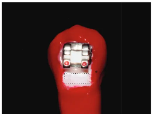
Figure 1: Photo showing a tooth with an orthodontic bracket and the enamel area that was exposed to the artiicial demineralization (dotted rectangle). he asterisk (∗) indicate the control area for QLF measurements, covered with nail varnish.
for 30 seconds and then thoroughly washed and dried. Trans-bond XT (3M, Monrovia, USA) was applied on the etched enamel and light-polymerized for 20 seconds. Brackets (Mini Diamond VS, Ormco, Orange, California, USA) were then placed 2 mm gingivally to the buccal cusp tip and in the mesiodistal center of the clinical crown and bonded with Transbond XT adhesive resin. Ater using a dental scaler to remove any residual adhesive around brackets, the resin was light-polymerized for 40 seconds (Elipar Freelight, 3M, Seefeld, Bavaria, Germany). Aterward, the dental wax was removed from each tooth.
he crowns and roots of the teeth were sealed with two coasts of acid resistant enamel (Colorama, Ceil, Com Exp Ind Ltda, S˜ao Paulo, SP, Brazil), leaving only a rectangular area measuring 2.5 mm ×2 mm exposed in the cervical region of the bracket for induction of the artiicial demineralization process [14] (Figure 1).
2.3. Microbiological Caries Induction. he teeth were im-mersed in a cariogenic solution (pH around 4.0) containing 3.7 g of brain heart infusion culture supplemented with 0.5 g of yeast extract (Becton Dikinson and Company), 1.0 g of glucose (Synth; LabSynth, S˜ao Paulo, SP, Brazil), and 2.0 g of sucrose (Synth; LabSynth) per 100 mL distilled water. his solution was autoclaved for 20 min at 121∘C and inoculated with young primary culture ofStreptococcus mutans(ATCC 25175; Tropical Culture Collection, Andre Tosello Research Foundation, Campinas, SP, Brazil). he teeth were incubated in a microaerophilic environment at 37∘C in a candle jar (BBL GasPak system, Becton-Dickinson, Franklin Lakes, USA) for 9 days. Every 48 hours, the teeth were transferred to another beaker containing a new artiicial caries solution without inoculation of new microorganisms [15–17].
he bioilm formed on tooth surfaces was removed with gauze and the nail varnish was removed manually with a scalpel blade. he teeth were copiously washed in deionized water, revealing a white spot lesion adjacent to orthodontic bracket.
analysis, which served as the gold standard for validation of mineral loss and lesion depth.
2.4. Groups. hirty teeth were randomly allocated to three groups (� = 10). In the luoride group (F) WSLs were treated with 5% NaF varnish (Duraphat, Colgate Palmolive, Hamburg, Germany). In the antimicrobial group (CHX), WSLs were treated with 2% chlorhexidine gel (Clorexal gel 2%, biodinˆamica, Ibipor˜a, PR, Brazil). Both agents (F or CHX) were applied using a swab, two times with an interval of one week between applications. Ater 24 hours the remaining luoride varnish or chlorhexidine was removed with a scalpel. In the control (CO) group, no professional treatment was performed. he teeth were only rubbed with cotton swabs imbibed with deionized water.
During the experiments the teeth were individually kept in 5 mL of artiicial saliva (1.45 mM CaCl2⋅2H2O, 5.4 mM KH2PO4, 0.1 M Tris bufer, 2.2 g/l porcine gastric mucin, pH 7.0) at 37∘C, which was changed every week.
2.5. QLF and TMR. he primary outcome was the change in luorescence measured using a QLF (Inspektor Dental Care BV, Amsterdam, he Netherlands), at the following time intervals: before and ater caries induction, 1 week ater each application, and 1, 2, and 3 months ater the last application ofFor CHX.
he QLF measurements were performed in an envi-ronment with low light. he handpiece of the device was positioned parallel to the buccal surface. he image was cap-tured and analyzed using the Inspektor Pro sotware program (version 2.0.0.32, Inspektor Dental Care BV, Amsterdam, he Netherlands) to delimit the WSL. he changes in luorescence values were determined by the percentage diference between sound and demineralized areas of each test site.ΔFvalue was then recorded, considering a 5% limited level [18].
At the end of the experimental time and when the measurements with QLF had been completed (3 months later) the teeth were prepared for microradiographic analysis. he brackets were carefully debonded from the tooth surface with a bracket remover. he teeth were sectioned once with a diamond band saw, perpendicularly to the lesion to obtain tooth slices with a thickness of approximately 500�m. he tooth slices were then manually ground with water-cooled silicon carbide discs (600-, and 1200-grade papers; Buehler, Lake Bluf, USA) to a thickness of 80–100�m.
he slices were ixed in a sample-holder together with an aluminum calibration step wedge with 11 steps. A microra-diograph was taken using an X-ray generator (Sotex, Tokyo, Japan) on the glass plate at 20 kV and 20 mA (at a distance of 42 cm) for 20 min. he glass plates were developed for 5 min, rinsed in deionized water, ixed for 3 min in a dark environ-ment, and then rinsed in running water for 10 min and air-dried (all procedures were performed at 20∘C). he developed plate was analyzed using a transmitted light microscope itted with a 20x objective (Zeiss, Germany), a CCD camera (Canon, Japan), and a computer. he images were taken using a data-acquisition sotware program (version 2012) and interpreted using calculation (version 2006) sotware programs from Inspektor Research System BV (Amsterdam,
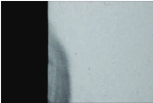
Figure 2: TMR image of a representative specimen ater microbio-logical caries induction.
Figure 3: QLF image showing demineralized area adjacent to orthodontic bracket.
he Netherlands). Five parameters were obtained: the lesion depth (LD), the integrated mineral loss (ΔZ), the average mineral loss over the lesion depth (R), the mean thickness of the “pseudointact” surface layer (SL), and the maximum mineral content of the surface layer (�max).
2.6. Statistical Analysis. he data were analyzed using Graph-Pad Instat version 4.0 (San Diego, USA) and BioEstat version 5.0 (Tef´e, AM, Brazil) sotware programs. he data set for each variable were evaluated concerning their distribution using the Kolmogorov-Smirnov test. he follow-upΔFwas analyzed using repeated-measures ANOVA and the post hoc Tukey test. he mineral loss (ΔZ) and lesion depth (LD) were compared using the Kruskal-Wallis test followed by Dunn’s multiple comparison test and ANOVA, respectively. To analyze a possible relationship betweenΔFand ΔZthe data were submitted to linear regression. he correlation between these measures was evaluated using Pearson’s cor-relation coeicient. he level of signiicance for all tests was set at 5%.
3. Results
he microbiological caries induction model was able to produce a subsurface lesion (Figure 2) that was detected by the QLF (Figure 3).Table 1 shows an overview of all TMR parameters ater microbiological caries induction.
he results of the QLF measurements are summarized
Table 1: Summary for all TMR parameters (mean±SD).
Mineral loss Lesion depth Ratio Lesion width hickness SS layer Lesion body
(Vol%,�m) (�m) (Vol%) (�m) (�m) (�m, Vol%) (�m, Vol%)
10174.4±2060.3 248.3±58.4 42.2±9.24 18.3±6.6 42.8±5.9 88.0±44.6 33.3±5.6
n= 10, ater microbiological caries induction.
Table 2: Mean and SD of the luorescence values for each group during the experimental time.
Group Baseline Ater artiicial induction of WSL
1 week ater the 1st application
1 week ater the
2nd application 1 month 2 months 3 months
� −7.04±0.83aA −13.03±3.77aB −8.77±2.01aA −6.92±0.52aA −7.82±2.25aA −7.60±1.88aA −8.03±1.89aA CHX gel−7.32±0.96aA −13.84±5.24aB −11.42±4.77aB −8.05±1.69abA −8.34±1.38aA −8.25±0.99aA −8.10±0.94Aa Control −6.84±1.47aA −12.42±2.45aB −10.98±3.89aB −11.28±3.19bB −10.12±1.78aB −10.20±1.73aB −8.58±0.063aA Diferent lower case letters within the same column show signiicant diferences among the treatments. Diferent capital letters within the same row show signiicant diferences among the periods of remineralization (repeated-measures ANOVA and Tukey’s tests).
Table 3: Summary and statistical comparison for TMR parameters ater WSL induction and 3 months ater the last application of�or CHX (mean±SD).
TMR parameter Ater artiicial induction of WSL F* CHX* Control*
Δ�(%vol/�m) 10174.4±2060.3a 7459±960.1b 7670±7699.6b 7608±7608b
Lesion depth (�m) 248.3±58.5A 224.43±76.3A 266.7±87.2A 208.9±92.8A Diferent superscript letters in the same line show signiicant diference among the groups (Kruskal-Wallis forΔ�and ANOVA for lesion depth).
*
Performed at the end of the experimental time interval, 3 months ater the last application of�or CHX.
signiicantly increased the luorescence values, which were kept constant throughout the experimental period. Whereas, the increase in luorescence values for CHX was observed 1 week ater the second application, which was kept constant throughout the experimental period. It was only in the third month that the luorescence values of the control group returned to being similar to those at baseline. When the groups were compared at baseline, ater artiicial WSL induc-tion, and, at 3 months, no statistically signiicant diferences were observed between them (� > 0.05).
Table 3 shows the comparison of the ΔZ (%vol/�m)
and lesion depth (�m) values obtained ater artiicial WSL induction and ater the third month. heΔZparameter values were statistically similar between GroupsF, CHX, and control ater 3 months, however, difering from the values presented at baseline. he lesion depth remained constant throughout the experimental period (Table 3) (� = 0.4212).
he Pearson correlation coeicient indicated moderate
(� = 0.63, 95% CI = 0.35–0.81) but statistically signiicant
positive correlation betweenΔZ(TMR) andΔF(QLF) (� =
0.0002) (Figure 4).
4. Discussion
Laboratory models have been widely applied in Cariology, allowing analysis of the diferent remineralizing treatments for WSLs [14, 19]. he microbial method is one of the protocols most used to induce caries lesion in dentin [15–
17]. his was the irst study to adapt this model to produce enamel WSL in permanent teeth. he main advantage of the microbial method in comparison with the abiotic types is the
4000 6000 8000 10000 12000 14000
M
ea
n
fl
u
o
res
cence loss, Q
L
F
−6
−7
−8
−9
−11 −10
TMR (vol%/𝜇m)
Figure 4: Correlation between QLF and TMR.
similarity to the clinical condition, considering the presence of bioilm and cariogenic challenges.
we found a correlation coeicient of 0.63, in agreement with the literature. herefore, QLF seems to be a valid method to quantify demineralization and monitor the treatment of WSLs [21,22].
Orthodontic patients develop signiicantly more WSLs than nonorthodontic patients, which might compromise the inal result of treatment. In addition, the progression of caries is faster in patients with full orthodontic appliances. WSLs can become noticeable around the brackets within 1 month ater bracket placement, although the formation of regular caries usually takes at least 6 months [23]. herefore, early diagnosis enables the clinician to implement minimally invasive treatments with the use of remineralizing therapies, with the goal of paralyzing lesion progression.
In our study, lesions treated withFvarnish showed faster remineralization than the other treatments. Remineralization with the application ofFvarnish was stable throughout the period of 3 months. he beneit of this application regime could be the precipitation of CaF2-like layer on the enamel, thus increasing the remineralization of predemineralized enamel [24]. Our results support those of previous studies [6, 25] in which quantitative analyses were performed, and it was shown that if the frequency of professional luoride application were increased, higher mineral contents could be obtained. It is important to note that the inhibition of enamel demineralization and the enhancement of remineralization are positively but not linearly related to the concentration of luoride [25]. We do, however, believe that since brack-ets favor bioilm accumulation, they could also favor the retention of varnish, thereby increasing the contact time with enamel. Consequently the brackets would prolong the reactivity of NaF with the tooth surface [26] and justify its use in orthodontic patients with active WSLs.
he fact that CHX is an agent frequently indicated for chemical bioilm control in patients with orthodontic appliances, led us to evaluating its possible efect on the remineralization of active white spot lesions. Our results showed that two applications of 2% CHX gel increased the luorescence values. hese results cannot be attributed exclusively to the antimicrobial property of CHX, which allows us to hypothesize that its efect on the remineralization of WSL may be due to electrostatic links with the phosphate groups present in the hydroxyapatite of the dental structure and artiicial saliva, which could favor the precipitation of phosphate salts on the reactive surface of demineralized enamel [27].
Although the results of this in vitro study were positive, the clinical situation is diferent, because remineralization without treatment (control group) is seldom achieved, par-ticularly when we consider the high risk of the orthodontic patient and the microbiological dynamics of the oral envi-ronment. We believe that the absence of cariogenic bioilm and carbohydrates, in addition to the presence of conditions favorable to remineralization (storage of teeth in artiicial saliva containing Ca and P ions) may have had an inluence on the increase in mineral content in the control group.
Further studies, using models more close to the in vivo condition, are necessary to understand the action of CHX. Furthermore, it is important to evaluate the efect of
frequency of application ofFor CHX, associated (or not) with oral hygiene instruction, on the remineralization of WSL in vivo, to establish a better clinical protocol, showing eiciency and a good cost-beneit for orthodontic patients with active WSL.
5. Conclusion
Two applications of luoride varnish or 2% chlorhexidine gel with a one-week interval were efective in controlling WSLs adjacent to orthodontic brackets. However, the F varnish showed a faster action, which might be an advantage in the clinical condition. QLF was efective in detecting dem-ineralization and remdem-ineralization adjacent to orthodontic brackets.
Conflict of Interests
he authors declare that there is no conlict of interests regarding the publication of this paper.
Acknowledgment
he authors thank the Fundac¸˜ao de Amparo `a Pesquisa do Estado de S˜ao Paulo, FAPESP (Grant 2011/21012-1) for funding the study. he funder had no role in the study design, data collection and analysis, decision to publish, or preparation of the paper.
References
[1] Y. Liu, Y. Zhang, L. Wang, Y. Guo, and S. Xiao, “Prevalence of porphyromonas gingivalis four rag locus genotypes in patients of orthodontic gingivitis and periodontitis,”PLoS ONE, vol. 8, no. 4, Article ID e61028, 2013.
[2] C. M. Moriyama, J. A. Rodrigues, A. Lussi, and M. B. Diniz, “Efectiveness of luorescence-based methods to detect in situ demineralization and remineralization on smooth surfaces,”
Caries Research, vol. 48, no. 6, pp. 507–514, 2014.
[3] J. A. Cury and L. M. A. Tenuta, “Enamel remineralization: controlling the caries disease or treating early caries lesions?”
Brazilian Oral Research, vol. 23, supplement 1, pp. 23–30, 2009.
[4] B. Øgaard, E. Larsson, T. Henriksson, D. Birkhed, and S. E. Bishara, “Efects of combined application of antimicrobial and luoride varnishes in orthodontic patients,”American Journal of
Orthodontics & Dentofacial Orthopedics, vol. 120, no. 1, pp. 28–
35, 2001.
[5] V. C. C. Marinho, H. V. Worthington, T. Walsh, and J. E. Clarkson, “Fluoride varnishes for preventing dental caries in children and adolescents,”he Cochrane Database of Systematic
Reviews, vol. 7, Article ID CD002279, 2013.
[6] J. J. Jardim, M. A. Pagot, and M. Maltz, “Artiicial enamel dental caries treated with diferent topical luoride regimes: an in situ study,”Journal of Dentistry, vol. 36, no. 6, pp. 396–401, 2008. [7] Y. Ren, M. A. Jongsma, L. Mei, H. C. van der Mei, and H.
J. Busscher, “Orthodontic treatment with ixed appliances and bioilm formation-a potential public health threat?”Clinical
Oral Investigations, vol. 18, no. 7, pp. 1711–1718, 2014.
[8] E. Sari and I. Birinci, “Microbiological evaluation of 0.2% chlorhexidine gluconate mouth rinse in orthodontic patients,”
[9] P. James, C. Parnell, and H. Whelton, “he caries-preventive efect of chlorhexidine varnish in children and adolescents: a systematic review,”Caries Research, vol. 44, no. 4, pp. 333–340, 2010.
[10] G. Rolla and B. Melsen, “On the mechanism of the plaque inhibition by chlorhexidine,”Journal of Dental Research, vol. 54, no. 2, supplement, pp. 57–62, 1975.
[11] O. Yazio˘glu and H. Ulukapi, “he investigation of non-invasive techniques for treating early approximal carious lesions: an in vivo study,”International Dental Journal, vol. 64, no. 1, pp. 1–11, 2014.
[12] M. H. Spiguel, M. F. Tovo, P. F. Kramer, K. S. Franco, K. M. Alves, and A. C. Delbem, “Evaluation of laser luorescence in the monitoring of the initial stage of the de-/remineralization process: an in vitro and in situ study,”Caries Research, vol. 43, no. 4, pp. 302–307, 2009.
[13] N. J. Cochrane, G. D. Walker, D. J. Manton, and E. C. Reynolds, “Comparison of quantitative light-induced luorescence, digital photography and transverse microradiography for quantiica-tion of enamel remineralizaquantiica-tion,”Australian Dental Journal, vol. 57, no. 3, pp. 271–276, 2012.
[14] S. M. Behnan, A. O. Arruda, C. Gonz´alez-Cabezas, W. Sohn, and M. C. Peters, “In-vitro evaluation of various treatments to prevent demineralization next to orthodontic brackets,”
Amer-ican Journal of Orthodontics and Dentofacial Orthopedics, vol.
138, no. 6, pp. 712.e1–712.e7, 2010.
[15] C. Motisuki, L. M. Lima, E. S. Bronzi, D. M. P. Spolidorio, and L. Santos-Pinto, “he efectiveness of alumina powder on carious dentin removal,”Operative Dentistry, vol. 31, no. 3, pp. 371–376, 2006.
[16] M. Marquezan, F. N. P. Corrˆea, M. E. Sanabe et al., “Artiicial methods of dentine caries induction: a hardness and morpho-logical comparative study,”Archives of Oral Biology, vol. 54, no. 12, pp. 1111–1117, 2009.
[17] H. A. Ricci, D. L. S. Schefel, C. A. de Souza Costa, F. J. dos Santos, J. Jafelicci, and J. Hebling, “Wettability of chlorhexidine treated non-carious and caries-afected dentine,” Australian
Dental Journal, vol. 59, no. 1, pp. 37–42, 2014.
[18] E. de Josselin de Jong, F. Sundstr¨om, H. Westerling, S. Tranaeus, J. J. ten Bosch, and B. Angmar-M˚ansson, “A new method for in vivo quantiication of changes in initial enamel caries with laser luorescence,”Caries Research, vol. 29, no. 1, pp. 2–7, 1995. [19] M. A. R. Buzalaf, A. R. Hannas, A. C. Magalh˜aes, D. Rios, H.
M. Hon´orio, and A. C. B. Delbem, “Ph-cycling models for in vitro evaluation of the eicacy of luoridated dentifrices for caries control: strengths and limitations,”Journal of Applied Oral
Science, vol. 18, no. 4, pp. 316–334, 2010.
[20] A. F. Hall, E. DeSchepper, M. Ando, and G. K. Stookey, “In vitro
studies of laser luorescence for detection and quantiication of mineral loss from dental caries,”Advances in Dental Research, vol. 11, no. 4, pp. 507–514, 1997.
[21] A. Aljehani, S. Tranæus, C.-M. Forsberg, B. Angmar-M˚ansson, and X.-Q. Shi, “In vitro quantiication of white spot enamel lesions adjacent to ixed orthodontic appliances using quan-titative light-induced luorescence and DIAGNOdent,” Acta
Odontologica Scandinavica, vol. 62, no. 6, pp. 313–318, 2004.
[22] J. Gomez, I. A. Pretty, R. P. Santarpia III et al., “Quantitative light-induced luorescence to measure enamel remineralization in vitro,”Caries Research, vol. 48, no. 3, pp. 223–227, 2014. [23] B. Øgaard, G. Rølla, and J. Arends, “Orthodontic appliances
and enamel demineralization. Part 1. Lesion development,”
American Journal of Orthodontics & Dentofacial Orthopedics,
vol. 94, no. 1, pp. 68–73, 1988.
[24] G. L. Vogel, “Oral luoride reservoirs and the prevention of dental caries,”Monographs in Oral Science, vol. 22, pp. 146–157, 2011.
[25] M. D. Lagerweij and J. M. Ten Cate, “Remineralisation of enamel lesions with daily applications of a high-concentration luoride gel and a luoridated toothpaste: an in situ study,”Caries
Research, vol. 36, no. 4, pp. 270–274, 2002.
[26] C. E. Fern´andez, L. M. A. Tenuta, P. Z´arate, and J. A. Cury, “Insoluble naf in duraphat may prolong luoride reactivity of varnish retained on dental surfaces,”Brazilian Dental Journal, vol. 25, no. 2, pp. 160–164, 2014.
[27] D. N. Misra, “Interaction of chlorhexidine digluconate with and adsorption of chlorhexidine on hydroxyapatite,”Journal
of Biomedical Materials Research, vol. 28, no. 11, pp. 1375–1381,
Submit your manuscripts at
http://www.hindawi.com
Hindawi Publishing Corporation
http://www.hindawi.com Volume 2014
Oral Oncology
Journal ofDentistry
International Journal ofHindawi Publishing Corporation
http://www.hindawi.com Volume 2014
Hindawi Publishing Corporation
http://www.hindawi.com Volume 2014 International Journal of
Biomaterials
Hindawi Publishing Corporation
http://www.hindawi.com Volume 2014
BioMed
Research International
Hindawi Publishing Corporation
http://www.hindawi.com Volume 2014
Case Reports in
Dentistry
Hindawi Publishing Corporation
http://www.hindawi.com Volume 2014
Oral Implants
Journal ofHindawi Publishing Corporation
http://www.hindawi.com Volume 2014
Anesthesiology Research and Practice
Hindawi Publishing Corporation
http://www.hindawi.com Volume 2014
Radiology
Research and Practice
Environmental and Public Health Journal of
Hindawi Publishing Corporation
http://www.hindawi.com Volume 2014
The Scientiic
World Journal
Hindawi Publishing Corporation
http://www.hindawi.com Volume 2014
Hindawi Publishing Corporation
http://www.hindawi.com Volume 2014
Dental Surgery
Journal ofDrug Delivery
Journal of Hindawi Publishing Corporationhttp://www.hindawi.com Volume 2014
Hindawi Publishing Corporation
http://www.hindawi.com Volume 2014
Oral Diseases
Journal ofHindawi Publishing Corporation
http://www.hindawi.com Volume 2014
Computational and Mathematical Methods in Medicine
Scientifica
Hindawi Publishing Corporation
http://www.hindawi.com Volume 2014
Pain
Research and Treatment
Hindawi Publishing Corporation
http://www.hindawi.com Volume 2014 Hindawi Publishing Corporation
http://www.hindawi.com Volume 2014
Endocrinology
International Journal ofHindawi Publishing Corporation
http://www.hindawi.com Volume 2014
Hindawi Publishing Corporation