Evaluation of antimicrobial activity of orthodontic
adhesive associated with chlorhexidine-thymol
varnish in bracket bonding
Carolina Freire de Carvalho Calabrich*, Marcelo de Castellucci e Barbosa**, Maria Regina Lorenzetti Simionato***, Rogério Frederico Alves Ferreira****
Objective: To assess the antimicrobial activity resulting from the association of an orthodon-tic adhesive with chlorhexidine-thymol varnish. Methods: Thirty-two extracted human pre-molars were used, divided into four groups. In Group 1, the control group, the adhesive used to bond the bracket was not associated with any antimicrobial agent. Groups 2, 3 and 4 were bonded with an adhesive system associated with chlorhexidine-thymol varnish. Groups 3 and 4 were stored in water for 7 days and 30 days, respectively, while the specimens from group 2 were, soon after bonding, placed on agar seeded with Streptococcus mutans for 48 hours, at 37º C. Results: The experimental groups, with the exception of the control group, showed antimicrobial activity whose action tended to decline commensurately with the amount of time that they remained immersed in water. Conclusions: The association of chlorhexidine-thymol varnish with an adhesive system used in orthodontics proved to be advantageous due to its antimicrobial activity.
Abstract
Keywords: Chlorhexidine. Adhesives. Antimicrobial agents.
* Orthodontist, Center of Orthodontics and Dentofacial Orthopedics Prof. José Édimo Soares Martins, UFBA. ** MSc in Dental Clinic, UFBA. Professor of Orthodontics, UFBA.
*** Professor of Oral Microbiology, USP.
**** MSc in Orthodontics, UNICAMP. Associate Professor of Orthodontics, UFBA.
INTRODUCTION
Nowadays, the use of orthodontic appliances is widespread. However, these appliances can be associated to difficulty in cleaning. During treat-ment, retentive areas are created that favor bio-film accumulation and bacterial growth. One of the greatest challenges in orthodontics consists in maintaining proper oral hygiene during treat-ment. Brackets, bands and other accessories fur-ther aggravate these condition by retaining dental plaque, which can lead to gingivitis and enamel
demineralization, causing white spots and caries.8
Thus, orthodontic treatment success lies in correcting occlusion in the best possible manner without, however, affecting the pre-existing health of teeth and supporting tissues. Otherwise, treatment benefits may be ques-tioned.30 Orthodontic practice undergoes
con-stant progress with the use of new techniques and materials that benefit both patients and practitioners.2 Attempts to inhibit the
devel-opment of carious lesions in orthodontic pa-tients have been focused on controlling the bacterial biofilm around the brackets.8 During
therapy, orthodontists are also responsible for caries prevention.30
In order to reduce the appearance of decal-cified areas around the brackets, authors have suggested the use of orthodontic bonding res-ins which either contain or are associated with antimicrobial agents.2,17
In orthodontics, composite materials are generally used for bonding brackets. These composites can act as a source of nutrition and agglomeration of opportunistic bacteria.8 Hahn
et al13 concluded that microorganisms
accu-mulate around restorative materials. Moreover, it has been previously reported that compos-ites do not exhibit antibacterial activity after polymerization.6 It would be convenient to
modify existing materials to perform addition-al functions. Considering that these materiaddition-als would already be present in the mouth, they could serve as reservoirs or platforms for the dispersal of therapeutic agents.21
According to Korbmacher et al,17
orthodon-tic bonding systems that release antimicrobial agents to adjacent areas are useful because they reduce the need for patient compliance and can potentially decrease decalcification.
It has been suggested that the incorpora-tion of chlorhexidine could impart antibacte-rial properties to composites. Chlorhexidine is a cationic clorophenylbiguanide with antimicro-bial properties and affinity for oral structures.
Its bactericidal activity results from coagulation of bacterial cytoplasm with subsequent rupture of cell membrane.24 This agent is considered the
gold standard compared to other substances designed to interfere with biofilm formation and development of gengivitis.3 Its spectrum is
broad, covering Gram-positive and Gram-neg-ative bacteria, yeasts, dermatophiles and some lipophilic viruses, in addition to having a selec-tive effect on Streptococcus mutans.25
However, composite resins are considered clinically insoluble since their components re-main trapped inside and experience great dif-ficulty in being released because the resin com-ponents restrict their displacement.6 Ribeiro
and Ericson,22 however, observed antimicrobial
activity after combining a composite resin with chlorhexidine to release antimicrobial compo-nents, although such activity decreased with time. Ehara et al,10 however, concluded that
res-ins that release antibacterial agents have certain drawbacks, since their effects are transitory and decrease over time, they also impair mechanical properties and are potentially toxic.9
Bishara et al1 and Damon et al7 found that a
combination of chlorhexidine and orthodontic resins yielded sufficient shear strength for use in orthodontics, provided that the varnish is pre-mixed with the resin, applied to the etched enamel and cured. Karaman and Uysal15 agreed
that shear strength becomes clinically accept-able when the varnish has been mixed with the resin in a 2:1 ratio, respectively.
The association of orthodontic bonding ma-terials with chlorhexidine is useful as it is an adjunctive method to prevent the appearance of white spot lesions and caries around the brack-ets. It could play an important role as an auxil-iary tool in preventing the demineralization of tooth enamel surfaces, thereby preserving the teeth during orthodontic treatment.
association of an orthodontic adhesive system with chlorhexidine-thymol varnish.
MATERIAL AND METHODS
An experimental, transverse laboratory study using 32 human premolars with healthy buccal surfaces, extracted for orthodontic pur-poses was conducted.
In this research, a modified version of the culture technique described by Ribeiro23 for
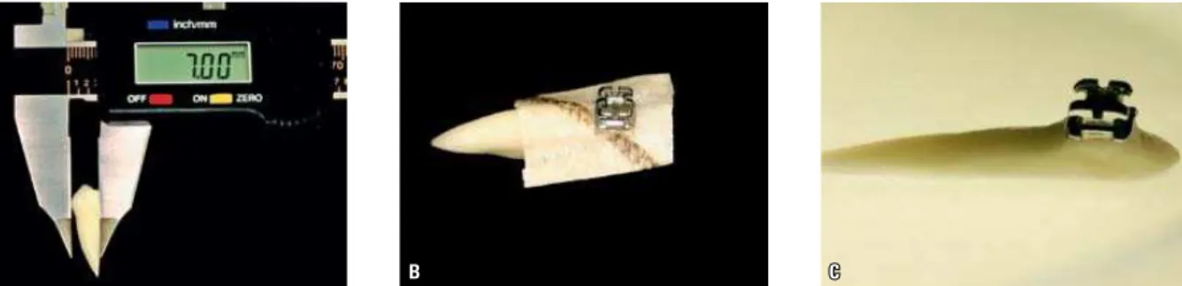
verification of growth inhibition was used. The lingual surfaces of the teeth were flat-tened, both crown and root, so that the teeth had a buccolingual width of about 7 mm (Fig 1, A), and could be laid on a flat surface with their buccal surfaces facing upward. The me-sial and distal surfaces of all teeth were also abraded down to 3.8 mm width, to match the width of the bracket to be bonded. The bond-ing area was demarcated with adhesive tape so that only the bracket base area was exposed and came into contact with the material being evaluated. The specimens were then subject-ed to a complete cycle of autoclaving (120ºC for 20 minutes). Then, the brackets (Morel-liTM, Brazil) were bonded in aseptic conditions
and in a laminar flow, after sterilization of the bonding materials. The center of the buccal surface of each tooth was etched with phos-phoric acid at 37% for 15 seconds, then washed with pyrogen-free sterile water and the etched enamel was dried with sterile filter paper.
This bonding protocol was described by Martinez,18 who achieved adequate shear
strength with this method. In group 1 (con-trol), 9µL of Ortho Primer MorelliTM was
ap-plied to each tooth, waiting up to 30 seconds for it to dry off slightly and curing it for 20 seconds. The metal brackets (MorelliTM) with
the composite on them (Transbond XT, 3M, USA) were seated on each tooth with the aid of orthodontic tweezers, positioned and cured for 10 seconds on each side.
In Groups 2, 3 and 4 (Table 1) a mixture of 6µL chlorhexidine varnish (Cervitec, Ivo-clar-VivadentTM, Swiss) and 3µL primer
(Or-tho Primer MorelliTM) at a ratio of 2:1 was
used. This ratio is advocated in the literature as providing adequate mechanical properties for clinical use in orthodontics.2,7,15,18,22 The
mixture was applied to the etched enamel surface and light-cured for 20 seconds. The metal brackets (MorelliTM) with the composite
on them (Transbond XT, 3M) were seated on each tooth with the aid of orthodontic twee-zers, positioned and light-cured for 10 seconds on each surface (Fig 1, B). After bonding the brackets, the adhesive tapes were removed.
The specimens in group 3 were kept sepa-rately in sterile pyrogen-free water for seven days in airtight jars. The specimens in group 4 were kept under the same conditions for 30 days and the water was replaced after 15 days. Groups 1 and 2 were bonded and cul-tured within up to four hours after bonding. All groups were cultured at the same time us-ing the same bacterial culture.
With the purpose of placing each specimen on sterile Petri plates (100 mm diameter by 15 mm height), about 40 mL of Tryptic Soy Agar (TSA - Difco) culture medium were added to each plate. Each specimen was then placed in one of the plates, according to each group, with the flattened lingual surface seated on the bot-tom of the plate. Consequently, the buccal sur-faces were facing upward, so that each bracket and the enamel adjacent to it were kept free from contact with the culture medium (Fig 1, C). After complete solidification, about 15 mL of the same molten culture medium, cooled to 50°C and seeded with Streptococcus mu-tans (ATCC 25175) was added. The inoculum consisted of a suspension of 8.0 X 10¹² CFU
A B C
An amount of agar with Streptococcus mutans culture that was sufficient to cover the base of the brackets and enamel surface surrounding the bonding area without covering the tie-wings was added to each plate. The medium was let to dry off and then the plates were placed in a bacteriological incubator for 48 hours at 37ºC. After culture, the presence or absence of a zone of inhibition of bacterial growth was evaluated. In cases where a zone of inhibition was formed, its diameter was measured with the aid of a bow divider and a millimeter ruler. The results were subjected to statistical analysis of variance (ANOVA) and Tukey’s test.
RESULTS
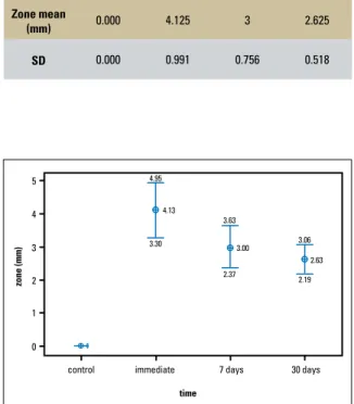
ANOVA demonstrated significant differences in the results obtained by the groups (p value = 0.000). The control group had no evidence of a zone of inhibition. In group 2, the mean value
was 4.125 mm, with standard deviation of 0.991. The mean value found in group 3 was 3 mm, with standard deviation of 0.756. In group 4, the mean value was 2.625 mm, with standard devia-tion of 0.518 (Table 2). Statistically significant differences were found between the results ob-tained in group 1 and other groups and between group 2 and other groups. Between groups 3 and 4, however, no statistically significant difference was found, although, as can be seen in Figure 2, values exhibited a declining trend.
DISCUSSION
A gold standard method is not yet available for in vitro evaluation of antimicrobial agents in bonding agents.26 Several in vitro studies have
evaluated the antimicrobial effectiveness of bonding materials by the agar diffusion meth-od.14,16 The agar diffusion test is an acceptable
method for differentiating the antimicrobial ac-tivity of substances at an early stage.29 The zones
of growth inhibition are dependent on the tox-icity of the material used against the bacteria and the diffusibility of the material inside the culture medium.11 In this study, it was used the
agar diffusion method to observe the activity of the agent against one of the most common bacteria associated with caries: Streptococcus mutans. These bacteria also feature considerable affinity for composite resins.19
FIGURE 1 - A) Abrasion of palatal surface. B) Bracket bonded to an area bounded by adhesive tape. C) Tooth fixed in agar without contact with
bracket tie-wings.
TABLE 1 - Description of study groups.
Group Storage time in
water Adhesive
Association with chlorhexidine and thymol (CervitecTM)
1 - Ortho Primer Absent
2 - Ortho Primer Present
3 7 days Ortho Primer Present
5
4
3
2
1
0
Group 1 Group 2 Group 3 Group 4
Zone mean
(mm) 0.000 4.125 3 2.625
SD 0.000 0.991 0.756 0.518
Cervitec varnish was used because it is a compound widely used as a source of chlorhexi-dine in many studies.1,7,18,23
This study further disclosed the antimicrobial action of chlorhexidine, whose effectiveness is well established in dentistry,5 where it is
associ-ated with bonding resins used in orthodontics. However, further in vitro and in vivo studies are needed to determine the clinical significance and duration of antimicrobial properties on a variety of oral cavity microorganisms involved in the pathogenicity of bacterial biofilms and caries.
Similarly to Ribeiro’s23 findings, it appears
that given the formation of a zone of inhibi-tion the combinainhibi-tion of chlorhexidine varnish and orthodontic bonding material enabled antimicrobial activity by releasing the anti-microbial substance into the culture medium,
thereby inhibiting in vitro bacterial growth in areas surrounding the bracket. It is likely that a small amount of chlorhexidine was released from the portion below the bracket since only a thin layer of adhesive associated with varnish was exposed to the culture medium.4
The reduction in the effects of chlorhexi-dine over time may be due to a reduction in the release rate or a reduction in the ac-tual amount of material present. According to Couto Júnior et al,6 although component
release seems larger at first, the decrease in this rate indicates that the components in the outer layer are depleted or dissolved in the water. On the other hand, the components trapped inside the resin mass are released with immense difficulty because resin components restrict such displacement.6 The literature
re-ports the sustained release, in aqueous envi-ronment, of compounds initially located with-in orthodontic adhesive reswith-ins for 150 days,28
or even up to two years.12
Often, the therapeutic agents of dental bio-materials are released from bio-materials and ex-hibit a decreasing release rate. The water in the oral cavity diffuses into the resin matrix. The agent trapped in the adhesive dissolves and disperses in ever smaller concentrations. Over time, the agent is released and extracted from an increasingly deeper matrix layer, which means that the time needed for diffusion to the exter-nal environment increases as the rate of release declines.21 This may also explain the absence
of statistically significant differences between the antimicrobial activity of the group that was stored in water for seven days and the group stored for thirty days. However, we observed a significant reduction in antimicrobial activ-ity between the group that was never stored in water and the group stored for seven days. It has been reported that immersion in water in the first three hours causes 50% of releasable components to be released from the resin.27
TABLE 2 - Obtained results (p = 0.000).
FIGURE 2 - Reduction tendency of the obtained zone when evaluated by the mean for each group.
control immediate
zo
n
e
(m
m
)
7 days 30 days
time
4.95
3.30
3.63
2.37
3.00 3.06
There is no way of telling how long the system will display antimicrobial activity, mainly in the oral environment. It is clear, however, that this is an association whose antimicrobial effects dis-play a decreasing trend, although it is probably an inexhaustible source of chlorhexidine. Therefore, these benefits do not last throughout the orth-odontic treatment and changes may occur in me-chanical properties after the release of the sub-stance. However, it is likely that this activity will last through the most critical period of biofilm accumulation, when proper oral hygiene is a key issue. This period spans from the time of orth-odontic appliance installation through the fol-lowing four months,20 thus justifying its benefits.
Damon et al7 and Bishara et al1 found that a
combination of chlorhexidine and orthodontic adhesives yielded sufficient shear strength for use in orthodontics when applied to the etched enamel and cured. Karaman and Uysal15 agreed
that shear strength becomes clinically accept-able when the varnish has been mixed with the
adhesive in a 2:1 ratio, respectively. Ribeiro23 and
Martinez,18 after evaluating the bond strength
of bonding systems whose adhesives had been pre-mixed with Cervitec chlorhexidine varnish, concluded that there was no statistically signifi-cant change in bond strength compared with adhesive alone. Further studies are needed to evaluate mechanical strength after the release of chlorhexidine, color stability, local and sys-temic cell and tissue compatibility, before the use of an adhesive/varnish combination in daily clinical practice is fully warranted.
CONCLUSIONS
Based on this study, it is possible to con-clude that the association of chlorhexidine varnish with an orthodontic adhesive showed antimicrobial activity in vitro, even after im-mersion in water for seven or thirty days. It was also possible to notice a decreasing trend in antimicrobial activity with the increase of immersion time in aqueous media.
1. Bishara SE, Vonwald L, Zamtua J, Damon PL. Effects of various methods of chlorhexidine application on shear bond strength. Am J Orthod Dentofacial Orthop. 1998 Aug;114(2):150-3. 2. Bishara SE, Damon PL, Olsen ME, Jakobsen JR. Effect of applying
chlorhexidine antibacterial agent on the shear bond strength of orthodontic brackets. Angle Orthod. 1996;66(4):313-6. 3. Bowen WH. Wither or whither caries research? Caries Res.
1999;33(1):1-3.
4. Chan DC, Swift EJ Jr, Bishara SE. In vitro evaluation of
a luoride-releasing orthodontic resin. J Dent Res. 1990
Sep;69(9):1576-9.
5. Cleghorn B, Bowden GH. The effect of pH on the sensitivity of species of Lactobacillus to chlorhexidine and the antibiotics minocycline and spiramycin. J Dent Res. 1989 Jul;68(7):1146-50.
6. Couto MP Jr, Nagem H Filho, Nagem HD, Couto MGP.
Determinação da taxa de lúor liberado por cinco
resinas compostas. Rev Facul Odontol Bauru. 2000 jan-jun;8(1/2):65-69.
REFERENCES
7. Damon PL, Bishara SE, Olsen ME, Jakobsen JR. Bond strength following the application of chlorhexidine on etched enamel. Angle Orthod. 1997;67(3):169-72.
8. Derks A, Katsaros C, Frencken JE, van’t Hof MA, Kuijpers-Jagtman AM. Caries-inhibiting effect of preventive measures
during orthodontic treatment with ixed appliances. Caries
Res. 2004 Sep-Oct;38(5):413-20.
9. Ebi N, Imazato S, Noiri Y, Ebisu S. Inhibitory effects of resin
composite containing bactericide-immobilized iller on plaque
accumulation. Dent Mater. 2001 Nov;17(6):485-91. 10. Ehara A, Torii M, Imazato S, Ebisu S. Antibacterial activities
and release kinetics of a newly developed recoverable controlled agent-release system. J Dent Res. 2000 Mar;79(3):824-8.
11. Estrela C, Estrela CRA, Moura J, Bammann LL. Testing calcium hydroxide antimicrobial potential by different methods. J Dent Res. 2000;79:529 (IADR Abstract 3081).
Contact address
Carolina Freire de Carvalho Calabrich Av. Araújo Pinho, nº 62, 7º andar, Canela CEP: 40.110-912 – Salvador / BA, Brazil E-mail: [email protected]
Submitted: August 2008
Revised and accepted: November 2008
13. Hahn R, Weiger R, Netuschil L, Brüch M. Microbial
accumulation and vitality on different restorative materials. Dent Mater. 1993 Sep;9(5):312-6.
14. Herrera M, Carrión P, Bravo M, Castillo A. Antibacterial activity of four dentin bonding systems. Int J Antimicrob Agents. 2000 Aug;15(4):305-9.
15. Karaman AI, Uysal T. Effectiveness of a hydrophilic primer when different antimicrobial agents are mixed. Angle Orthod. 2004 Jun;74(3):414-9.
16. Karanika-Kouma A, Dionysopoulos P, Koliniotou-Koubia E, Kolokotronis A. Antibacterial properties of dentin bonding
systems, polyacid-modiied composite resins and composite
resins. J Oral Rehabil. 2001 Feb;28(2):157-60.
17. Korbmacher HM, Huck L, Kahl-Nieke B. Fluoride-releasing and antimicrobial self-etching primer effects on the shear bond strength of orthodontic brackets. Angle Orthod. 2006 Sep;76(5):845-50.
18. Martinez TP. Avaliação da resistência ao cisalhamento de
bráquetes, colados com sistemas adesivos associados a diferentes
agentes antimicrobianos. [dissertação]. Salvador (BA).Faculdade de Odontologia, Universidade Federal da Bahia; 2006. 19. Pedrini D, Gaetti-Jardim E Jr, Vasconcelos AC. Retention
of oral microorganisms on conventional and resin-modiied glass-ionomer cements. Pesqui Odontol Bras. 2001
jul-set;15(3):196-200.
20. Petersson LG, Maki Y, Twetman S, Edwardsson S. Mutans streptococci in saliva and interdental spaces after topical applications of an antibacterial varnish in school children. Oral Microbiol Immunol. 1991 Oct;6(5):284-7.
21. Rawls HR. Preventive dental materials: sustained delivery of
luoride and other therapeutic agents. Adv Dent Res. 1991
Dec;5:50-5.
22. Ribeiro J, Ericson D. In vitro antibacterial effect of chlorhexidine added to glass-ionomer cements. Scand J Dent Res. 1991 Dec;99(6):533-40.
23. Ribeiro JLO. Avaliação da resistência adesiva e da atividade antimicrobiana de diferentes sistemas de colagem de
bráquetes associados à clorexidina e ao lúor. [dissertação].
Salvador (BA): Universidade Federal da Bahia; 2006. 24. van Rijkom HM, Truin GJ, van ‘t Hof MA. A meta-analysis of
clinical studies on the caries-inhibiting effect of chlorhexidine treatment. J Dent Res. 1996 Feb;75(2):790-5.
25. Rosa OPS, Rocha RSS. Clorexidina e cárie dentária. CECADE News. 1993 jan-ago;1(1/2):1-24.
26. Schmidlin OA, Zehnder M, Schmidlin PR. Effectiveness of dentine bonding agents against cariogenic bacteria in vitro: a comparison of two methods. Oral Microbiol Immunol. 2003 Jun;18(3):140-3.
27. Tanaka K, Taira M, Shintani H, Wakasa K, Yamaki M. Residual monomers (TEGDMA and Bis-GMA) of a set visible-light-cured dental composite resin when immersed in water. J Oral Rehabil. 1991 Jul;18(4):353-62.
28. Thompson LR, Miller EG, Bowles WH. Leaching of
unpolymerized materials from orthodontic bonding resin. J Dent Res. 1982 Aug;61(8):989-92.
29. Tobias RS. Antibacterial properties of dental restorative materials: a review. Int Endod J. 1988 Mar;21(2):155-60.
30. Zimmer BW, Rottwinkel Y. Assessing patient-speciic decalciication risk in ixed orthodontic treatment and its