The
Schistosoma mansoni
Cytochrome P450
(CYP3050A1) Is Essential for Worm Survival
and Egg Development
Peter D. Ziniel1¤a, Bhargava Karumudi2, Andrew H. Barnard1, Ethan M. S. Fisher1¤b, Gregory R. J. Thatcher2, Larissa M. Podust3, David L. Williams1*
1Department of Immunology & Microbiology, Rush University Medical Center, Chicago, Illinois, United States of America,2Department of Medicinal Chemistry and Pharmacognosy, University of Illinois College of Pharmacy, University of Illinois at Chicago, Chicago, Illinois, United States of America,3Skaggs School of Pharmacy and Pharmaceutical Sciences, University of California, San Diego, La Jolla, California, United States of America
¤a Current address: Department of Microbiology and Immunology, Uniformed Services University of the Health Sciences, Bethesda, Maryland, United States of America
¤b Current address: Illinois Mathematics and Science Academy, Aurora, Illinois, United States of America *[email protected]
Abstract
Schistosomiasis affects millions of people in developing countries and is responsible for more than 200,000 deaths annually. Because of toxicity and limited spectrum of activity of alternatives, there is effectively only one drug, praziquantel, available for its treatment. Recent data suggest that drug resistance could soon be a problem. There is therefore the need to identify new drug targets and develop drugs for the treatment of schistosomiasis. Analysis of theSchistosoma mansonigenome sequence for proteins involved in detoxifica-tion processes found that it encodes a single cytochrome P450 (CYP450) gene. Here we report that the 1452 bp open reading frame has a characteristic heme-binding region in its catalytic domain with a conserved heme ligating cysteine, a hydrophobic leader sequence present as the membrane interacting region, and overall structural conservation. The high-est sequence identity to human CYP450s is 22%. Double stranded RNA (dsRNA) silencing ofS.mansoni(Sm)CYP450 in schistosomula results in worm death. Treating larval or adult worms with antifungal azole CYP450 inhibitors results in worm death at low micromolar con-centrations. In addition, combinations ofSmCYP450-specific dsRNA and miconazole show additive schistosomicidal effects supporting the hypothesis thatSmCYP450 is the target of miconazole. Treatment of developingS.mansonieggs with miconazole results in a dose dependent arrest in embryonic development. Our results indicate thatSmCYP450 is essen-tial for worm survival and egg development and validates it as a novel drug target. Prelimi-nary structure-activity relationship suggests that the 1-(2,4-dichlorophenyl)-2-(1H-imidazol-1-yl)ethan-1-ol moiety of miconazole is necessary for activity and that miconazole activity and selectivity could be improved by rational drug design.
OPEN ACCESS
Citation:Ziniel PD, Karumudi B, Barnard AH, Fisher EMS, Thatcher GRJ, Podust LM, et al. (2015) The
Schistosoma mansoniCytochrome P450 (CYP3050A1) Is Essential for Worm Survival and Egg Development. PLoS Negl Trop Dis 9(12): e0004279. doi:10.1371/journal.pntd.0004279
Editor:John Pius Dalton, McGill University, CANADA
Received:August 18, 2015
Accepted:November 13, 2015
Published:December 29, 2015
Copyright:© 2015 Ziniel et al. This is an open access article distributed under the terms of the
Creative Commons Attribution License, which permits unrestricted use, distribution, and reproduction in any medium, provided the original author and source are credited.
Data Availability Statement:The full-length sequence of Schistosoma mansoni cytochrome P450 was submitted to GenBank and assigned the accession number KT072747.
Funding:Peter Ziniel received a Burroughs Wellcome Fund travel grant. Peter Ziniel received a stipend from the Graduate College at Rush University Medical Center. The funders had no role in study design, data collection and analysis, decision to publish, or preparation of the manuscript.
Author Summary
Over 600 million people in endemic countries are at risk of contracting schistosomiasis, which results in over 200,000 deaths each year and significant illness to most people that are infected. There are concerns that the drug widely used for the treatment of schistoso-miasis, praziquantel, may be losing efficacy due to evolution of drug resistant worms. Since the disease mainly affects the poor in developing countries, pharmaceutical compa-nies have little interest in developing new drugs and none are currently being tested. In this paper we focus on a novel parasite protein, cytochrome P450, which we propose to be a new drug target. Worms are unusual in having only one cytochrome P450 gene; humans have 57 cytochrome P450 genes. By using reverse genetic and chemical approaches we found that the schistosome cytochrome P450 is essential for worm survival and egg devel-opment and, therefore, is an essential and druggable target. Drugs that target fungal cyto-chrome P450s and are already in use for treating several human diseases were identified as potential hits for further development for schistosomiasis treatment.
Introduction
Schistosomiasis is a helminthiasis caused by trematode worms of three main schistosome spe-cies,Schistosoma mansoni,S.haematobium, andS.japonicum. The disease is responsible for approximately 280,000 deaths annually and significant morbidity in more than 200 million people [1,2]. Schistosomiasis belongs to a class of neglected tropical diseases whose control has been given limited attention by the pharmaceutic industry because they affect poor people in developing nations. Currently, praziquantel (PZQ) is the only treatment for schistosomiasis [3]. However, studies indicate that PZQ-resistant laboratory strains can be isolated and clinical isolates with increased PZQ resistance have been reported [4]. Therefore, it is a matter of time before resistance fully evolves. In addition, PZQ is much less active against juvenile worms and often results in incomplete cures [5–8] and its mechanism of action, including its biotransfor-mation are not fully understood [3].
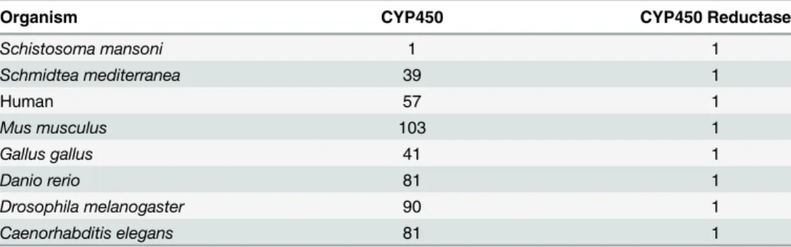
CYP450s are heme-containing monooxygenases. In concert with NADPH CYP450 reduc-tases, the heme group of CYP450s serves as a terminal oxidase, i.e., a source of electrons to split molecular oxygen, with one oxygen atom added to the substrate and the other atom accepting reducing equivalents from NADPH to form water [14]. Characterized CYP450 reductase pro-teins are well conserved and occur as single copy genes in individual organisms. However, the CYP450 proteins are quite diverse, with most organisms having multiple CYP450 genes (Table 1) [9,15]. Analysis of theS.mansonigenome database has identified only one potential CYP450 gene [16]. In a previous study, extracts of adultS.mansoniandS.haematobiumwere shown to metabolize some typical CYP450 substrates and immunoblotting experiments with an anti-rat CYP450 antibody had cross-reactivity with bothS.mansoniandS.haematobium homogenates with a specific band at ~50 kDa, well within typical CYP450 molecular weight range [17].
In addition to biotransformation activities, CYP450 proteins are involved in the metabolism of many essential endobiotic compounds. Synthesis of membrane sterols, cholesterol and ergosterol depends on CYP450s as does synthesis and degradation of steroid hormones [18,19]. Cellular levels of retinoic acid, the active metabolite of vitamin A, which is essential for embryonic development, postnatal survival, and germ cell development, are regulated and metabolized by several CYP450 proteins [18]. Other CYP450s are involved in the metabolism of prostaglandins, prostacyclins, and leukotrienes [19], all derivatives of fatty acids and impor-tant for cell signaling and immune response. InCaenorhabditis elegansCYP450 proteins are thought to be involved in meiosis, egg polarization, and egg shell development [20].
In this study, we hypothesize that the single CYP450 gene present in schistosomes is essen-tial for worm survival and that blocking its function would lead to worm death and/or interfer-ence in parasite development. We used both genetic and pharmacological approaches to test this hypothesis. Treating larval parasites withSmCYP450-specific double-stranded RNA led to significant decreases in CYP450 mRNA and resulted in worm death. Screening a collection of CYP450 inhibitors (Fig 1) we found that low micromolar concentrations of imidazole antifun-gal CYP450 inhibitors had schistosomicidal activity against adult and larval worms and blocked embryonic development in the egg. We conclude thatSmCYP450 is essential for para-site survival and egg development, and it is proposed as a novel target for antischistosomal drug development, with miconazole analogs as starting points in drug discovery.
Materials and Methods
Ethics statement
In all of the experiments involving the use of animals, maintenance and use of these animals were performed in accordance with protocols approved by the Institutional Animal Care and
Table 1. Comparison of the number of CYP450 and CYP450 reductase genes from different species compiled from Nelson et al. [15]
Organism CYP450 CYP450 Reductase
Schistosoma mansoni 1 1
Schmidtea mediterranea 39 1
Human 57 1
Mus musculus 103 1
Gallus gallus 41 1
Danio rerio 81 1
Drosophila melanogaster 90 1
Caenorhabditis elegans 81 1
Use Committee (IACUC) at Rush University Medical Center (IACUC number 14–080; DHHS animal welfare assurance number A3120-01). Animals were euthanized with a lethal dose of Nembutal.
Chemicals and reagents
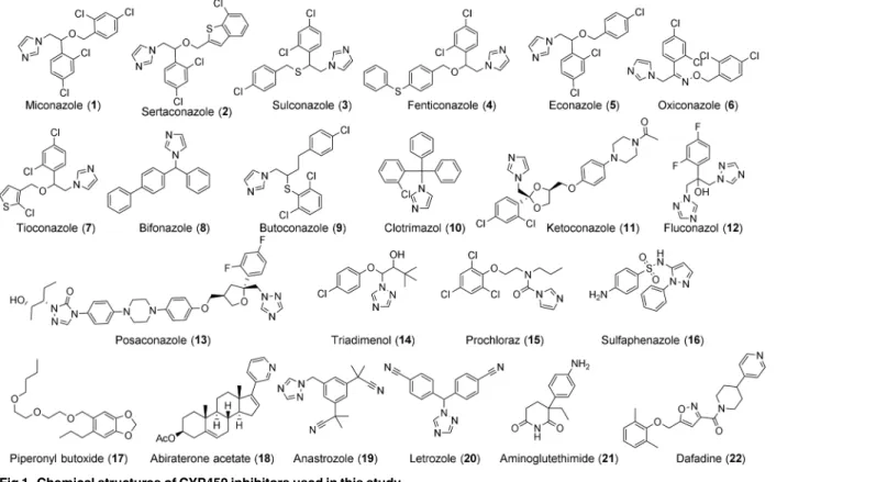
CYP450 inhibitors (Fig 1) were purchased from Sigma Aldrich (miconazole, clotrimazole, ketoconazole, posaconazole, triadimenol, sertaconazole, bifoconazole, econazole, butoconazole, dafadine, fluconazole), Santa Cruz Biotechnology (piperonyl butoxide, tioconazole, fenticona-zole, prochloraz, sulconafenticona-zole, oxiconafenticona-zole, anastrofenticona-zole, letrofenticona-zole, aminoglutethimide), and Cayman Chemical Company (abiraterone acetate). Sulfaphenazole was synthesized according to published procedures [21,22].
Experimental organisms
A Puerto Rican strain ofS.mansonimaintained inBiomphalaria glabratasnails and the same strain ofS.mansonimaintained in NIH Swiss mice was supplied by the Biomedical Research Institute (Rockville, Maryland, USA). All adult worms, schistosomula, and egg cultures were incubated in Basch’s Media 169 [23]. Basal Medium Eagle was from Life Technologies; glucose and fungizone were from Fisher Scientific; hypoxanthine, serotonin, insulin, hydrocortisone, triiodothyronine were from Sigma Aldrich; MEM vitamins, Schneider’s Drosophila Medium, and gentamicin were from Gibco; HEPES buffer from Mediatech, Inc.; penicillin/streptomycin from Cellgro; and fetal bovine serum was from HyClone Laboratories, Inc.
Cercariae were shed from infectedBiomphalaria glabratasnails and mechanically trans-formed to schistosomula as described [24]. To collect liver-stage, juvenile parasites mice were
Fig 1. Chemical structures of CYP450 inhibitors used in this study.
perfused 23 days post infection and to collect adult worms mice were perfused 6–7 weeks after infection with Dulbecco’s modified Eagle’s medium (Gibco) using methods described previ-ously [24]. Live worms were washed thoroughly with DMEM. Eggs were obtained from the liv-ers of the mice 7 weeks post infection. Livliv-ers were placed in ice-cold PBS and stored at 4°C overnight and processed the following day as described [24]. Parasite material was stored at -80°C for later use in stage specificSmCYP450 mRNA quantitation.
Analysis of the
Sm
CYP450 sequence and investigation of sequence
variation
The CYP450 open reading frame was amplified from adult mixed cDNA using P450_5' and P450_3' (all primers listed inTable 2) and GoTaq Flexi DNA Polymerase (Promega). PCR product was cloned into pCRII (Invitrogen) and plasmids were purified (Plasmid Mini Kit (QIAGEN) and sequenced at the University of Illinois-Chicago Core Sequencing Center (UIC). Alignment of the obtained open reading frame with the genome sequence was done using the Needleman-Wunsch Global Sequence Alignment Tool (http://blast.ncbi.nlm.nih. gov/Blast.cgi). Prediction of the molecular weight of the encoded protein was done at the Swiss Institute of Bioinformatics Resource Portal (http://web.expasy.org/compute_pi/).
Internal Coordinate Mechanics (ICM) homology modeling tool (http://www.molsoft.com) [25,26] was used to generate a CYP3050A1 model based on the CYP2C5 (PDB ID 1nr6) tem-plate and the structure-superimposition-guided sequence alignments performed using the iter-ative dynamic programming and superimposition steps implemented in the ICM Homology Modeling module [27]. Alignments were further adjusted manually to preserve integrity of the a-helices and b-sheets, patterns of positive (blue) and negative (red) charges, aromatic (purple) and hydrophobic (green) functionalities, and finally, proline (ochre) and cysteine (yellow) side chains. Global optimization was performed using the Biased Probability Monte Carlo (BPMC)
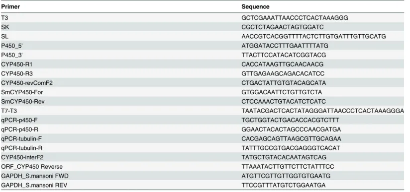
Table 2. List of primers used for PCR, RT-PCR and qRT-PCR.
Primer Sequence
T3 GCTCGAAATTAACCCTCACTAAAGGG
SK CGCTCTAGAACTAGTGGATC
SL AACCGTCACGGTTTTACTCTTGTGATTTGTTGCATG
P450_5' ATGGATACCTTTGAATTTTATG
P450_3' TTACTTCCATACATCGGTACG
CYP450-R1 CACCATAAGTTGCAACAACG
CYP450-R3 GTTGAGAAGCAGACACATCC
CYP450-revComF2 CTGACTATTGTGTACAGCATA
SmCYP450-For GTGGACAATTCTGTTGTCTA
SmCYP450-Rev CTCCAAACTGTACATCTCATC
T7-T3 TAATACGACTCACTATAGGGATTAACCCTCACTAAAGGGA
qPCR-p450-F TGCTGGTACTGACACCACGTCTTT
qPCR-p450-R GGAACTACACTAGCCCAACGATGA
qPCR-tubulin-F CACGAGCAGTTAAGCGTTGCAGAA
qPCR-tubulin-R TATTTGCCGTGACGAGGGTCACAT
CYP450-interF2 TATGCTGTACACAATAGTCAG
ORF_CYP450 Reverse TTAAATACTTGTTCTTCTATTTCC
GAPDH_S.mansoni FWD ATGTTCGTTGTTGGTGTGAATG
GAPDH_S.mansoni REV TTCCGTTTATGTCTGGAATGA
conformational search combined with the electrostatic energy term [28]. Loop search and side chain refinement was conducted for up to 100,000 iterations, which included full energy mini-mization at each step, to result in a model with satisfactory local strain parameters [29].
To determine if a subset of CYP450 mRNAs was trans-spliced, theS.mansonitrans-spliced leader sequence was used in PCR with either CYP450-R1 or CYP450-R3 specific internal prim-ers (Table 2). A modified 5’rapid amplification of cDNA ends (RACE) with Q5 DNA polymer-ase (New England Biolabs) was done in a nested PCR using an adult cDNA library (kindly provided by Dr. Philip LoVerde) as the template and vector primer T3 + gene-specific
CYP450-revComF2 in the first stage and the vector primer SK + gene-specific SmCYP450-Rev (Table 2) for the second stage. The product of the second PCR was cloned into pCR4 (Invitro-gen). To determine if theSmCYP450 mRNA is alternatively spliced, the complete ORF was amplified using Q5 DNA polymerase from adult male, adult female, and egg cDNA (synthe-sized as described below) with P450_5' and P450_3'. PCR products were cloned into pCR4. Plasmid DNAs were isolated (GeneJET Plasmid Miniprep Kit, Thermo Scientific) and sequenced at the UIC sequencing core.
RNA interference (RNAi)
Plasmid Construction. A 566 bpSmCYP450 sequence close to the N-terminal region of theSmCYP450 gene was amplified using SmCYP450-For and SmCYP450-Rev and cloned into a pCRII vector (Invitrogen) according to the manufacturer’s protocol. The sequence was veri-fied through Sanger sequencing at the UIC sequencing core. A new primer (T7-T3) was designed flanking the 566 bp sequence so that it included the full T7 promoter primer followed by part of the T3 primer sequence. PCR was carried out using T7 and T7-T3 primers with Taq DNA polymerase (Thermo Scientific) at 96°C 2 min, followed by 40 cycles at 94°C, 1 min; 48°C, 2 min; 72°C, 1.5 min; then 72°C, 7 min. The PCR product was run on a 1% agarose gel containing ethidium bromide to verify the insert size. The PCR product was cut out from the gel and cleaned with Gel Extraction kit (Qiagen) and the concentration determined.
CYP450 dsRNA Synthesis. Both a published method [30] using T7 RNA polymerase (New England BioLabs) and the MEGAclear kit (Life Technologies) were used to synthesize SmCYP450 dsRNA. In the first method, synthesis was carried out in a 100μl reaction mix
using 100μg/ml BSA (NEB), 500μM each of rNTPs (NEB), 1 x RNA Pol reaction buffer (40
mM Tris-Cl, 6 mM MgCl2, 10 mM dithiothreitol), and 800 units/ml RNase inhibitor (NEB) at
40°C for 4.5 hours. The resultant product was treated with RNAase free DNase I (NEB) at 37°C for 10 min and cleaned using Zymogen DNA-free RNA kit and eluted with DNase/RNase free water, or the DNase I treated samples were precipitated in 75% DEPC treated ethanol and 4 M LiCl and re-suspended in DEPC treated water. The concentration of the cleaned ssRNA was determined using a Nanodrop spectrophotometer. Synthesis using the MEGAclear kit followed manufacturer’s protocols with RNA products cleaned as described above. The RNAs were annealed to form dsRNA by incubating at 75°C, 50°C, and 37°C for 3 min each, and the con-centration was determined by Nanodrop spectrophotometry. A negative control dsRNA was synthesized as described above from theccdBandcamR- bacterial gene insert of pJC53.2 plas-mid obtained from Dr. James Collins (UIUC) [30].
RNAi Cultures. Freshly prepared schistosomula (300–400) were placed in each well con-taining 1 ml Basch’s media in a 24-well plate and incubated overnight in a 37°C and 5% CO2.
The following daySmCYP450 dsRNA or control irrelevant dsRNA was added to each well to a final concentration of 10 or 30μg/ml. Treatments were done in duplicate. Over several days
Inhibitor treatment
To determine the activity of CYP450 inhibitors, 10 worm pairs in 5 ml Basch’s media per well in 6-well plates were cultured overnight at 37°C and 5% CO2and the following day CYP450
inhibitors (Fig 1) were added to each well. The media were replenished every 48 hr with fresh media and inhibitors. Dead worms were identified as those that showed no motility when observed for several minutes. For larval worms, 300–400 freshly prepared schistosomula were placed in each well in a 24-well plate containing 1 ml Basch’s Media and incubated overnight at 37°C and 5% CO2. The following day compounds were added to each well and the parasites
observed for several days without changing the media or adding fresh compounds. Live and dead parasites were classified as before.
To monitor the effects of miconazole on egg development we followed a recently published method [32]. Freshly perfused adult worm pairs were incubated in Basch’s media overnight. The following day worms were removed and miconazole (5 or 10μM) or an equal volume of
DMSO was added to the eggs produced. Eggs were further incubated a total of 72 hr in the presence of miconazole. Each group of treated eggs was then collected and centrifuged (500 x g, 5 min) and the supernatant discarded. The egg pellets were each washed in excess PBS and centrifuged. The eggs were then fixed in 100% methanol at room temperature for 10 min. After removing the methanol the eggs were incubated in DAPI (4’6-diamidino-2-phenylindole) Fluoromount-G (SouthernBiotech) overnight at 4°C for nuclear staining. Images were cap-tured using Zeiss Axiovert Z1 imaging microscope and analyzed with AxioVision software LE (release 4.8.2 SP3, 2013).
Combined inhibitor and RNA interference
To see if their activities had additive effects, schistosomula were treated with dsCYP450 RNA at a concentration that alone did not kill schistosomula (10μg/mL) and miconazole at
concen-trations that resulted in minimal killing (2.5 or 5μM) or each alone. Schistosomula cultures
were set up as described above. A control experiment was set up with irrelevant dsRNA with and without 5μM miconazole. Parasites were observed as described above.
Total RNA isolation and cDNA synthesis
Total RNA was isolated from frozen worm and egg samples using the TRIzol Reagent (Life Technologies) per the manufacturer’s recommendation in a 2 ml Lysis Matrix Tubes (MP Bio-medicals) containing 500μl TRIzol reagent. Tubes were shaken three times for 20 seconds each
using a tissue homogenizer (FastPrep-24 5G Instrument, MP Biomedicals). The samples were incubated on ice for 5 minutes in between each lysis process. After lysis, another 500μl TRIzol
Quantitative RT-PCR (qPCR) and semi-quantitative RT-PCR
Primers used for qPCR are shown inTable 2.α–tubulin (GenBank accession M80214) was used to normalize the results. The reactions were each carried out in a 20μl reaction using
ROX Passive Reference Dye (Bio-Rad) according to the manufacturer’s protocol. The amplifi-cation was monitored in a 7900HT Fast Real-Time PCR Machine (Applied Biosystems) under the following cycle conditions: (stage 1, 95°C 30 sec, stage 2, 95°C 5 sec, 60 C 30 sec) x 50, plus a one cycle dissociation curve. Fold differences were calculated using the 2-ΔΔCTas described [33] withα–tubulin transcript levels serving as the internal standard. Reactions were done in triplicate. Semi-quantitative RT-PCR was used to assess the relative abundance ofSmCYP450 mRNA after RNAi silencing using Platinum Taq DNA polymerase (Life Technologies. Glycer-aldehyde 3-phosphate dehydrogenase (GenBank accession M92359) was used as a control gene (primers GAPDH_S.mansoni FWD and GAPDH_S.mansoni REV) andSmCYP450 cDNA was amplified with primers CYP450-interF2 and ORF_CYP450 Reverse.
Results
The
Sm
CYP450 coding sequence is similar to CYP450 proteins in other
organisms
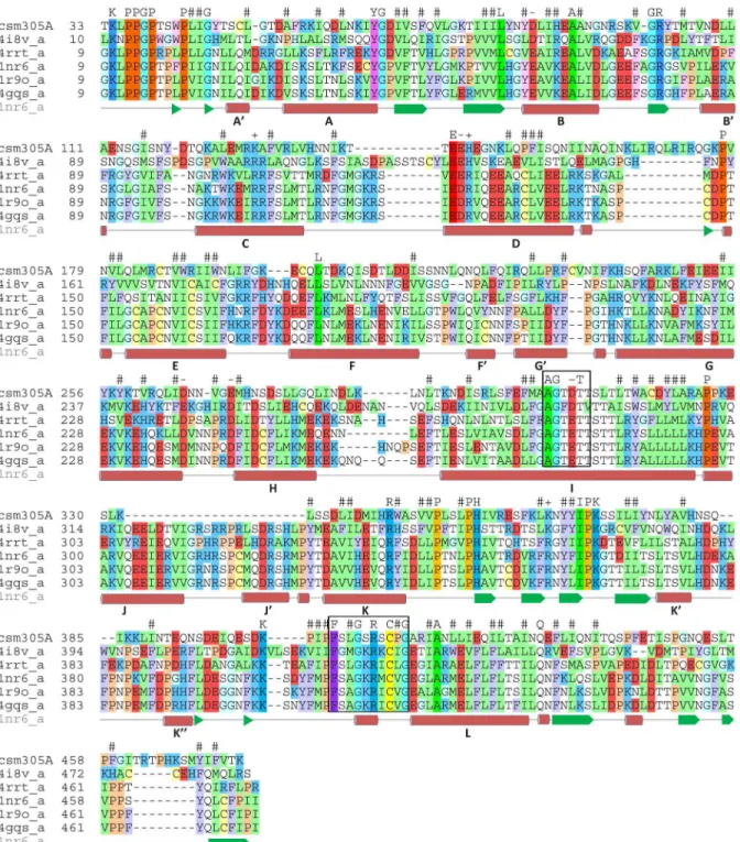
Cloning and sequence analysis shows that theSmCYP450 coding sequence is 1452 base pairs encoding a protein of 483 amino acids with a predicted molecular weight of 55.28 kDa. The family assignment as CYP3050A1 was made by Dr. David R. Nelson according to the CYP450 nomenclature [34,35]. The sequence was found to be longer than the sequence reported in GeneBank (Smp_156400, 1245 base pairs,) due to a miscalled junction of the 5thintron/6th exon during genome annotation. The sequence obtained was submitted to GenBank with the accession number KT072747. The gene is composed of 7 exons and 6 introns spanning 15,378 base pairs (not including 5’and 3’noncoding sequences). Sequence analysis shows it to be comparable to CYP450 proteins from other organisms. The signature heme-binding motif [14,36], [FW]-[SGNH]-x-[GD]-{F}-[RKHPT]-{P}-C-[LIVMFAP]-[GAD], is present (the bold, underlined residues are present inSmCYP450) (Fig 2). The‘P450-signature’sequence, [AG]-G-X-[DE]-T-[TS], which forms a channel for electron transfer [36], is also present in the SmCYP450 peptide. The protein has an N-terminal membrane spanning region followed by the poly-proline domain, which is important for protein folding and structural integrity [37]. The turns by the poly-proline region provide a junction between the transmembrane region and the main catalytic domain typical for most CYP450 proteins [37]. The organization of the predicted secondary structure of theSmCYP450 protein sequence follows other CYP450 pro-teins, beginning from helix A in the N-terminal region of the protein sequence and ending with helix L, which contains the heme-binding sequence (Fig 2). Likewise, with the exception of the absence of the J and J’helices, the tertiary structure ofSmCYP450 protein is predicted to be similar to known CYP450 proteins (Fig 3).
There is a single CYP450 protein in
S
.
mansoni
Fig 2. Comparison ofSchistosoma mansoniCYP450 protein (Sman) with CYP450 proteins from other species.Multiple alignment of CYP450 proteins fromS.mansoni(csm305A); rabbit CYP450 2C5 (1nr6_a); human CYP450 2C9 (1r9o_a); human CYP450 2C19 (4gqs_a); human CYP450 1A1 (4i8v_a); and human CYP450 2b6 (4rrt_a). The residues are shown in one letter code and colored by type: red- negatively charged, blue—positively charged, yellow—Cys, green—hydrophobic, cyan—Gly, ochre—Pro, purple—aromatic. The residues are shown in brighter colors for conserved positions. The‘P450-signature’sequence, which forms a channel for electron transfer, and the CYP450 consensus motif responsible for heme-binding and interaction with molecular oxygen and the relevant substrates are boxed. Predicted helices in the secondary structure based on homology modelling of SmCYP450 are indicated by the bold letters A-L based on rabbit CYP450 2C5 [38].
Therefore, we found no evidence for alternative splicing or other sequence variations. PCR with the spliced leader sequence and two different internal CYP450-specific primers resulted in no PCR products; therefore, the SmCYP450 mRNA does not appear to be trans-spliced. Therefore, it appears that theSmCYP450 gene encodes a single CYP450 protein.
S
.
mansoni
CYP450 is differentially expressed during parasite
development in the mammalian host
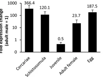
Using qRT-PCR we found thatSmCYP450 mRNA was present at all developmental stages investigated and that it is differentially present during development (Fig 4). Eggs, the larval stages of development (cercariae and schistosomula) and adult female worms had higher mRNA levels than adult male worms. Liver stage parasites had the lowestSmCYP450 mRNA expression levels, about 50% that of adult males.
Fig 3. Structural modeling ofS.mansoniCYP450 (CYP3050A1) and comparison to the structure determined for rabbit CYP450 2C5 (1nr6_a) [38].The heme is shown is each model as a space-filling projection. The J and J’helices in rabbit CYP450 2C5, which are absent inS.mansoniCYP450, are highlighted in yellow.
doi:10.1371/journal.pntd.0004279.g003
Fig 4. CYP450 messenger RNA abundance during the lifecycle ofSchistosoma mansoni.Whole RNA was extracted from different stages ofS.mansoni(cercariae, 1-day old schistosomula; juvenile liver worms (23 days post infection), adult males (49 days post infection), adult females (49 days post infection) and eggs) using TRIzol reagent and chloroform/ethanol extraction protocol. cDNA was synthesized from whole RNA and used for qRT-PCR, with reactions done in triplicate. Adult males (= 1) were used as calibrator stage and mRNA abundance was normalized toα-tubulin. Error bars indicate standard error of the mean with n3 biological replicates. Numbers indicate fold change relative to adult males and all values are significantly different from adult males; p<0.05; student t-test. The results indicate thatS.mansoniCYP450 is expressed in all stages investigated and that its expression is developmentally regulated.
S
.
mansoni
CYP450 dsRNA treatment leads to schistosomula death
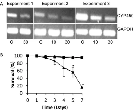
To determine ifSmCYP450 is essential for schistosomula survival we used RNAi to silence SmCYP450 expression. Treating worms with 10μg/mL or 30μg/mLSmCYP450 specificdsRNA for two or three days resulted in a dose-dependent reduction inSmCYP450 message (Fig 5A). No change was seen inSmCYP450 mRNA after treatment with 30μg/mL irrelevant
dsRNA or in GAPDH mRNA abundance after treatment with either dsRNA (Fig 5A). Treat-ment with 30μg/mLSmCYP450 specific dsRNA resulted in 80% schistosomula survival by day
3, 40% survival by day 5, and 15% survival by day 7. In contrast, 95% and 94.5% of schistoso-mula were alive on day 7 after treatment with 30μg/mL irrelevant dsRNA or 10μg/mL
SmCYP450 specific dsRNA, respectively (Fig 5B).
The imidazole subgroup of azole antifungal CYP450 inhibitors is active
against
S
.
mansoni
CYP450 enzymes are inhibited by numerous anti-infective and anticancer agents. We next asked if clinically relevant CYP450 inhibitors (Fig 1) affected parasite survival. Several gal imidazoles (miconazole, clotrimazole, ketoconazole) but not closely related triazole antifun-gals (fluconazole, posaconazole and triadimenol) were active against both larval and adult
Fig 5. Effect of silencingSchistosoma mansoniCYP450 in cultured larval worms.Freshly prepared schistosomula (300–400) were placed in each well containing 1 ml Basch’s Media in a 24-well plate and overnight in a 37°C with 5% CO2. The following day schistosomula were treated with 10 or 30μg/mlS.
mansoniCYP450 dsRNA or 30μg/ml negative control dsRNA. Over several days worms were observed for
dead (dark, granular appearance) or alive (translucent). (A) mRNA expression patterns in schistosomula treated withS.mansoniCYP450 specific dsRNA or negative control dsRNA control after 3 days of treatment (Experiments 1 and 2) or 2 days treatment (Experiment 3). The control gene for cDNA input isS.mansoni
glyceraldehyde 3-phosphate dehydrogenase (GAPDH). C, schistosomula treated with 30μg/mL irrelevant
dsRNA; 10, schistosomula treated with 10μg/mLSmCYP450 dsRNA; 30, schistosomula treated with 30μg/
mLSmCYP450 dsRNA. (B) Effect ofS.mansoniCYP450 dsRNA on schistosomula survival in cultures with 30μg/mL negative control dsRNA (black square), 10μg/mLS.mansoniCYP450-specific ds RNA (open
triangle), and 30μg/mLS.mansoniCYP450-specific ds RNA (black triangle). Treatments were done in
triplicate and repeated 3 times. Error bars indicate standard error of the mean;*, p<0.05; student t-test.
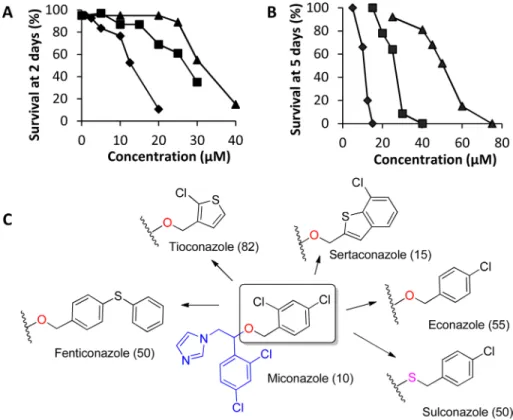
worms (Fig 6A and 6BandTable 3). Miconazole, clotrimazole, and ketoconazole had ED50
(Effective Dose producing 50% worm death) values of 10μM, 20μM, and 40μM, after 5 day
treatments against adult worms and 12.5μM, 27.5μM, and 30μM after 2 day treatments
against schistosomula, respectively. Other CYP450 inhibitors, such as prochloraz, sulfaphena-zole, piperonyl butoxide, dafadine, letrosulfaphena-zole, aminoglutethimide, abiraterone acetate, and ana-strozole had no significant schistosomicidal activity against either larval or adult worms (Table 3). Expansion of the anti-fungal imidazole series was done to generate preliminary structure activity relationships of this compound series. Our studies revealed that imidazoles that retained the 1-(2,4-dichlorophenyl)-2-(1H-imidazol-1-yl)ethan-1-ol moiety of miconazole had significant schistosomicidal activity against both larval and adult worms, while those which lacked this moiety had much reduced or no schistosomicidal activity (Table 3,Fig 6C).
Miconazole targets
Sm
CYP450
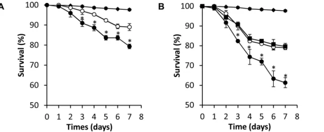
Does the potent schistosomicidal activity of miconazole act through inhibition of worm CYP450 or does it have other targets in the worm? To address this question we tested low doses of miconazole against worms treated with 10μg/mL dsRNA CYP450, which caused no
significant worm death itself. While 5μM miconazole alone resulted in 80% survival after 6
days, combinations of 5μM miconazole and 10μg/mL SmCYP450-specific dsRNA resulted in
60% survival (p = 0.0042). Combining 2.5μM miconazole (90% survival alone) and 10μg/mL
Fig 6. Activity of anti-fungal imidazole CYP450 inhibitors on larval and adultSchistosoma mansoni
worms.Survival of schistosomula (A) after 2 d culture and adult worms (B) after 5 d culture for miconazole (black diamond), clotrimazole (black square), and ketoconazole (black triangle). (C) In house SAR on known miconazole analogs against adult worms. Numbers in the parenthesis are survival (%) of adult worms on day 7 in 10μM of respective compound.
Table 3. Results with selected cytochrome P450 inhibitors used in this study.
Entry Compound Survival at 7 days (%) Function and CYP450 class inhibited
Adult Schistosomula
5μM 10μM 5μM 10μM
1 Miconazole 57 10 72 22 antifungal CYP51
2 Sertaconazole 95 15 65 6
3 Sulconazole 100 50 90 56
4 Fenticonazole 75 50 92 79
5 Econazole 100 55 87 66
6 Oxiconazole 100 60 89 71
7 Tioconazole 100 85 86 82
8 Bifoconazole 100 100 80 79
9 Butoconazole 100 80 77 60
10 Clotrimazole n.d.1 100 97 90
11 Ketoconazole n.d. 100 98 97
12 Fluconazole 100 100 98 94
13 Posaconazole 100 100 94 88
14 Triadimenol 100 100 99 99
15 Prochloraz 100 100 94 82
16 Sulfaphenazole 100 100 99 93 antibacterial CYP2C9
17 Piperonyl butoxide 100 100 100 100 pesticide CYP6D1
18 Abiraterone acetate 100 100 100 100 prostate cancer CYP17A1
19 Anastrozole 100 100 100 100 breast cancer CYP19A1
20 Letrozole 100 100 100 100
21 Aminoglutethimide 100 100 100 100
22 Dafadine 100 100 100 100 CYP27A1
1
n.d., not determined.
doi:10.1371/journal.pntd.0004279.t003
Fig 7. Combinations of miconazole and RNAi have increased killing activity, suggesting that they function through inhibition of the same target.
(A) Schistosomula cultured with 10μg/mlS.mansoniCYP450 dsRNA (black diamond); 2.5μM miconazole (open circle); or 10μg/mlS.mansoniCYP450 dsRNA and 2.5μM miconazole (black circle). (B) Schistosomula cultured with 10μg/mlS.mansoniCYP450 dsRNA (black diamond); 5μM miconazole
(black square); 5μM miconazole plus 30μg/ml irrelevant dsRNA and 5μM miconazole (open circle); or 5μM miconazole plus 10μg/mlS.mansoniCYP450 dsRNA and 5μM miconazole (black circle). All experiments were done in triplicate. Error bars indicate standard error of mean;*, p<0.05; student t-test).
SmCYP450-specific dsRNA resulted in 75% survival (p = 0.007) (Fig 7A). Addition of 30μg/
mL irrelevant dsRNA treatment had no effect on killing by 5μM miconazole (Fig 7B). These
results strongly suggest that miconazole schistosomicidal activity is specific forSmCYP450.
Miconazole treatment results in impaired schistosome egg development
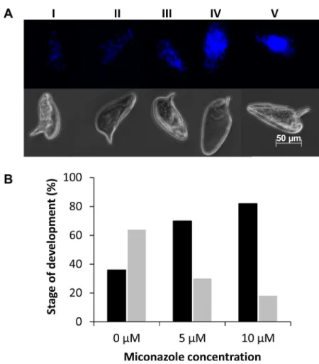
To determine if miconazole interferes with egg development and maturation we treated eggs deposited by freshly perfused adult worm pairs with miconazole and monitored embryo devel-opment using a recently described method [32,39]. Egg development was scored based on the number and arrangement of cell nuclei (Fig 8A). Our results indicate that there is a general interference of egg development and accumulation of early embryonic stages (I, II and III) and decrease in late stage embryos (IV and V) in the miconazole treatments compared to the DMSO controls. Only 30% (18/62) of eggs treated with 5μM miconazole and 18% (10/56)treated with 10μM miconazole reached the latter stages of egg development (stages IV and V)
compared to 64% (35/55) in DMSO control (Fig 8B). These results indicate that miconazole affects embryonic development.
Fig 8. Effect of miconazole on egg development.(A) Example of egg development scoring scheme. Upper panel shows fluorescent images of eggs representative of each developmental stage scored; the bottom panel shows brightfield images of the same eggs. (B) Scoring of egg development in cultured eggs treated with 0, 5, or 10μM miconazole. The percentage of eggs scored at developmental stages I-III (black bars) and
eggs scored at developmental stages IV-V (gray bars) are indicated. For 0μM miconazole, n = 55 eggs
scored; for 5μM miconazole, n = 56 eggs scored; for 10μM miconazole, n = 62 eggs scored.
Discussion
Because schistosomiasis control relies on a single drug and there is field evidence for the evolu-tion of drug resistance [3,4], there is an urgent need to identify new, druggable worm targets. In this study we present the first detailed characterization of the CYP450 fromS.mansoniand provide strong evidence that it is an essential and druggable target in the worm.
TheSmCYP450 exists as a single copy gene in theS.mansonigenome [16]. This is in stark contrast to humans, which have 57 genes and alternative splicing and genetic variations that can lead to the production of many more distinct protein species [40,41], and to the free-living flatwormSchmidtea mediterranea, which has at least 39 CYP450 genes [15]. The loss of CYP450 family members in parasitic helminths has been noted previously [42]. However, the fact that parasitic flatworms have retained one CYP450 signifies that it plays an important and perhaps essential function. We add here that inS.mansonithere appears to be no post-tran-scriptional modifications (alternative or trans-splicing, RNA editing) to the mRNA. Therefore, it is likely that a single protein product is produced from theSmCYP450 gene. Since there was no evidence for alternative splicing to insert different leader sequences at the N-terminus, the protein product is likely only targeted to the endoplasmic reticulum.
The predicted protein has generally low sequence identity with the other CYP450s; the high-est identity to human CYP450 proteins is 22% to CYP2C9. Importantly, the CYP450 consensus motif responsible for heme-binding and interaction with molecular oxygen and the relevant substrates and the‘P450-signature’sequence are conserved in theSmCYP450 protein sequence. Curiously,SmCYP450 lacks a number of motifs found in many characterized CYP450. The majority of CYP450s contain an‘EXXR motif’in helix K. The glutamic acid and arginine residues form a charge pair with a third amino acid more distant in the meander region. This is frequently an arginine in the so-called‘PERF motif’. Putative functions of the EXXR motif and PERF motif may be to associate heme with the newly synthesized CYP450 polypeptide and/or to maintain the CYP450 tertiary architecture [43]. This is key to the struc-tural fold of CYP450s and previous studies in which mutagenesis directed at the side-chains of glutamic acid or arginine in the EXXR motif or at the invariant cysteine in the L-helix resulted in completely inactive and misfolded proteins [44]. However, these motifs are not present in CYP450s from parasitic Trematodes (e.g.,Schistosoma,Clonorchis sinensis) and Cestodes (e.g., Echinococcus multilocularis) [42]. Their absence is not without precedent as the EXXR motif is also absent in most members of a CYP157 subfamily inStreptomycesspp [45]. The Trematode CYP450 proteins also lack the J and J’helices, which occur to the N-terminal side of and include the EXXR motif. How these differences affect protein structure and function remains to be determined.
Unlike the only previously characterized trematode CYP450, which showed highest expres-sion in adult hermaphrodites [42],SmCYP450 is expressed at the highest levels in larval and egg stages. It is important to note that the developmental cycles and tissue locations of these organisms are significantly different. After active host localization and penetration,S.mansoni has extensive interactions with host skin, lungs, liver, and vascular epithelia, whileOpisthorchis worms reside in the bilary ducts after excysting from metacercariae in the duodenum. As sequence identity betweenS.mansoniandO.felineusCYP450 proteins is only 37% it is quite possible that CYP450s have different functions in the worms.
The function of theSmCYP450 is not yet known. Different development stages may require different CYP450 metabolites and/or experience different immunological stresses. For instance larval parasites penetrate the skin of human host and begin migration through the skin and other tissue and may encounter different stress and immunological responses than adult worms in the mesenteric system. Larval schistosomes synthesize and secrete eicosanoids [48– 53], which are signaling molecules derived from arachidonic acid, some of which are produced by CYP450s. The eicosanoids produced by schistosomes may down modulate host immune function [54,55]. Eicosanoids produced by adult worms may control other functions such as vasodilator activity, and/or vasoconstrictive action [55].
Other potential functions ofSmCYP450 are in the metabolism of cholesterol and steroid hormones. Adult worms have been shown to convert cholesterol into several metabolites including pregnenolone, the first committed metabolite in steroid hormone biosynthesis [56,57]. Male worms transfer cholesterol and uncharacterized cholesterol metabolites to female worms [56] and synthetic steroids have been shown to affect worm egg productionin vivo[56]. More recently, a catechol-estrogen conjugate (downstream products of CYP450 metabolism of estradiol and estrone), which has anti-estrogen affects, was identified in schistosome worm extracts and in the serum of infected humans [58]. Retinoic acid is essential for embryonic development in all metazoan organisms investigated, including free-living flatworms [59]. Ret-inoic acid activity is controlled through its tightly regulated synthesis from vitamin A (all-trans retinol) in a 2-step process by retinol dehydrogenases to all-trans retinal and by retinaldehyde dehydrogenases to all-trans-retinoic acid and is terminated via its breakdown by CYP450s [18,60]. Although retinoic acid signaling or metabolism in schistosomes is largely unknown, they have enzymes involved in retinoic acid metabolism (10 retinol dehydrogenases and 2 reti-naldehyde dehydrogenases) and nuclear receptors related to retinoic acid receptors [61–64]. Ecdysteroids are hormones involved in insect molting and development and CYP450s are involved in their synthesis and transformation from farnesyl diphosphate and cholesterol. Ecdysteroids have been detected in schistosomes and their levels shown to vary during devel-opment [65,66].S.mansonisynthesizes ecdysone and 20-OH ecdysone, which were shown to be potent stimulators of growth and vitellogenesis [67].β-Ecdysterone was found to be effective in stimulating host location activities inS.mansonimiracidia [68]. Worms have two nuclear receptors related to insect ecdysone receptors, but their function in ecdysteroid signaling has not been determined [69,70]. Identification of the function ofSmCYP450 will be targeted in future studies.
Sertaconazole, which results from substitution with a (7-chlorobenzo[b]thiophen-2-yl) metha-nol group, was equipotent to miconazole against adult and larval worms. Replacement with (4-chlorophenyl) methanol group results in econazole. Replacement of the oxygen by a sulfur in the econazole led to sulconazole. Modification of the econazole by substitution of a phe-nylthio group for the 4-chloro led to fenticonazole. Replacement with an oxime moiety into the miconazole gave oxiconazole. Econazole, sulconazole, fenticonazole and oxiconazole were less potent than miconazole. Substitution with (2-chlorothiophen-3-yl) methanol moiety results tioconazole, which is much less active. Our results indicate that miconazole constitutes a prom-ising scaffold for targeting schistosome worms. Evidence that schistosomicidal activity of miconazole and analogs resides in the 1-(2,4-dichlorophenyl)-2-(1H-imidazol-1-yl)ethan-1-ol moiety of miconazole suggests routes to improved activity by rational drug design in future studies.
Miconazole had previously been included in a medium throughput phenotypic screen against schistosomula in an effort to repurpose approved drugs [71]. In, this study, compounds were screened at 1μM against schistosomula and miconazole was found to be inactive, which
is consistent with our results. However, for our screening purposes we tested compounds at higher concentrations and therefore, identified the schistosomicidal activity of this class of compounds. Although the concentrations required for worm killing activityin vitromay not be attainedin vivodue to low biological availability, improved pharmacological properties can be incorporated into miconazole analogs to overcome these limitations. Our results indicate that the schistosomicidal activity of miconazole is due to inhibition ofSmCYP450. Low con-centrations of miconazole alone resulted in low schistosomicidal activity and partial reduction ofSmCYP450 mRNA alone resulted in no larval worm death. However, combination treat-ments produced more than an additive response: 10% death in 2.5μM miconazole alone
increased to 20% with partial mRNA silencing and 20% death in 5μM miconazole alone
increased to 40% with partial mRNA silencing. The simplest explanation for this effect is that partial mRNA silencing results in decreases inSmCYP450 protein, which although it is not lethal to the worms itself, results in increased activity of miconazole due to a reduction in its protein target abundance. This strongly suggests that bothSmCYP450 dsRNA and miconazole target the same pathway.
Schistosomiasis remains a challenging disease to people living in endemic areas. In spite of many years of praziquantel use, the prevalence of infection remains high. The specter of evolv-ing resistance to praziquantel, the only drug available for disease treatment, calls for the identi-fication of new protein targets, the discovery of lead compounds and the development of new drugs for the treatment of the disease. TheS.mansoniCYP450 exists as a single gene in the par-asite genome. Our work shows that it is essential for parpar-asite survival and could be an ideal drug target. In addition, select anti-fungal azoles could be promising starting points for future studies towards identifying new therapies for schistosomiasis.
Acknowledgments
We thank Dr. David R. Nelson, University of Tennessee Health Science Center, for helpful dis-cussions and Dr. Ruben Abagyan, UCSD, for building the CYP3050A1 structure model. Schis-tosome-infected mice and snails were provided by the NIAID Schistosomiasis Resource Center at the Biomedical Research Institute (Rockville, MD) through NIH-NIAID Contract
HHSN272201000005I for distribution through BEI Resources.
Author Contributions
Conceived and designed the experiments: PDZ BK GRJT LMP DLW. Performed the experi-ments: PDZ BK AHB EMSF DLW. Analyzed the data: PDZ BK AHB EMSF LMP DLW. Con-tributed reagents/materials/analysis tools: BK GRJT LMP. Wrote the paper: PDZ BK GRJT DLW.
References
1. van der Werf MJ, de Vlas SJ, Brooker S, Looman CW, Nagelkerke NJ, Habbema JD, et al. Quantifica-tion of clinical morbidity associated with schistosome infecQuantifica-tion in sub-Saharan Africa. Acta Trop 2003 May; 86(2–3):125–139. PMID:12745133
2. King CH. Health metrics for helminth infections. Acta Trop 2015 Jan; 141(Pt B):150–160. doi:10.1016/ j.actatropica.2013.12.001PMID:24333545
3. Cioli D, Pica-Mattoccia L, Basso A, Guidi A. Schistosomiasis control: praziquantel forever? Mol Bio-chem Parasitol 2014 Jun; 195(1):23–29. doi:10.1016/j.molbiopara.2014.06.002PMID:24955523 4. Wang W, Wang L, Liang YS. Susceptibility or resistance of praziquantel in human schistosomiasis: a
review. Parasitol Res 2012 Nov; 111(5):1871–1877. doi:10.1007/s00436-012-3151-zPMID: 23052781
5. Aragon AD, Imani RA, Blackburn VR, Cupit PM, Melman SD, Goronga T, et al. Towards an understand-ing of the mechanism of action of praziquantel. Mol Biochem Parasitol 2009 Mar; 164(1):57–65. doi:10. 1016/j.molbiopara.2008.11.007PMID:19100294
6. Sabah AA, Fletcher C, Webbe G, Doenhoff MJ. Schistosoma mansoni: chemotherapy of infections of different ages. Exp Parasitol 1986 Jun; 61(3):294–303. PMID:3086114
7. Xiao SH, Catto BA, Webster LT Jr. Effects of praziquantel on different developmental stages of Schisto-soma mansoni in vitro and in vivo. J Infect Dis 1985 Jun; 151(6):1130–1137. PMID:3998507
8. Pica-Mattoccia L, Cioli D. Sex- and stage-related sensitivity of Schistosoma mansoni to in vivo and in vitro praziquantel treatment. Int J Parasitol 2004 Mar 29; 34(4):527–533. PMID:15013742
9. Cvilink V, Lamka J, Skalova L. Xenobiotic metabolizing enzymes and metabolism of anthelminthics in helminths. Drug Metab Rev 2009; 41(1):8–26. doi:10.1080/03602530802602880PMID:19514969 10. Brophy PM, Barrett J. Glutathione transferase in helminths. Parasitology 1990 Apr; 100 Pt 2:345–349.
PMID:2189115
11. Mo AX, Agosti JM, Walson JL, Hall BF, Gordon L. Schistosomiasis elimination strategies and potential role of a vaccine in achieving global health goals. Am J Trop Med Hyg 2014 Jan; 90(1):54–60. doi:10. 4269/ajtmh.13-0467PMID:24402703
13. Greenberg RM. Schistosome ABC multidrug transporters: From pharmacology to physiology. Int J Parasitol Drugs Drug Resist 2014 Sep 26; 4(3):301–309. doi:10.1016/j.ijpddr.2014.09.007PMID: 25516841
14. Ortiz de Montellano PR editor. Cytochrome P450. 3rd ed. New York: Kluwer Academic/Plenum Pub-lishers; 2005.
15. Nelson DR. The cytochrome p450 homepage. Hum Genomics 2009 Oct; 4(1):59–65. PMID:19951895 16. Berriman M, Haas BJ, LoVerde PT, Wilson RA, Dillon GP, Cerqueira GC, et al. The genome of the
blood fluke Schistosoma mansoni. Nature 2009 Jul 16; 460(7253):352–358. doi:10.1038/nature08160 PMID:19606141
17. Saeed HM, Mostafa MH, O'Connor PJ, Rafferty JA, Doenhoff MJ. Evidence for the presence of active cytochrome P450 systems in Schistosoma mansoni and Schistosoma haematobium adult worms. FEBS Lett 2002 May 22; 519(1–3):205–209. PMID:12023046
18. Ross AC, Zolfaghari R. Cytochrome P450s in the regulation of cellular retinoic acid metabolism. Annu Rev Nutr 2011 Aug 21; 31:65–87. doi:10.1146/annurev-nutr-072610-145127PMID:21529158 19. Tomaszewski P, Kubiak-Tomaszewska G, Pachecka J. Cytochrome P450 polymorphism—molecular,
metabolic, and pharmacogenetic aspects. II. Participation of CYP isoenzymes in the metabolism of endogenous substances and drugs. Acta Pol Pharm 2008 May-Jun; 65(3):307–318. PMID:18646550 20. Benenati G, Penkov S, Muller-Reichert T, Entchev EV, Kurzchalia TV. Two cytochrome P450s in Cae-norhabditis elegans are essential for the organization of eggshell, correct execution of meiosis and the polarization of embryo. Mech Dev 2009 May-Jun; 126(5–6):382–393. doi:10.1016/j.mod.2009.02.001 PMID:19368796
21. Ha-Duong NT, Dijols S, Marques-Soares C, Minoletti C, Dansette PM, Mansuy D. Synthesis of sulfa-phenazole derivatives and their use as inhibitors and tools for comparing the active sites of human liver cytochromes P450 of the 2C subfamily. J Med Chem 2001 Oct 25; 44(22):3622–3631. PMID: 11606127
22. Ha-Duong NT, Marques-Soares C, Dijols S, Sari MA, Dansette PM, Mansuy D. Interaction of new sulfa-phenazole derivatives with human liver cytochrome p450 2Cs: structural determinants required for selective recognition by CYP 2C9 and for inhibition of human CYP 2Cs. Arch Biochem Biophys 2001 Oct 15; 394(2):189–200. PMID:11594733
23. Basch PF. Cultivation of Schistosoma mansoni in vitro. I. Establishment of cultures from cercariae and development until pairing. The Journal of Parasitology 1981; 67(2):179–85. PMID:7241277
24. Lewis F. Schistosomiasis. Curr Protoc Immunol 2001 May;Chapter 19:Unit 19.1.
25. Abagyan R, Totrov M, Kuznetsov D. ICM: A new method for protein modeling and design: Applications to docking and structure prediction from the distorted native conformation. Journal of Computational Chemistry 1994; 15(5):488–506.
26. Abagyan R, Batalov S, Cardozo T, Totrov M, Webber J, Zhou Y. Homology modeling with internal coor-dinate mechanics: deformation zone mapping and improvements of models via conformational search. Proteins 1997;Suppl 1: :29–37. PMID:9485492
27. Marsden B, Abagyan R. SAD—a normalized structural alignment database: improving sequence-struc-ture alignments. Bioinformatics 2004 Oct 12; 20(15):2333–2344. PMID:15087320
28. Abagyan R, Totrov M. Biased probability Monte Carlo conformational searches and electrostatic calcu-lations for peptides and proteins. J Mol Biol 1994 Jan 21; 235(3):983–1002. PMID:8289329
29. Maiorov V, Abagyan R. Energy strain in three-dimensional protein structures. Fold Des 1998; 3(4):259–
269. PMID:9710569
30. Collins JJ 3rd, Hou X, Romanova EV, Lambrus BG, Miller CM, Saberi A, et al. Genome-wide analyses reveal a role for peptide hormones in planarian germline development. PLoS Biol 2010 Oct 12; 8(10): e1000509. doi:10.1371/journal.pbio.1000509PMID:20967238
31. Stefanic S, Dvorak J, Horn M, Braschi S, Sojka D, Ruelas DS, et al. RNA interference in Schistosoma mansoni schistosomula: selectivity, sensitivity and operation for larger-scale screening. PLoS Negl Trop Dis 2010 Oct 19; 4(10):e850. doi:10.1371/journal.pntd.0000850PMID:20976050
32. Toh S. Haem Biosynthesis and Uptake in Schistosoma mansoni School of Veterinary Science, The University of Queensland; 2014.
33. Schefe JH, Lehmann KE, Buschmann IR, Unger T, Funke-Kaiser H. Quantitative real-time RT-PCR data analysis: current concepts and the novel "gene expression's CT difference" formula. J Mol Med (Berl). 2006; 84(11):901–10.
35. Nelson DR, Koymans L, Kamataki T, Stegeman JJ, Feyereisen R, Waxman DJ, et al. P450 superfam-ily: Update on new sequences, gene mapping, accession numbers and nomenclature. Pharmacoge-netics 1996; 6(1):1–42. PMID:8845856
36. Werck-Reichhart D, Feyereisen R. Cytochromes P450: a success story. Genome Biol 2000; 1(6): REVIEWS3003. PMID:11178272
37. Kemper B. Structural basis for the role in protein folding of conserved proline-rich regions in cyto-chromes P450. Toxicol Appl Pharmacol 2004 Sep 15; 199(3):305–315. PMID:15364546 38. Wester MR, Johnson EF, Marques-Soares C, Dijols S, Dansette PM, Mansuy D, et al. Structure of
mammalian cytochrome P450 2C5 complexed with diclofenac at 2.1 A resolution: evidence for an induced fit model of substrate binding. Biochemistry 2003 Aug 12; 42(31):9335–9345. PMID:12899620 39. You H, Gobert GN, Duke MG, Zhang W, Li Y, Jones MK, et al. The insulin receptor is a transmission
blocking veterinary vaccine target for zoonotic Schistosoma japonicum. Int J Parasitol 2012 Aug; 42 (9):801–807. doi:10.1016/j.ijpara.2012.06.002PMID:22771861
40. Turman CM, Hatley JM, Ryder DJ, Ravindranath V, Strobel HW. Alternative splicing within the human cytochrome P450 superfamily with an emphasis on the brain: The convolution continues. Expert Opin Drug Metab Toxicol 2006 Jun; 2(3):399–418. PMID:16863442
41. Nelson DR, Zeldin DC, Hoffman SM, Maltais LJ, Wain HM, Nebert DW. Comparison of cytochrome P450 (CYP) genes from the mouse and human genomes, including nomenclature recommendations for genes, pseudogenes and alternative-splice variants. Pharmacogenetics 2004 Jan; 14(1):1–18. PMID:15128046
42. Pakharukova MY, Ershov NI, Vorontsova EV, Katokhin AV, Merkulova TI, Mordvinov VA. Cytochrome P450 in fluke Opisthorchis felineus: identification and characterization. Mol Biochem Parasitol 2012 Feb; 181(2):190–194. doi:10.1016/j.molbiopara.2011.11.005PMID:22115821
43. Hasemann CA, Kurumbail RG, Boddupalli SS, Peterson JA, Deisenhofer J. Structure and function of cytochromes P450: a comparative analysis of three crystal structures. Structure 1995 Jan 15; 3(1):41–
62. PMID:7743131
44. Hatae T, Hara S, Yokoyama C, Yabuki T, Inoue H, Ullrich V, et al. Site-directed mutagenesis of human prostacyclin synthase: Alteration of Cys441 of the Cys-pocket, and Glu347 and Arg350 of the EXXR motif. FEBS Lett 1996 Jul 8; 389(3):268–272. PMID:8766713
45. Rupasinghe S, Schuler MA, Kagawa N, Yuan H, Lei L, Zhao B, et al. The cytochrome P450 gene family CYP157 does not contain EXXR in the K-helix reducing the absolute conserved P450 residues to a sin-gle cysteine. FEBS Lett 2006 Nov 27; 580(27):6338–6342. PMID:17092500
46. Girardini JE, Khayath N, Amirante A, Dissous C, Serra E. Schistosoma mansoni: ferredoxin-NADP(H) oxidoreductase and the metabolism of reactive oxygen species. Exp Parasitol 2005 Jun; 110(2):157–
161. PMID:15888298
47. Girardini JE, Dissous C, Serra E. Schistosoma mansoni ferredoxin NADP(H) oxidoreductase and its role in detoxification. Mol Biochem Parasitol 2002 Sep-Oct; 124(1–2):37–45. PMID:12387848 48. Abdel Baset H, O'Neill GP, Ford-Hutchinson AW. Characterization of arachidonic-acid-metabolizing
enzymes in adult Schistisoma mansoni. Mol Biochem Parasitol 1995 Jul; 73(1–2):31–41. PMID: 8577345
49. Angeli V, Faveeuw C, Roye O, Fontaine J, Teissier E, Capron A, et al. Role of the parasite-derived prostaglandin D2 in the inhibition of epidermal Langerhans cell migration during schistosomiasis infec-tion. J Exp Med 2001 May 21; 193(10):1135–1147. PMID:11369785
50. Fusco AC, Salafsky B, Kevin MB. Schistosoma mansoni: eicosanoid production by cercariae. Exp Parasitol 1985 Feb; 59(1):44–50. PMID:3917929
51. Nevhutalu PA, Salafsky B, Haas W, Conway T. Schistosoma mansoni and Trichobilharzia ocellata: comparison of secreted cercarial eicosanoids. J Parasitol 1993 Feb; 79(1):130–133. PMID:8437054 52. Nirde P, De Reggi ML, Capron A. Fundamental aspects and potential roles of ecdysteroids in
schisto-somes an update overview. J Chem Ecol 1986 Aug; 12(8):1863–1884. doi:10.1007/BF01022389 PMID:24305901
53. Salafsky B, Fusco AC. Schistosoma mansoni: a comparison of secreted vs nonsecreted eicosanoids in developing schistosomulae and adults. Exp Parasitol 1987 Dec; 64(3):361–367. PMID:2824233 54. Mebius MM, van Genderen PJ, Urbanus RT, Tielens AG, de Groot PG, van Hellemond JJ. Interference
with the host haemostatic system by schistosomes. PLoS Pathog 2013; 9(12):e1003781. doi:10.1371/ journal.ppat.1003781PMID:24385897
56. Silveira AM, Friche AA, Rumjanek FD. Transfer of [14C] cholesterol and its metabolites between adult male and female worms of Schistosoma mansoni. Comp Biochem Physiol B 1986; 85(4):851–857. PMID:3816158
57. Briggs MH. Metabolism of steroid hormones by schistosomes. Biochim Biophys Acta 1972 Nov 30; 280 (3):481–485. PMID:4643348
58. Correia da Costa JM, Vale N, Gouveia MJ, Botelho MC, Sripa B, Santos LL, et al. Schistosome and liver fluke derived catechol-estrogens and helminth associated cancers. Front Genet 2014 Dec 23; 5:444. doi:10.3389/fgene.2014.00444PMID:25566326
59. Romero R, Bueno D. Disto-proximal regional determination and intercalary regeneration in planarians, revealed by retinoic acid induced disruption of regeneration. Int J Dev Biol 2001 Jun; 45(4):669–673. PMID:11461003
60. Napoli JL. Retinoic acid biosynthesis and metabolism. FASEB J 1996 Jul; 10(9):993–1001. PMID: 8801182
61. Freebern WJ, Osman A, Niles EG, Christen L, LoVerde PT. Identification of a cDNA encoding a retinoid X receptor homologue from Schistosoma mansoni. Evidence for a role in female-specific gene expres-sion. J Biol Chem 1999 Feb 19; 274(8):4577–4585. PMID:9988692
62. de Mendonca RL, Escriva H, Bouton D, Zelus D, Vanacker JM, Bonnelye E, et al. Structural and func-tional divergence of a nuclear receptor of the RXR family from the trematode parasite Schistosoma mansoni. Eur J Biochem 2000 Jun; 267(11):3208–3219. PMID:10824105
63. Fantappie MR, Freebern WJ, Osman A, LaDuca J, Niles EG, LoVerde PT. Evaluation of Schistosoma mansoni retinoid X receptor (SmRXR1 and SmRXR2) activity and tissue distribution. Mol Biochem Parasitol 2001 Jun; 115(1):87–99. PMID:11377743
64. Qiu C, Fu Z, Shi Y, Hong Y, Liu S, Lin J. A retinoid X receptor (RXR1) homolog from Schistosoma japo-nicum: its ligand-binding domain may bind to 9-cis-retinoic acid. Mol Biochem Parasitol 2013 Mar; 188 (1):40–50. doi:10.1016/j.molbiopara.2013.02.002PMID:23485353
65. Torpier G, Hirn M, Nirde P, De Reggi M, Capron A. Detection of ecdysteroids in the human trematode, Schistosoma mansoni. Parasitology 1982 Feb; 84(1):123–130. PMID:7063249
66. Foster JM, Mercer JG, Rees HH. Analysis of ecdysteroids in the trematodes, Schistosoma mansoni and Fasciola hepatica. Trop Med Parasitol 1992 Dec; 43(4):239–244. PMID:1293728
67. Nirde P, Torpier G, De Reggi ML, Capron A. Ecdysone and 20 hydroxyecdysone: new hormones for the human parasite schistosoma mansoni. FEBS Lett 1983 Jan 24; 151(2):223–227. PMID:6832354 68. Shiff CJ, Dossaji SF. Ecdysteroids as regulators of host and parasite interactions: a study of interrela-tionships between Schistosoma mansoni and the host snail, Biomphalaria glabrata. Trop Med Parasitol 1991 Mar; 42(1):11–16. PMID:2052849
69. De Mendonca RL, Bouton D, Bertin B, Escriva H, Noel C, Vanacker JM, et al. A functionally conserved member of the FTZ-F1 nuclear receptor family from Schistosoma mansoni. Eur J Biochem 2002 Nov; 269(22):5700–5711. PMID:12423370
70. Wu W, Tak EY, LoVerde PT. Schistosoma mansoni: SmE78, a nuclear receptor orthologue of Drosoph-ila ecdysone-induced protein 78. Exp Parasitol 2008 Jun; 119(2):313–318. doi:10.1016/j.exppara. 2008.03.001PMID:18430421
71. Abdulla MH, Ruelas DS, Wolff B, Snedecor J, Lim KC, Xu F, et al. Drug discovery for schistosomiasis: hit and lead compounds identified in a library of known drugs by medium-throughput phenotypic screening. PLoS Negl Trop Dis 2009 Jul 14; 3(7):e478. doi:10.1371/journal.pntd.0000478PMID: 19597541
72. Moore DV, Sandground JH. The relative egg producing capacity of Schistosoma mansoni and Schisto-soma japonicum. Am J Trop Med Hyg 1956 Sep; 5(5):831–840. PMID:13362750
73. Pellegrino J, Coelho PM. Schistosoma mansoni: wandering capacity of a worm couple. J Parasitol 1978 Feb; 64(1):181–182. PMID:627964
74. Jurberg AD, Goncalves T, Costa TA, de Mattos AC, Pascarelli BM, de Manso PP, et al. The embryonic development of Schistosoma mansoni eggs: proposal for a new staging system. Dev Genes Evol 2009 May; 219(5):219–234. doi:10.1007/s00427-009-0285-9PMID:19415326
75. Mathieson W, Wilson RA. A comparative proteomic study of the undeveloped and developed Schisto-soma mansoni egg and its contents: the miracidium, hatch fluid and secretions. Int J Parasitol 2010 Apr; 40(5):617–628. doi:10.1016/j.ijpara.2009.10.014PMID:19917288