and Inactivation of Human Serine Protease Inhibitors by
the Bacterial SPATE Protease EspP
a
from
Enterohemorrhagic
E. coli
Andre´ Weiss, Hanna Joerss, Jens Brockmeyer*
Institute of Food Chemistry, Westfa¨lische Wilhelms-Universita¨t Mu¨nster, Mu¨nster, Germany
Abstract
EspPaand EspI are serine protease autotransporters found in enterohemorrhagicEscherichia coli. They both belong to the SPATE autotransporter family and are believed to contribute to pathogenicity via proteolytic cleavage and inactivation of different key host proteins during infection. Here, we describe the specific cleavage and functional inactivation of serine protease inhibitors (serpins) by EspPaand compare this activity with the related SPATE EspI. Serpins are structurally related
proteins that regulate vital protease cascades, such as blood coagulation and inflammatory host response. For the rapid determination of serpin cleavage sites, we applied direct MALDI-TOF-MS or ESI-FTMS analysis of coincubations of serpins and SPATE proteases and confirmed observed cleavage positions using in-gel-digest of SDS-PAGE-separated degradation products. Activities of both serpin and SPATE protease were assessed in a newly developed photometrical assay using chromogenic peptide substrates. EspPa cleaved the serpins a1-protease inhibitor (a1-PI), a1-antichymotrypsin, angiotensinogen, anda2-antiplasmin. Serpin cleavage led to loss of inhibitory function as demonstrated fora1-PI while EspPaactivity was not affected. Notably, EspPashowed pronounced specificity and cleaved procoagulatory serpins such as
a2-antiplasmin while the anticoagulatory antithrombin III was not affected. Together with recently published research, this underlines the interference of EspPa with hemostasis or inflammatory responses during infection, while the observed interaction of EspI with serpins is likely to be not physiologically relevant. EspPa-mediated serpin cleavage occurred always in flexible loops, indicating that this structural motif might be required for substrate recognition.
Citation:Weiss A, Joerss H, Brockmeyer J (2014) Structural and Functional Characterization of Cleavage and Inactivation of Human Serine Protease Inhibitors by the Bacterial SPATE Protease EspPafrom EnterohemorrhagicE. coli. PLoS ONE 9(10): e111363. doi:10.1371/journal.pone.0111363
Editor:Stefan Bereswill, Charite´-University Medicine Berlin, Germany
ReceivedJuly 7, 2014;AcceptedOctober 1, 2014;PublishedOctober 27, 2014
Copyright:ß2014 Weiss et al. This is an open-access article distributed under the terms of the Creative Commons Attribution License, which permits unrestricted use, distribution, and reproduction in any medium, provided the original author and source are credited.
Data Availability:The authors confirm that all data underlying the findings are fully available without restriction. All relevant data are within the paper and its Supporting Information files.
Funding:The study was supported by the Deutsche Forschungsgemeinschaft (DFG) (http://dfg.de), grant BR 4258/1-1. The funder had no role in study design, data collection and analysis, decision to publish, or preparation of the manuscript.
Competing Interests:The authors have declared that no competing interests exist. * Email: [email protected]
Introduction
Enterohemorrhagic Escherichia coli (EHEC) cause severe diseases in humans worldwide. Shiga toxins are regarded as their main virulence factor. However, EHEC possess various further virulence factors that mediate adherence or interfere with host defense [1,2]. One of these additional virulence factors is the plasmid-encoded extracellular serine protease EspP which belongs to the serine protease autotransporter of Enterobacteriaceae
(SPATE) family [3]. Five subtypes of EspP have been described (EspPa-EspPe[4,5], from which the translocation-competent and proteolytically active subtype EspPa(Uniprot Accession Number: Q7BSW5) is associated with highly virulent strains and isolates from patients with severe disease [4,6]. EspPa exhibits serine protease activity. In addition to porcine pepsin A and EHEC-Hemolysin [3,7], EspPa cleaves the human plasma proteins apolipoprotein A-I, the complement factors C3 and C5, and coagulation factor V [3,8,9]. EspPa-mediated cleavage of com-plement factors has been demonstrated to significantly reduce complement activation [9]. In addition, the degradation of factor
V has been suggested to interfere with blood coagulation possibly leading to prolonged bleeding during EHEC infection [3].
TheE.colisecreted protease, island-encoded (EspI) is a further member of the SPATE family and is secreted by Shiga toxin-producingE.coli(STEC) [8]. Notably, EspI has been found in less pathogenicE.coliserotypes [8,10,11]. The physiological function of EspI is yet unknown and to date only two substrates have been identified, namely porcine pepsin A and human apolipoprotein A-I [8].
Inhibitor (a1-PI, Uniprot Accession Number: P01009) is the archetypal member of the serpin family and the most abundant serpin in human plasma. Its main physiological target is neutrophil elastase [15]. a1-antichymotrypsin, (a1-AC, Uniprot Accession Number:P01011) which is closely related toa1-PI, [16,17] mainly inhibits cathepsin G and mast cell chymases [15,18]. a 2-antiplasmin (a2-AP, Uniprot Accession Number: P08697) is the main physiological inhibitor of plasmin and thus influences fibrinolysis following blood coagulation [19,20]. Antithrombin III (ATIII, Uniprot Accession Number: P01008) inhibits thrombin, FIXa, and FXa proteases of the blood coagulation pathway -which is considerably faster in the presence of its cofactor heparin [21–24]. Angiotensinogen (AGT, Uniprot Accession Number: P01019) is a non-inhibitory serpin that does not target proteases [25]. Via proteolytic processing by renin, AGT releases the vasopressor peptide angiotensin I which is further converted to angiotensin II [26,27]. An overview of serpin functions and nomenclature is given in Table 1.
Serpins are therefore highly relevant concerning their regula-tory function as pseudosubstrates that inactivate serine proteases by formation of serpin-enzyme-complexes. In addition, cleavage of serpins without formation of an inhibitory complex has been described in literature for different metalloproteases. The human matrix metalloproteinase-3, e.g., cleaves a1-AC, a2-AP, and plasminogen activator inhibitor-1 [28,29] while human matrix metalloproteinase-9 cleaves a1-PI [30]. The bacterial 56-kDa proteinase from Serratia marcescens also cleaves a1-PI, a2-AP, ATIII, and C1 esterase inhibitor (C1-INH) [31,32]. C1-INH is also specifically cleaved by StcE, a metalloprotease found in highly pathogenic EHEC [33]. Surprisingly, interference of StcE with C1-INH also results in enhanced inhibition of complement-mediated lysis irrespective of cleavage of this serpin [34,35]. Interference with serpin function in the human host during bacterial infection is therefore a further pathogenicity mechanism. Notably, we describe here the specific cleavage of various serpins from human plasma by the bacterial serine protease EspPa and compare this activity with the related SPATE EspI. Presented data further support the hypothesis that EspPamediates virulence by interaction with key regulatory proteins of host defense and blood coagulation. In addition, we developed a photometrical assay for the analysis of serpin activity and applied matrix assisted laser desorption ionization-time of flight-mass spectrometry (MALDI-TOF-MS) and electrospray ionisation-fourier transform
mass spectrometry (ESI-FTMS) for the direct elucidation of proteolytic cleavage sites.
Materials and Methods
Pseudonymized residual sample material from voluntary blood donations from the Transfusion medicine of the University Clinics Mu¨nster was used. Blood donors approved prior to donation that residual sample material can be used for scientific studies. The Ethics Committee of the Medical Faculty of the University of Mu¨nster was informed and approved the study design.
Proteins
EspPa was purified from clone HB101 (WB4–5k) containing
espPfromE.coliO157:H7 strain EDL933 [3]. The inactive EspP mutant S263A served as a negative control [36] and EspI was purified in the same way from clone DH5a/pZH4 containingespI
fromE.coliO91:H2strain 4797/97 [4,8]. Protein precipitation from culture supernatants was performed as described previously [4]. Briefly, protein pellets were dissolved in 20 mM Tris buffer containing 50 mM NaCl (pH 6.5). Proteins were purified using HiPrep 16/10 DEAE FF, HiTrap Benzamidine FF (HS), and HiPrep 16/60 Sephacryl S-200 HR columns (GE Healthcare) according to the manufacturers instructions. Protein preparations were diluted to 1mg/mL with phosphate buffered saline (PBS,
100 mM NaCl, 4.5 mM KCl, 7.0 mM Na2HPO4, 3.0 mM
KH2PO4, pH 7.4).
Purified serpins were purchased from Merck Millipore and dissolved according to the manufacturers instructions in the following buffers:a1-PI, 30 mM Na3PO4, 300 mM NaCl, pH 6.5,
a1-AC, 20 mM Tris, 250 mM NaCl, 4.5 mM KCl, 7.0 mM
Na2HPO4, 3.0 mM KH2PO4, pH 7,4, a2-AP, 20 mM Bis-Tris,
200 mM NaCl, pH 6.4, ATIII, 100 mM NaCl, 4.5 mM KCl, 7.0 mM Na2HPO4, 3.0 mM KH2PO4, pH 7.4, AGT, 50 mM
Na3PO4, 150 mM NaCl, pH 7.0.
Plasma fractionation
Plasma samples (fresh frozen plasma, FFP) were stabilized with 17–23% (v/v) citrate-phosphate-dextrose (CPD) and were derived from whole blood donations using standard separation procedures for blood banks.
Plasma was diluted with 20 mM Na3PO4buffer (pH 7.0) and
depleted using HiTrap Protein A FF and HiTrap Blue HP (GE
Table 1.Serpins used in this study.
Serpin
Systematic name
Main Target
proteases Function Reference
a1-Protease Inhibitor
SERPINA1 neutrophil elastase
Protection of tissue during inflammation, deficiency results in emphysema
[15,56–58]
a1-Antichymotrypsin SERPINA3 Cathepsin
G, mast cell chymases
Deficiency may result in emphysema, possible contribution to Alzheimer
[15,18,59–61]
Angiotensinogen SERPINA8 - Non-inhibitory, renin
substrate, release of angiotensin I
[25,62]
a2-Antiplasmin SERPINF2 plasmin Regulation of fibrinolysis [19,45] Antithrombin III SERPINC1 thrombin,
FIXa, FXa
Most important inhibitor of the coagulation pathway
[21–24]
Healthcare) according to the manufacturers instructions. The depleted plasma was further fractionated using HiPrep 16/10 DEAE FF via gradient elution ranging from 100% buffer A (20 mM Tris, 50 mM NaCl, pH 8.0) to 70% buffer B (20 mM Tris, 500 mM NaCl, pH 8.0). The protein fraction eluting from 15–40% buffer B was used for further experiments.
Cleavage of Substrates
To determine cleavage of substrates by EspPa or EspI, fractionated plasma (25mg) or serpins (5mg or 10mg) were incubated (15 h, 37uC) with 1.5mg of purified protease in 30mL PBS buffer. ATIII was incubated in the same way after addition of 25mg/mL (4.8 units/mL) unfractionated heparin (Merck). Pro-teins were either separated via sodium dodecyl sulfate-polyacryl-amide gel electrophoresis (SDS-PAGE), digested in-gel and analyzed using matrix assisted laser desorption ionization-time of flight-mass spectrometry (MALDI-TOF-MS) or subjected directly to MS analysis.
SDS-PAGE
After denaturation, proteins were separated on a 7.5% SDS-PAGE gel using a glycine (19.2 mM) containing buffer [37] or on
a 13.3% SDS-PAGE gel using a tricine (100 mM) containing buffer [38] and stained with Coomassie Blue.
In-gel-digestion
In-gel-digestion was performed as described before [39]. Briefly, gel pieces were cut out, proteins were reduced using dithiothreitol (10 mM), alkylated with iodoacetamide (55 mM) and digested (15 h, 37uC) with trypsin (13 ng/mL, Promega). Peptides were extracted and desalted using ZipTip C18 Pipette Tips (Merck
Millipore) according to the manufacturers instructions. Peptides were eluted with 40% acetonitrile (MeCN)/1% formic acid (FA) and 70% MeCN/1% FA (5mL each) and eluates were combined.
Mass spectrometric analysis
In-gel-digests or incubation mixtures (0.5mL) were mixed with 0.5mLa-cyano-4-hydroxycinnamic acid (Sigma-Aldrich, 10mg/
mL in 50% MeCN/1% trifluoroacetic acid) and 0.5mL of the mixture were spotted on a MALDI target (MTP 384 target plate ground steel, Bruker). Samples were analyzed using a Bruker autoflex speed in positive mode.
To determine the accurate masses of the largest a2-AP fragment, the incubation mixture was desalted using ZipTip C18
Pipette Tips as described before and measured using a Thermo
Figure 1. Identification of substrates in plasma.Fractionated plasma (25mg) was incubated (15 h, 37uC) with EspPaor S263A (1.5mg) and separated via SDS-PAGE using a glycine buffer. M, molecular weight marker, *, EspPaautodegradation product,a, a1-PI,a*, a1-PI degradation product.
LTQ Orbitrap XL in positive static nanospray mode (sheath gas flow rate 15 arb.u., aux gas flow rate 10 arb.u., sweep gas flow rate 5 arb.u.).
Determination of EspPaanda1-PI activity
Potential functional consequences of the interaction between a1-PI and EspPa were analyzed by measuring the activities of both proteins after coincubation. To investigate effects ofa1-PI on EspPa protease activity, both proteins were incubated together (15 h, 37uC) at equimolar concentrations. Preincubated (15 h, 37uC) EspPa or a1-PI were used as controls. The remaining EspPa protease activity was then determined by the incubation (15 h, 37uC) of an aliquot containing 1mg EspPa (either preincubated alone or witha1-PI) with 2 mM of the chromogenic peptide substrate Suc-Ala-Ala-Pro-Leu-pNA (Bachem) in 100mL PBS (pH 7.4) and 5% dimethyl sulfoxide (DMSO). Active EspPa releases para-nitroaniline (pNA) from the peptide which is detected at 405 nm using a FLUOstar Optima plate reader (BMG Labtech). PBS was used as a buffer control.
The effect of EspPa-mediated cleavage ona1-PI serpin activity was determined by coincubation (15 h, 37uC) ofa1-PI and EspPa in a molar ratio of 4:1. Again, incubations (15 h, 37uC) of EspPa ora1-PI alone were used as controls. To assess remaining serpin activity of a1-PI, the coincubation mixture and controls were incubated (5 h, 37uC) with trypsin (Promega) at a molar ratio of a1-PI and trypsin of 4:1. Active a1-PI inhibits trypsin protease activity. The remaining serpin activity was therefore assessed indirectly by determination of reduced trypsin activity using aliquots of coincubation mixtures and controls containing 0.25mg trypsin and incubation (2 h, 37uC) with 2 mM of the chromogenic peptide Bz-Arg-pNA (Bachem) in 100mL PBS (pH 7.4) containing 5% DMSO. Active trypsin releases pNA and absorbance was measured at 405 nm using a FLUOstar Optima plate reader. PBS was used as a buffer control.
Results and Discussion
Purification of EspPaand S263A
EspPa and the inactive EspPa mutant S263A were purified from culture supernatants using ammonium sulfate precipitation and liquid chromatography. Purity was verified via SDS-PAGE (Fig. 1, lane 5 and 6). EspPa shows a band at ,104 kDa
representing the intact EspPaand a band at,80 kDa which was identified by MALDI-TOF-MS as autoproteolysis product. S263A samples showed a pronounced protein band at,104 kDa and a
weaker band at,85 kDa which was identified as a truncated form
of S263A. The autoproteolyis product of EspPa remains active even after long term incubation (Figure S1). Proteolytic activity of purified EspPa and the inactive S263A were assessed using a chromogenic oligopeptide substrate. As expected, all EspPa samples were proteolytically active while S263A showed no proteolytic activity (Fig. S1).
Identification of EspPasubstrates in plasma
To identify physiological relevant substrates of EspPa, fraction-ated plasma was incubfraction-ated either with EspPa or the EspPa negative control S263A (Fig. 1, lane 3 and 4). Incubation with EspPa resulted in loss of a pronounced 50 kDa band in plasma and the occurrence of a degradation product with a molecular weight of,45 kDa in SDS-PAGE. The according protein band
was digested in-gel and subjected to MALDI-TOF-MS analysis and unambiguously identified as a1-PI (Aldente score 235.7, sequence coverage 69% toa1-PI (UniProtKB: P01009)).
EspPa cleaves various serpins
To determine if further serpins are cleaved by EspPa, different serpins were incubated with EspPa or S263A and cleavage was monitored by SDS-PAGE. EspPadegradesa1-PI,a1-AC, and the Figure 2. Cleavage of various serpins by EspPa.Serpins (5mg)
non-inhibitory serpin AGT into a large (.40 kDa) and a small (, 10 kDa) fragment (Fig. 2 a–f), while incubation ofa2-AP leads to several degradation products (Fig. 2 g, h). None of the incubations
led to pronounced formation of an inhibitory serpin-enzyme complex. Interestingly, the anticoagulatory serpin ATIII was not degraded by EspPa(Fig. 2i, j).
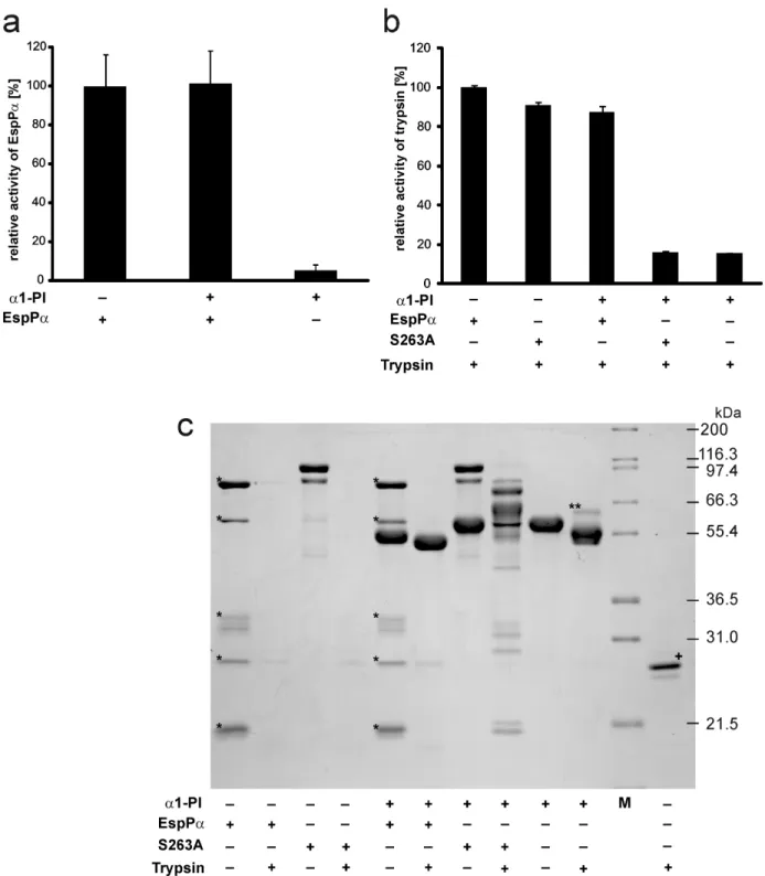
Figure 3. Activity of EspPaanda1-PI after coincubation.a, Determination of EspPaactivity. EspPaanda1-PI were preincubated (15 h, 37uC) at equimolar concentrations and remaining activity of EspPawas analyzed by incubation of an aliquot of the mixture with the chromogenic substrate Suc-Ala-Ala-Pro-Leu-pNA. Activity was measured via releasedpara-nitroaniline and normalized to EspPa. n = 9 for EspPaand EspPa+a1-PI or n = 6 for a1-PI, respectively. b,a1-PI activity (measured as inhibitory potential on trypsin) after incubation with EspPa. a1-PI and EspPaor S263A were preincubated at a molar ratio of serpin:enzyme = 4:1. Remaining inhibitory activity ofa1-PI on trypsin was analyzed by incubation at a molar ratio of a1-PI:trypsin = 4:1. Trypsin activity was measured via release ofpara-nitroaniline from the chromogenic substrate Bz-Arg-pNA. c, SDS-PAGE analysis of conincubations.a1-PI, EspPa, S263A, and trypsin were incubated as in b) and mixtures were separated via SDS-PAGE (12% SDS-PAGE gel, glycine buffer). M, molecular weight marker, *, EspPaautodegradation product, **, inhibitory complex ofa1-PI and trypsin,+, trypsin was directly subjected to SDS-PAGE without incubation.
Activity ofa1-PI and EspPaafter incubation
We next determined the functional consequences of the coincubation of serpin and SPATE protease by use of the bona fide serpin a1-PI and EspPa. The remaining EspPa-activity following incubation with a1-PI was assessed in a photometrical
assay using the chromogenic EspPa substrate
Suc-Ala-Ala-Pro-Leu-pNA. Incubation with a1-PI had no influence on the
proteolytic activity of EspPa (Fig. 3a), demonstrating that a1-PI does not target EspPa.
Figure 4. Peptide mapping of EspPacleavage products ofa1-PI.a1-PI fragments were subjected to tryptic in-gel-digest and generated peptides were analyzed via MALDI-TOF-MS. a, Sequence coverage ofa1-PI fragments. Peptides of the large fragment are given in bold and numbered 1–25. Peptides of the small fragment are given in italics and numbered 19-69. Note the newly formedN-terminus of the small fragment (SIPPEVK, underlined). b, MALDI-TOF-MS spectrum of the large fragment ofa1-PI. Inset: SDS-PAGE gel, glycine buffer. Fragment used for peptide mapping is marked by arrow. c, MALDI-TOF-MS spectrum of the small fragment ofa1-PI. Inset: SDS-PAGE gel, tricine buffer. Fragments used for peptide mapping are marked by arrow.a1-PI peptides are numbered according to a, T, trypsin autoproteolysis products, E, EspPaautoproteolysis products. doi:10.1371/journal.pone.0111363.g004
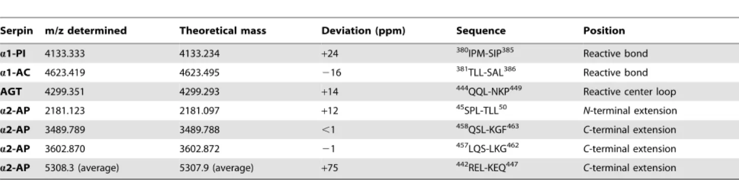
Table 2.Serpin cleavage sites determined by MALDI-TOF-MS.
Serpin m/z determined Theoretical mass Deviation (ppm) Sequence Position
a1-PI 4133.333 4133.234 +24 380IPM-SIP385 Reactive bond
a1-AC 4623.419 4623.495 216 381TLL-SAL386 Reactive bond
AGT 4299.351 4299.293 +14 444QQL-NKP449 Reactive center loop
a2-AP 2181.123 2181.097 +12 45SPL-TLL50 N-terminal extension
a2-AP 3489.789 3489.788 ,1 458QSL-KGF463 C-terminal extension
a2-AP 3602.870 3602.872 21 457LQS-LKG462 C-terminal extension
a2-AP 5308.3 (average) 5307.9 (average) +75 442REL-KEQ447 C-terminal extension
Given are masses determined by MALDI-TOF-MS directly after incubation of serpin with EspPa, theoretical masses, mass deviation, according sequence, and position inside the serpin sequence. Numeration is according to the serpin precursor.
The remaining inhibitory potential ofa1-PI following incuba-tion with EspPa was analyzed using trypsin as a serpin target. Although neutrophil elastase is the physiological target fora1-PI, trypsin also forms an irreversible inhibitory complex with the serpin and can therefore be used as an indicator fora1-PI activity [40]. Active a1-PI inhibits the proteolytic activity of trypsin and consequently loss ofa1-PI serpin activity results in high proteolytic activity in the assay. Trypsin activity was determined by photometrical detection of the cleavage of the trypsin substrate Bz-Arg-pNA.
Incubation of trypsin witha1-PI or a1-PI preincubated with S263A resulted in nearly complete loss of trypsin activity (Fig. 3b), demonstrating that the employeda1-PI shows high serpin activity and that the inactive EspPamutant S263A does not affecta1-PI. In contrast,a1-PI preincubated with EspPadid not reduce trypsin activity in the following assay (Fig. 3b). This demonstrates that EspPa-mediated a1-PI cleavage leads to loss of the inhibitory serpin activity. Corresponding results were obtained using SDS-PAGE (Fig. 3c). Incubation of a1-PI with trypsin leads to the formation of a serpin-enzyme-complex (Fig. 3c, lane 10). After incubation with EspPa,a1-PI is not able to form this complex with trypsin. Instead, the largea1-PI fragment is further degraded by trypsin (Fig. 3c, lane 6). EspPaas well as S263A were completely degraded when incubated with trypsin, demonstrating that neither EspPanor S263A directly interfere with trypsin activity (Fig. 3c, lanes 2 and 4). In addition,a1-PI does not interact with S263A (no serpin enzyme complex) (Fig. 3c, lane 7) but is cleaved by EspPa (Fig. 3c, lane 5). The addition of trypsin to the mixture ofa1-PI and S263A led to incomplete degradation and occurrence of several degradation bands in SDS-PAGE. This is due to the fact that degradation of S263A by trypsin and the inhibition of trypsin by a1-PI occur in parallel resulting in only incomplete S263A degradation (Fig. 3c, lane 8).
EspPacleaves inside the reactive center loop
The loss of activity ofa1-PI but not EspPais based on cleavage of a1-PI without formation of an inhibitory serpin-enzyme-complex. To further understand how EspPa-mediated cleavage affects the inhibitory function, we determined the cleavage sites in a1-PI and the other serpins included in this study. To this end,
large and small fragments of cleaved serpins were separated using SDS-PAGE, in-gel-digested and subjected to MALDI-TOF-MS analysis. Figure 4 shows the peptide mapping of EspPacleavage products of a1-PI. The large a1-PI fragment consists of the N -terminal part of the serpin (Fig. 4a and b), while theC-terminal part from residue 383 to 418 forms the small fragment (Fig. 4 a, and c). EspPa cleavage occurs at the active site of the serpin between382Met and383Ser as demonstrated by the occurrence of
the non-tryptic peptide 19(SIPPEVK) and the complete sequence coverage for the small fragment (Fig. 4c). Sequence coverage of degradation products of the other serpins are given in Figure S2.
Direct MALDI-TOF-MS analysis of small fragments
Not all cleavage sites can be identified via in-gel-digest. Tryptic peptides might be too small when cleavage occurs close to lysine or arginine residues or when several cleavage sites are in close proximity to each other. As all small fragments formed by EspPa -cleavage show a molecular weight below 10 kDa, we applied direct MALDI-TOF-MS analysis to determine the exact mass of the small serpin fragments to elucidate and confirm cleavage sites (Fig. 5). For the smalla1-PI fragment we observed a signal for the proton adduct of thea1-PI sequence383
Ser-418Lys (m/z 4133.333) confirming the cleavage site determined via in-gel-digest. In addition, signals representing the Na+
adduct and the oxidized Na+
adduct of the according a1-PI fragment sequence were observed (Fig. 5a). a1-AC shows a similar spectrum with a pronounced signal at m/z 4623.419 demonstrating cleavage C-terminal of383Leu at the reactive bond (Fig. 6b), which is in good
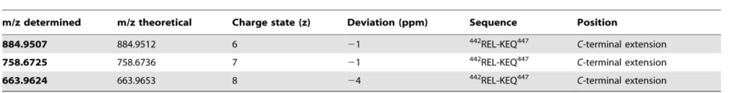
accordance with data from in-gel-digest (Figure S2). For AGT, we already observed three bands in SDS-PAGE (intact AGT and two non-proteolytic fragments) when incubated without protease (Fig. 2e). Accordingly, signals of two small AGT fragments were observed in MALDI-TOF-MS (Fig. 5c, right lane). Incubation with EspPaled to degradation of intact AGT and occurrence of the corresponding small fragment in MALDI-TOF-MS (Fig. 2e and Fig. 5c, left lane). Fora2-AP, proteolytic cleavage into several fragments is observed in SDS-PAGE (see Fig. 2g and 2h and Fig. 5d) after incubation with EspPa. Four distinct signals are seen in the MS spectrum indicating 4 cleavage sites. As the resolution for the signal at m/z 5308.3 is too low to determine the monoisotopic mass, we measured this sample in addition via nanospray-ESI-FTMS. Table 2 summarizes EspPa cleavage sites and their positions within the respective serpin. Measurement of a2-AP after incubation with EspPa via nanospray ESI-FTMS is described in Table 3.
a1-PI anda1-AC are cleaved at their reactive bonds (position of
reactive sites are described in [41,42]), leading to loss of serpin function. In both molecules the reactive bonds are exposed in the RCL and serve as pseudosubstrates for the targeted proteases. In case of EspPa, the serpins are not able to form a stable inhibitor-enzyme-complex and therefore release the intact EspPa after cleavage. Although AGT as non-inhibitory serpin does not contain a reactive bond, it is structurally closely related to the other serpins and is also cleaved in the RCL, indicating that a reactive bond is not necessary for EspPa-mediated serpin degradation. This is further underlined fora2-AP, which is cleaved at four positions outside the RCL (for RCL position see [43]). Cleavage sites are located at theN- andC-terminal extensions 25 aa downstream the
N-terminus and 46, 31, and 30 aa upstream theC-terminus (see Table 2). Intriguingly, both theN- andC-terminal extensions are vital for the functional relevant binding ofa2-AP to other proteins [19,44,45].
Table 3.a2-AP cleavage site determined by ESI-FTMS.
m/z determined m/z theoretical Charge state (z) Deviation (ppm) Sequence Position
884.9507 884.9512 6 21 442REL-KEQ447 C-terminal extension
758.6725 758.6736 7 21 442REL-KEQ447 C-terminal extension
663.9624 663.9653 8 24 442REL-KEQ447 C-terminal extension
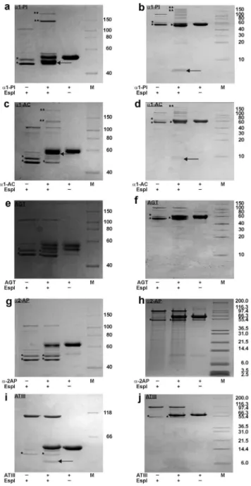
Cleavage of serpins by EspI
Purified EspI samples showed a protein band at ,110 kDa
(intact EspI) as well as two EspI autoproteolysis products at,50
and 45 kDa, respectively. Similar to EspPa, autoproteolysis products remain active. Serpins were incubated with purified
EspI in the same way as described for EspPa. Incubation ofa1-PI anda1-AC with EspI led to degradation of these serpins. Notably, EspI also forms a pronounced inhibitory complex with both protease inhibitors resulting in only incomplete serpin degradation (Fig. 6 a-d). In contrast to EspPa, EspI does not cleavea2-AP and Figure 5. Direct analysis of the small cleavage product of serpins via MALDI-TOF-MS.Serpins were incubated with EspPaand directly analyzed via MALDI-TOF-MS. a, MALDI-TOF-MS spectrum ofa1-PI fragment. Inset: Detailed view of the signal representing the smalla1-PI fragment. b, MALDI-TOF-MS spectrum ofa1-AC. c, MALDI-TOF-MS spectrum of AGT. Left lane: Spectrum after incubation with EspPa, right lane: Spectrum after incubation of AGT without EspPa. *, signals represent non-proteolytic fragments also found after incubation of AGT without EspPa. d, MALDI-TOF-MS spectrum ofa2-AP. Inset: Detailed view of the m/z window 2160–2260 representing signals (M H), (M Na), (M Na+O) of the cleavage site in the N-terminal extension ofa2-AP are exemplarily shown. (M H), proton adduct of small serpin fragment, (M Na), Na adduct of small serpin fragment, (M Na+O), Na adduct oxidized at one methionine residue.
AGT (Fig. 6e–h). Cleavage of ATIII occurred only with very low efficiency (Fig. 6i) and might not be relevant under physiological conditions.
To determine the cleavage sites of a1-PI and a1-AC, we subjected incubation mixtures of serpins and EspI to direct MALDI-TOF-MS analysis. Serpin cleavage occurred at the reactive bond leading to signals at m/z 4155.400 (a1-PI, 20 ppm deviation according to calculated m/z) and 4623.509
(a1-AC, 19 ppm deviation according to calculated m/z), respec-tively (data not shown).
Conclusions
EspPais an EHEC virulence factor that belongs to the SPATE family. As suggested for SPATEs in general, EspPa most likely mediates its virulence via cleavage and inactivation of host proteins. Here, we present a method for the rapid determination of EspPa-mediated cleavage sites in various human plasma serpins via MALDI-TOF-MS as well as a photometrical assay to analyze serpin functionality after proteolytic cleavage. Concerning the functional consequences, degradation of a2-AP might lead to bleeding disorders. This serpin is the primary physiological inhibitor of plasmin and deficiency has been shown to result in uncontrolled fibrinolysis and severe hemorrhagic complication [44,45].a2-AP harbors a 42 aaN-terminal and a 55 aaC-terminal extension [19,46]. While theN-terminal extension is cross-linked to fibrin, the veryC-terminal491Lys residue mediates binding to plasmin [47]. EspPa cleaves between47
Leu and48Thr releasing part of theN-terminal extension and at three different sites inside the C-terminal extension leading to release of a polypeptide containing 491Lys. Together, this most likely leads to loss of function ofa2-AP. The role ofa1-PI in thrombosis is not well understood. However,a1-PI is able to inhibit activated protein C. In pediatric ischemic stroke patients elevated levels ofa1-PI have been found and were discussed to contribute to this thrombotic disease in children [48,49]. ATIII is the main anticoagulatory serpin. Although it is able to interfere with virtually all proteolytic coagulation factors, its main targets are thrombin, FIXa, and FXa. Intriguingly, it is the only serpin in this study that is not cleaved by EspPa. Despite the structural similarity of serpins, EspPa specifically cleaves only selected serpins. More specific, procoagu-latory serpins sucha2-AP anda1-PI are efficiently degraded while the anticoagulatory ATIII is not affected at all. Together with data demonstrating that EspPa cleaves coagulation factor V [3], this underlines the hypothesis that interference with blood coagulation (and possibly also inflammatory host responses) [50] might be one of the major functions of EspPa which might contribute to formation of hemorrhages observed during EHEC infection.
Having a closer look at EspPacleavage sites, it is notable that more than 70% (5 of 7) of cleavage sites identified in this study occur after Leu. This is in good accordance to already reported EspPacleavage sites [3,9,7,51], indicating that substrate cleavage is most favorable C-terminal to Leu. In a2-AP, cleavage also occurs after459Ser. This residue, however, is positioned next to
460
Leu after which EspPa cleaves, too. The second non-Leu cleavage site is C-terminal to382Met ina1-PI. The382Met-383Ser bond, however, is the reactive bond exposed in the RCL and required to react with target proteases. Similarly,a1-AC is cleaved at the reactive bond that consists of a Leu-Ser motif which is also located in the exposed RCL. Cleavage of the non-inhibitory AGT shows that a reactive bond is not strictly required for substrate recognition by EspPa but cleavage also occurs inside the corresponding reactive center loop. In contrast, a2-AP is not cleaved in the RCL but inside theN- andC-terminal extensions which are vital for a2-AP functionality. Though the crystal structure of a2-AP has only been solved for a N-terminally truncated murine form, it seems that the C-terminal extension consists of a flexible loop because it could not be modeled into electron density maps [52]. Perhaps, this structural flexibility seen in the reactive center loops and in theC-terminal extension ofa 2-AP is required for substrate recognition by EspPa. Figure 7 shows crystal structures of the serpins that are cleaved by EspPa[52–55].
Figure 6. Cleavage of serpins by EspI. Serpins (5mg) were incubated (15 h, 37uC) with EspI (1.5mg). Degradation products were separated via SDS-PAGE using a glycine buffer (a, c, e, g, i) or a tricine buffer (b, d, f, h, j). a, ba1-PI is cleaved into two fragments (,45 kDa and,4 kDa), c, da1-AC is cleaved into two fragments, e, f AGT is not cleaved by EspI, g, ha2-AP is not cleaved by EspI, i, j ATIII is cleaved only with very low efficiency. Note the formation of inhibitory serpin-enzyme-complexes after incubation witha1-PI anda1-AC. M, molecular weight marker, *, autodegradation product of EspI, **, inhibitory serpin-EspI-complex. Serpin fragments are indicated by an arrow.
EspI shows significant differences in substrate specificity compared to EspPa.a1-PI anda1-AC are also cleaved at their reactive bonds which should lead to loss of function of these serpins. However, serpin cleavage and release of the protease is not complete for EspI, most probably due to the pronounced formation of an inhibitory serpin-enzyme-complex of EspI with a1-PI anda1-AC. In contrast, EspPa completely degrades both
serpins and forms only small amounts of the inhibitory complex only witha1-PI which does not significantly reduce EspPaactivity. In addition, AGT and a2-AP, which are degraded by EspPa at positions other than the reactive bond, are not degraded by EspI. Concerning the functional differences of both SPATE proteases,
EspPais able to cleave serpins specifically within accessible loop structures and is notably not inhibited by the analyzed serpins, while EspI is only able to interact with the reactive bond ofa1-PI anda1-AC. The latter interactions show equilibria between EspI inhibition and serpin degradation. Taking into account the high amounts of serpins such asa1-PI in plasma, EspI activity might be strongly reduced in this milieu in vivo, while serpin degradation and inactivation might be a relevant function of EspPaalso during infection.
In summary, we established a rapid method to determine cleavage sites of small proteolytic fragments via MALDI-TOF-MS. Functional implications have been investigated in a newly
Figure 7. Crystal structures of serpins cleaved by EspPa.Serpins are shown as cartoons. RCL is indicated in black, approximate cleavage sites are encircled. Non-resolved parts of the crystal structures are indicated by dots (c, RCL of AGT, d, RCL ofa2-AP and theN- andC-terminal extension of a2-AP). a, humana1-PI, b, cleaved humana1-AC, the RCL is indicated by dots, c, human angiotensinogen, d, murine truncateda2-APD43, the
developed photometrical assay using chromogenic peptide sub-strates. EspPa degrades and thereby inactivates different plasma serpins which, in case ofa2-AP, might lead to bleeding disorders or in case ofa1-PI anda1-AC might interfere with the acute phase reaction during inflammatory host response. Cleavage occurs in flexible regions most favorableC-terminal to Leu. Comparison of EspPaand EspI indicate different functions of this SPATE also in vivo.
Supporting Information
Figure S1 Activity of EspPaand S263A.a, Determination of
EspPa and S263A activity directly after purification. EspPa or S263A was incubated (15 h, 37uC) with the chromogenic substrate Suc-Ala-Ala-Pro-Leu-pNA. Activity was measured via released
para-nitroaniline and normalized to EspPa. PBS was used as control. n = 2, b, Determination of EspPaactivity after preincu-bation. Purified EspPa was preincubated for 15 h at 37uC resulting in the formation of autoproteolysis products (see Fig. 3c, lane1). To assess remaining proteolytic activity of autoproteolysis products the preincubated sample was incubated with the
chromogenic substrate Suc-Ala-Ala-Pro-Leu-pNA (15 h, 37uC). Again, activity was measured via releasedpara-nitroaniline and normalized to EspPa. PBS was used as control. n = 2.
(TIFF)
Figure S2 Peptide mapping of EspPacleavage products of the serpins.Serpin fragments were subjected to in-gel-digest and analyzed via MALDI-TOF-MS. Peptides of the large fragment are given in bold. Peptides of the small fragments are given in italics, a, sequence coverage of a1-AC fragments, b, sequence coverage of AGT, c, sequence coverage ofa2-AP. Note that in the small fragments of AGT anda2-AP no serpin peptides were found.
(TIF)
Author Contributions
Conceived and designed the experiments: AW HJ JB. Performed the experiments: AW HJ. Analyzed the data: AW HJ JB. Contributed reagents/materials/analysis tools: AW JB. Contributed to the writing of the manuscript: AW JB.
References
1. Law D (2000) Virulence factors of Escherichia coli O157 and other Shiga toxin-producingE.coli. Journal of Applied Microbiology 88(5): 729–745. 2. Karmali MA (2004) Infection by Shiga toxin-producing Escherichia coli: an
overview. Molecular Biotechnology 26(2): 117–122.
3. Brunder W, Schmidt H, Karch H (1997) EspP, a novel extracellular serine protease of enterohaemorrhagic Escherichia coli O157:H7 cleaves human coagulation factor V. Molecular Microbiology 24(4): 767–778.
4. Brockmeyer J, Bielaszewska M, Fruth A, Bonn ML, Mellmann A, et al. (2007) Subtypes of the plasmid-encoded serine protease EspP in Shiga toxin-producing Escherichia coli: distribution, secretion, and proteolytic activity. Applied and Environmental Microbiology 73(20): 6351–6359.
5. Bielaszewska M, Stoewe F, Fruth A, Zhang W, Prager R, et al. (2009) Shiga toxin, cytolethal distending toxin, and hemolysin repertoires in clinical Escherichia coliO91 isolates. Journal of Clinical Microbiology 47(7): 2061–2066. 6. Khan AB, Naim A, Orth D, Grif K, Mohsin M, et al. (2009) Serine protease espP subtype alpha, but not beta or gamma, of Shiga toxin-producing Escherichia coli is associated with highly pathogenic serogroups. Int Journal of Medical Microbiology 299(4): 247–254.
7. Brockmeyer J, Aldick T, Soltwisch J, Zhang W, Tarr PI, et al. (2011) Enterohaemorrhagic Escherichia coli haemolysin is cleaved and inactivated by serine protease EspPalpha. Environmental Microbiology 13(5): 1327–1341. 8. Schmidt H, Zhang WL, Hemmrich U, Jelacic S, Brunder W, et al. (2001)
Identification and characterization of a novel genomic island integrated at selC in locus of enterocyte effacement-negative, Shiga toxin-producing Escherichia coli. Infection and Immunity 69(11): 6863–6873.
9. Orth D, Ehrlenbach S, Brockmeyer J, Khan AB, Huber G, et al. (2010) EspP, a serine protease of enterohemorrhagic Escherichia coli, impairs complement activation by cleaving complement factors C3/C3b and C5. Infection and Immunity 78(10): 4294–4301.
10. dos Santos LF, Irino K, Vaz TM, Guth BE (2010) Set of virulence genes and genetic relatedness of O113: H21Escherichia coli strains isolated from the animal reservoir and human infections in Brazil. Journal of Medical Microbiology 59(Pt 6): 634–640.
11. Toszeghy M, Phillips N, Reeves H, Wu G, Teale C, et al. (2012) Molecular and phenotypic characterisation of Extended Spectrum beta-lactamase CTX-M Escherichia coli from farm animals in Great Britain. Research in Veterinary Science 93(3): 1142–1150.
12. Gettins PG (2002) Serpin structure, mechanism, and function. Chemical Reviews 102(12): 4751–4804.
13. Rau JC, Beaulieu LM, Huntington JA, Church FC (2007) Serpins in thrombosis, hemostasis and fibrinolysis. Journal of Thrombosis and Haemostasis 5 Suppl 1: 102–115.
14. Huntington JA, Read RJ, Carrell RW (2000) Structure of a serpin-protease complex shows inhibition by deformation. Nature 407(6806): 923–926. 15. Beatty K, Bieth J, Travis J (1980) Kinetics of association of serine proteinases
with native and oxidized alpha-1-proteinase inhibitor and alpha-1-antichymo-trypsin. The Journal of Biological Chemistry 255(9): 3931–3934.
16. Marshall CJ (1993) Evolutionary relationships among the serpins. Philosophical Transactions of the Royal Society of London. Series B, Biological Sciences 342(1300): 101–119.
17. Chandra T, Stackhouse R, Kidd VJ, Robson KJ, Woo SL (1983) Sequence homology between human alpha 1-antichymotrypsin, alpha 1-antitrypsin, and antithrombin III. Biochemistry 22(22): 5055–5061.
18. Schechter NM, Sprows JL, Schoenberger OL, Lazarus GS, Cooperman BS, et al. (1989) Reaction of human skin chymotrypsin-like proteinase chymase with plasma proteinase inhibitors. Journal of Biological Chemistry 264(35): 21308– 21315.
19. Coughlin PB (2005) Antiplasmin: the forgotten serpin? The FEBS Journal 272(19): 4852–4857.
20. Favier R, Aoki N, de Moerloose P (2001) Congenital alpha(2)-plasmin inhibitor deficiencies: a review. British Journal of Haematology 114(1): 4–10. 21. Rogers SJ, Pratt CW, Whinna HC, Church FC (1992) Role of thrombin exosites
in inhibition by heparin cofactor II. Journal of Biological Chemistry 267(6): 3613–3617.
22. Mauray S, de Raucourt E, Talbot JC, Dachary-Prigent J, Jozefowicz M, et al. (1998) Mechanism of factor IXa inhibition by antithrombin in the presence of unfractionated and low molecular weight heparins and fucoidan. Biochimica et Biophysica Acta 1387(1–2): 184–194.
23. Izaguirre G, Zhang W, Swanson R, Bedsted T, Olson ST (2003) Localization of an antithrombin exosite that promotes rapid inhibition of factors Xa and IXa dependent on heparin activation of the serpin. Journal of Biological Chemistry 278(51): 51433–51440.
24. Olson ST, Swanson R, Raub-Segall E, Bedsted T, Sadri M, et al. (2004) Accelerating ability of synthetic oligosaccharides on antithrombin inhibition of proteinases of the clotting and fibrinolytic systems. Comparison with heparin and low-molecular-weight heparin. Thrombosis and Haemostasis 92: 929–39. 25. Stein PE, Tewkesbury DA, Carrell RW (1989) Ovalbumin and angiotensinogen
lack serpin S-R conformational change. Biochemical Journal 262(1): 103–107. 26. Arakawa K, Minohara A, Yamada J, Nakamura M (1968) Enzymatic
degradation and electrophoresis of human angiotensin I. Biochimica et Biophysica Acta 168(1): 106–112.
27. Lentz KE, Skeggs LT, Jr., Woods KR, Kahn JR, Shumway NP (1956) The amino acid composition of hypertensin II and its biochemical relationship to hypertensin I. The Journal of Experimental Medicine 104(2): 183–191. 28. Mast AE, Enghild JJ, Nagase H, Suzuki K, Pizzo SV, et al. (1991) Kinetics and
physiologic relevance of the inactivation of alpha proteinase inhibitor, alpha 1-antichymotrypsin, and antithrombin III by matrix metalloproteinases-1 (tissue collagenase), -2 (72-kDa gelatinase/type IV collagenase), and -3 (stromelysin). The Journal of biological chemistry 266: 15810–15816.
29. Lijnen HR, Arza B, Van Hoef B, Collen D, Declerck PJ (2000) Inactivation of plasminogen activator inhibitor-1 by specific proteolysis with stromelysin-1 (MMP-3). The Journal of biological chemistry 275: 37645–37650.
30. Lijnen HR, Van Hoef B, Collen D (2001) Inactivation of the serpin alpha(2)-antiplasmin by stromelysin-1. Biochimica et biophysica acta 1547: 206–213. 31. Liu Z, Zhou X, Shapiro SD, Shipley JM, Twining SS, et al. (2000) The serpin
alpha1-proteinase inhibitor is a critical substrate for gelatinase B/MMP-9 in vivo. Cell 102: 647–655.
32. Virca GD, Lyerly D, Kreger A, Travis J (1982) Inactivation of human plasma alpha 1-proteinase inhibitor by a metalloproteinase from Serratia marcescens. Biochimica et biophysica acta 704: 267–271.
33. Molla A, Akaike T, Maeda H (1989) Inactivation of various proteinase inhibitors and the complement system in human plasma by the 56-kilodalton proteinase from Serratia marcescens. Infection and immunity 57: 1868–1871.
35. Lathem WW, Bergsbaken T, Welch RA (2004) Potentiation of C1 esterase inhibitor by StcE, a metalloprotease secreted by Escherichia coli O157:H7. The Journal of experimental medicine 199: 1077–1087.
36. Brockmeyer J, Spelten S, Kuczius T, Bielaszewska M, Karch H (2009) Structure and Function Relationship of the Autotransport and Proteolytic Activity of EspP from Shiga Toxin-ProducingEscherichia coli. PLoS ONE 4(7): e6100. 37. Laemmli UK (1970) Cleavage of structural proteins during the assembly of the
head of bacteriophage T4. Nature 227(5259): 680–685.
38. Schagger H, von Jagow G (1987) Tricine-sodium dodecyl sulfate-polyacrylamide gel electrophoresis for the separation of proteins in the range from 1 to 100 kDa. Analytical Biochemistry 166(2): 368–379.
39. Shevchenko A, Tomas H, Havlis J, Olsen JV, Mann M (2006) In-gel digestion for mass spectrometric characterization of proteins and proteomes. Nature Protocols 1(6): 2856–2860.
40. Thelwell C, Marszal E, Rigsby P, Longstaff C (2011) An international collaborative study to establish the WHO 1st international standard for alpha-1-antitrypsin. Vox Sanguinis 101(1): 83–89.
41. Johnson D, Travis J (1978) Structural evidence for methionine at the reactive site of human alpha-1-proteinase inhibitor. Journal of Biological Chemistry 253(20): 7142–7144.
42. Morii M, Travis J (1983) Amino acid sequence at the reactive site of human alpha 1-antichymotrypsin. Journal of Biological Chemistry 258(21): 12749– 12752.
43. Potempa J, Shieh BH, Travis J (1988) Alpha-2-antiplasmin: a serpin with two separate but overlapping reactive sites. Science 241(4866): 699–700. 44. Collen D, Wiman B (1979) Turnover of antiplasmin, the fast-acting plasmin
inhibitor of plasma. Blood 53(2): 313–324.
45. Aoki N (1984) Genetic abnormalities of the fibrinolytic system. Seminars in Thrombosis and Hemostasis 10(1): 42–50.
46. Holmes WE, Nelles L, Lijnen HR, Collen D (1987) Primary structure of human alpha 2-antiplasmin, a serine protease inhibitor (serpin). Journal of Biological Chemistry 262(4): 1659–1664.
47. Hortin GL, Gibson BL, Fok KF (1988) Alpha 2-antiplasmin’s carboxy-terminal lysine residue is a major site of interaction with plasmin. Biochemical and Biophysical Research Communications 155(2): 591–596.
48. Heeb MJ, Griffin JH (1988) Physiologic inhibition of human activated protein C by alpha 1-antitrypsin. Journal of Biological Chemistry 263(24): 11613–11616.
49. Burghaus B, Langer C, Thedieck S, Nowak-Gottl U (2006) Elevated alpha1-antitrypsin is a risk factor for arterial ischemic stroke in childhood. Acta Haematologica 115(3–4): 186–191.
50. Weiss A, Brockmeyer J,(2012) Prevalence, biogenesis, and functionality of the serine protease autotransporter EspP. Toxins 5(1): 25–48.
51. Dutta PR, Cappello R, Navarro-Garcia F, Nataro JP (2002) Functional Comparison of Serine Protease Autotransporters of Enterobacteriaceae. Infection and Immunity 70(12): 7105–7113.
52. Law RH, Sofian T, Kan WT, Horvath AJ, Hitchen CR, et al. (2008) X-ray crystal structure of the fibrinolysis inhibitor alpha2-antiplasmin. Blood 111(4): 2049–2052.
53. Elliott PR, Pei XY, Dafforn TR, Lomas DA (2000) Topography of a 2.0 A structure of alpha1-antitrypsin reveals targets for rational drug design to prevent conformational disease. Protein Science 9(7): 1274–1281.
54. Pearce MC, Powers GA, Feil SC, Hansen G, Parker MW, et al. (2010) Identification and characterization of a misfolded monomeric serpin formed at physiological temperature. Journal of Molecular Biology 403(3): 459–467. 55. Zhou A, Carrell RW, Murphy MP, Wei Z, Yan Y, et al. (2010) A redox switch in
angiotensinogen modulates angiotensin release. Nature 468(7320): 108–111. 56. Lomas DA, Evans DL, Finch JT, Carrell RW (1992) The mechanism of Z alpha
1-antitrypsin accumulation in the liver. Nature 357(6379): 605–607. 57. Lomas DA, Finch JT, Seyama K, Nukiwa T, Carrell RW (1993) Alpha
1-antitrypsin Siiyama (Ser53–.Phe). Further evidence for intracellular loop-sheet polymerization. Journal of Biological Chemistry 268(21): 15333–15335. 58. Lomas DA, Elliott PR, Sidhar SK, Foreman RC, Finch JT, et al. (1995) alpha
1-Antitrypsin Mmalton (Phe52-deleted) forms loop-sheet polymers in vivo. Evidence for the C sheet mechanism of polymerization. Journal of Biological Chemistry 270(28): 16864–16870.
59. Abraham CR, Selkoe DJ, Potter H (1988) Immunochemical identification of the serine protease inhibitor alpha 1-antichymotrypsin in the brain amyloid deposits of Alzheimer’s disease. Cell 52(4): 487–501.
60. Kamboh MI, Sanghera DK, Ferrell RE, DeKosky ST (1995) APOE*4-associated Alzheimer’s disease risk is modified by alpha 1-antichymotrypsin polymorphism. Nature Genetics 10(4): 486–488.
61. Nielsen HM, Minthon L, Londos E, Blennow K, Miranda E, et al. (2007) Plasma and CSF serpins in Alzheimer disease and dementia with Lewy bodies. Neurology 69(16): 1569–1579.