RESEARCH ARTICLE
High Resolution Melting Analysis Targeting
hsp70
as a Fast and Efficient Method for the
Discrimination of
Leishmania
Species
Ricardo Andrade Zampieri1, Maria Fernanda Laranjeira-Silva1, Sandra Marcia Muxel1, Ana Carolina Stocco de Lima2, Jeffrey Jon Shaw3, Lucile Maria Floeter-Winter1*
1Physiology Department, Biosciences Institute, São Paulo University, São Paulo, São Paulo, Brazil,
2Pathology Department, Medical Faculty, São Paulo University, São Paulo, São Paulo, Brazil,
3Parasitology Department, Biomedical Institute, São Paulo University, São Paulo, São Paulo, Brazil
Abstract
Background
Protozoan parasites of the genusLeishmaniacause a large spectrum of clinical manifesta-tions known as Leishmaniases. These diseases are increasingly important public health problems in many countries both within and outside endemic regions. Thus, an accurate dif-ferential diagnosis is extremely relevant for understanding epidemiological profiles and for the administration of the best therapeutic protocol.
Methods/Principal Findings
Exploring the High Resolution Melting (HRM) dissociation profiles of two amplicons using real time polymerase chain reaction (real-time PCR) targeting heat-shock protein 70 coding gene (hsp70) revealed differences that allowed the discrimination of genomic DNA samples of eightLeishmaniaspecies found in the Americas, includingLeishmania (Leishmania) infan-tum chagasi,L.(L.) amazonensis,L.(L.) mexicana,L.(Viannia) lainsoni,L.(V.) braziliensis,
L.(V.) guyanensis,L.(V.) naiffiandL.(V.) shawi, and three species found in Eurasia and Africa, includingL.(L.) tropica,L.(L.) donovaniandL.(L.) major. In addition, we tested DNA samples obtained from standard promastigote culture, naturally infected phlebotomines, experimentally infected mice and clinical human samples to validate the proposed protocol.
Conclusions/Significance
HRM analysis ofhsp70amplicons is a fast and robust strategy that allowed for the detection and discrimination of allLeishmaniaspecies responsible for the Leishmaniases in Brazil and Eurasia/Africa with high sensitivity and accuracy. This method could detect less than one parasite per reaction, even in the presence of host DNA.
OPEN ACCESS
Citation:Zampieri RA, Laranjeira-Silva MF, Muxel SM, Stocco de Lima AC, Shaw JJ, Floeter-Winter LM (2016) High Resolution Melting Analysis Targeting
hsp70as a Fast and Efficient Method for the Discrimination ofLeishmaniaSpecies. PLoS Negl Trop Dis 10(2): e0004485. doi:10.1371/journal. pntd.0004485
Editor:Alain Debrabant, US Food and Drug Administration, UNITED STATES
Received:October 1, 2015
Accepted:February 2, 2016
Published:February 29, 2016
Copyright:© 2016 Zampieri et al. This is an open access article distributed under the terms of the
Creative Commons Attribution License, which permits unrestricted use, distribution, and reproduction in any medium, provided the original author and source are credited.
Data Availability Statement:All relevant data are within the paper and its Supporting Information files.
Author Summary
The different clinical forms of the Leishmaniases range from cutaneous to visceral infec-tions and are caused by organisms belonging to the genusLeishmania. Controversy over the validity of different molecular methods to correctly identify a species hinders the asso-ciation of a given species with different clinical forms, complicating the prognosis and the development of suitable treatment protocols. A correct identification leads to a better understanding of the action and consequent development of new drugs and immunologi-cal reactions. It also provides important information about the relationship of each species with its hosts (humans, animal reservoirs and sandflies) in different geographical areas and ecological situations, helping to design control strategies. Today, PCR is the most commonly used method forLeishmaniaidentification, but even though several targets have been described, no simple and direct protocol has emerged. In this paper, we coupled hsp70real-time PCR with the determination of amplicon melting profiles in order to explore polymorphic regions by HRM analysis. This methodology yielded discriminatory melting temperature (Tm) values for Brazilian and Eurasian/AfricanLeishmaniaspecies. The protocol has proven to be 100% reliable with both clinical and experimental samples. The major advantage of the presently described method is that it is simple, less expensive, highly sensitive and easily automated.
Introduction
Leishmaniases are a major worldwide public health problem and manifest themselves as a spec-trum of diseases that may be exacerbated by other infections, such as human immunodefi-ciency virus. According to the World Health Organization, these diseases are endemic in 98 countries on 5 continents, with more than 350 million people at risk [1,2]. Clinically, Leish-maniases can be broadly divided as either cutaneous or visceral, but neither form is exclusively linked to a particular species. Although cutaneous manifestations of the diseases are not life threatening, these manifestations can result in obstruction or destruction of the pharynx, lar-ynx and nose in their final stages [2]. The visceral form is the most severe form, characterized by fever, loss of weight, splenomegaly, hepatomegaly, lymphadenopathies and anaemia, often with fatal outcomes if not timely treated [3].
The severity of the disease and its therapeutic responses are variable and depend on the patient’s immune response, theLeishmaniaspecies and even the parasite strain [4]. In this sce-nario, the development of optimized protocols for discriminating between the different Leish-maniaspecies is extremely useful and important in clinical management and treatment. The ability to evaluate the most appropriate species-specific treatments also supports the elucida-tion of the mechanisms of acelucida-tion of new drugs and the establishment of new species-specific treatment protocols. Furthermore, the identification of these parasites allows the generation of important data for clinical, epidemiological and ecological studies.
There are very few publications addressing a Leishmaniasis diagnosis using a High Resolu-tion Melting (HRM) analysis, a methodology that detects differences in the nucleotide compo-sition of a specific real-time PCR product. The method is based on thermodynamic differences in the dissociation curve profiles of amplicons generated from real-time PCR. The generated curves are specific signatures that identify polymorphisms due to small differences in nucleo-tide composition. In spite of the paucity of papers on the HRM method, some workers have already used it to discriminateLeishmaniausing targets against 7SL RNA [5,6],haspb[7], the Competing Interests:The authors have declared
rRNA ITS sequence [8,9], the rRNA ITS sequence coupled tohsp70[10,11] and a FRET-based assay using MPI and 6PGD [12].
Amongst several targets described forLeishmaniaidentification, the heat-shock protein 70 coding gene (hsp70) has proven to be useful in identifying many species of different geographi-cal origins [13–17].
In this work, we propose a more efficient protocol using HRM analyses targeting thehsp70 sequence for the discrimination of seven BrazilianLeishmaniaspecies, as well as three Eurasian and African species. This methodology was validated with DNA from reference strains, experi-mental infections in mice, human clinical samples and naturally infected phlebotomine sand flies.
Materials and Methods
Organisms
Promastigotes ofL.(L.) tropica(MHOM/SU/60/OD),L.(L.) donovani(MHOM/IN/80/DD8), L.(L.) infantum chagasi(MCER/BR/1981/M6445),L.(L.) major(MHOM/IL/81/Friedlin), L.(L.) amazonensis(MHOM/BR/1973/M2269),L.(L.) mexicana(MNYC/BZ/62/M379),L. (L.) lainsoni(MHOM/BR/81/M6426),L.(V.) braziliensis(MHOM/BR/1975/M2903),L.(V.) guyanensis(MHOM/BR/1975/M4147),L.(V.) naiffi(MDAS/BR/1979/M5533) andL.(V.) shawi(MCEB/BR/84/M8408) were grown at 25°C in M199 medium with 10% fetal bovine serum (Life Technologies, Carlsbad, CA, USA). Procyclic forms ofTrypanosoma cruzi(Y strain) andT.brucei(427 strain) were grown at 28°C in liver-infusion-tryptose medium and SDM-79, respectively, with 10% fetal bovine serum (Life Technologies). Human DNA, FMUS-P-IOF-2016, obtained from USP Medical School, was used in specificity tests.
Trypanosomatids DNA
DNA samples from reference strains were purified by a salting-out procedure using an adapta-tion of the protocol described by Miller et al. 1988 [18]. Approximately 2.5 x 10⁹promastigotes in stationary growth culture were centrifuged at 3000 x g for 10 min at 25°C. The cells were resuspended in 6 mL of lysis buffer (10 mM Tris-HCl, pH 7.4; 400 mM NaCl; 2 mM EDTA) and lysed by the addition of 600μL of 10% SDS. After overnight digestion with 1 mg of
pro-teinase K at 37°C, 2 mL of saturated NaCl solution was added to lysate, and then, the lysate was vigorously mixed for 15 seconds and centrifuged for 15 minutes at 25°C for the removal of pre-cipitated proteins. Two volumes of cold absolute ethanol were added to the supernatant, and the precipitated DNA was washed with 70% ethanol and resuspended in 1 mL of TE buffer (10 mM Tris, pH 7.4; 1 mM EDTA).
Clinical, experimental and natural DNA samples/ethical statements
DNA from samples obtained from fresh humans biopsies, collected by doctors at Clinical Hos-pital of Medical Faculty USP, or fixed and paraffin-embedded samples from the collection of Instituto Evandro Chagas, (Belem-Para) were used in accordance to the norms established by the National Committee of Ethics in Research (Comissão Nacional de Ética em Pesquisa, CONEP/CNS), resolution 196/96 with the approval of the Ethics in Research Committees of the Institutions of origin (CAPPesq no. 0804/07, IEC n°. 0029/2007).Fresh experimentally infected BALB/c mice samples ofL.(L.) amazonensisorL.(V.) brazi-liensiswere obtained 6 weeks after infection when the animals were sacrificed and tissues were collected and DNA was obtained as described below; the procedures involving the use of BALB/c mice had the approval of the Ethical Committee for use of Animals of Biomedical
Sciences Institute of University of São Paulo (CEUA-ICB-USP), under protocol #145 of Octo-ber 20th, 2011, according to Brazilian Federal Law 11.794 of October 8th2008.
DNA from infected phlebotomines captured in nature were purified using the commercial DNeasy Tissue & Blood kit (QIAGEN, Hilden, Germany), according to the manufacturer´s manual. Paraffin-embedded samples were prepared according to de Lima et al. 2011 [19]. The DNA concentration was measured by spectrophotometry.
PCR assays
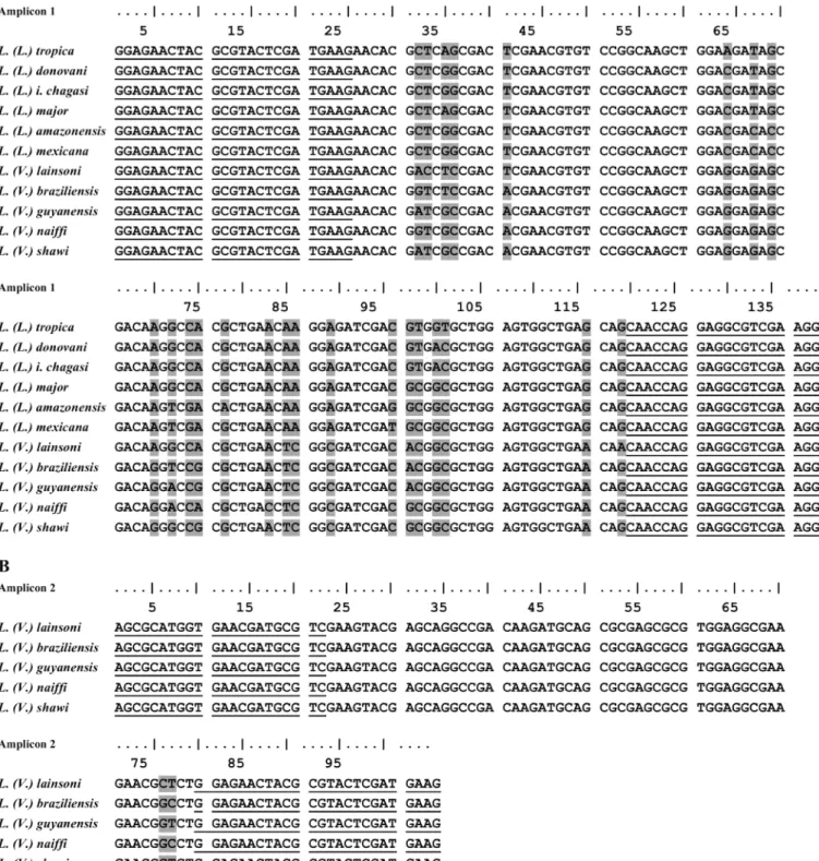
Initially, we amplified thehsp70234 bp fragments for all species analyzed in this study using the primers described by Graça et al. [17]. The alignment of the nucleotide sequence of those fragments was used to design primers for HRM analysis. Oligonucleotides used in the PCR assays to amplify a 144 bp fragment ofhsp70(amplicon 1) werehsp70C reverse, previously described by Graça et al. 2012 [17], and a new forward oligonucleotide designed and named hsp70F2 (5’–GGAGAACTACGCGTACTCGATGAAG–3’). For the amplification of a 104 bp fragment ofhsp70(amplicon 2) specific to the species from theL.(Viannia)subgenus, the oli-gonucleotideshsp70F1 (5’–AGCGCATGGTGAACGATGCGTC–3’) andhsp70R1 (5’–CTTCA TCGAGTACGCGTAGTTCTCC–3’) were designed. Thehsp70amplicon sequences are shown inFig 1and indicate the position of the primers. Conventional PCR reactions were performed on a Mastercycler Gradient Thermocycler (Eppendorf, Hamburg, Germany) with TopTaq Master Mix (QIAGEN) in a final volume of 25μL with 200 nM of each primer and 50 ng of
genomic DNA as a template. The thermal cycling conditions were as follows: an initial dena-turation step of 94°C for 5 min, followed by 40 cycles of denadena-turation at 94°C for 1 min, anneal-ing at 60°C for 30 sec and extension at 72°C for 30 sec, with a final extension at 72°C for 10 min. Real-time PCR reactions were performed using MeltDoc Master Mix for HRM with the fluorophore SYTO9 (Life Technologies) in a final volume of 20μL with 200 nM of each primer
and 50 ng of genomic DNA. The real time amplification conditions were as follows: an initial denaturation step at 94°C for 5 min, followed by 40 cycles of denaturation at 94°C for 30 sec and annealing/extension at 60°C for 30 sec, with the acquisition of fluorescent signals at the end of each extension step, followed by the dissociation curve for HRM analysis in Thermocy-cler PikoReal96 (Thermo Fisher Scientific, Walthman, MA, USA).
Cloning and sequencing
The 234 bphsp70fragment produced by conventional PCR, as described by Graça et al. 2012 [17], from eachLeishmaniaspecies used in this study was purified and cloned into a pGEM-T vector using the pGEM-T Easy Vector System (Promega, Madison, WI, USA) andE.coliSURE competent cells. The recombinant plasmids from at least three colonies were purified, and they were sequenced with T7 and SP6 primers and the BigDyeTerminator v3.1 Cycle Sequencing Kit (Applied Biosystems, Foster City, CA, USA). The sequencing was performed on an ABI 3130 XL Platform (Life Technologies).
Target quantification with standard curves
Fig 1. Nucleotide sequence ofhsp70amplicons and primer localization.Alignment of the nucleotide sequence of amplicon 1 (A) and amplicon 2 (B) of eachLeishmaniaspecies used in the HRM analysis. The underlined sequences indicate the position of the primers used in real-time PCR assays; the grey boxed nucleotides represent the variable regions found among reference strains ofLeishmania.
doi:10.1371/journal.pntd.0004485.g001
High resolution melting (HRM)
HRM assays were performed at the end of each real-time PCR. The amplicon dissociation anal-ysis was performed by capturing fluorescence signals in 0.2°C intervals and holding for 10 sec-onds in each range of the melting curve (between 60°C to 95°C). The acquisition of
fluorescence data and the construction of dissociation profiles were performed using PikoR-eal96 software. HRM software normalizes melting curves relatively to values from pre- and post-melting point assigned as 100% and 0%, respectively. Then the software determines the normalized difference that means the signal-to-noise ratio difference of each sample versus a user-defined sequence that can be any. The call efficiency is the benchmark measured in per-centage of the similarity between two dissociation profiles using fluorescence and Tm values as parameters. The software performs a paired comparison between the profile of the sample of unknown identity and each standard and chooses the standard that has the closest value. The
“call”identity refers to the designation allotted to the sample being identified based on that of the closest standard.
The graphs containing the means and standard deviations of the Tm values obtained by the HRM analyses were made in GraphPad PRISM v. 6.02 software.
Results
Polymorphic sites on the hsp70 gene
Thehsp70sequences deposited in GenBank forL.(L.) tropica(FN395025.1),L.(L.) donovani (AY702003.1),L.(L.) infantum(HF586351.1),L.(L.) major(HF586346.1),L.(L.) amazonensis (EU599090.1),L.(L.) mexicana(EU599091.1),L.(L.) infantum chagasi(FN395036.1),L.(V.) braziliensis(GU071173.1),L.(V.) guyanensis(EU599093.1),L.(V.) lainsoni(GU071174.1), L.(V.) naiffi(GU071183.1) andL.(V.) shawi(GU071177.1) were used for oligonucleotide design. DNA from allLeishmaniareference strains analyzed in this study was used as templates in conventional PCR, and the amplicons were cloned and sequenced to confirm the sequences to those deposited in GenBank. The obtainedhsp70amplicon sequences were then aligned, and we chose regions containing polymorphic sites to be used in HRM methodology (Fig 1).
The two pairs of oligonucleotides depicted in the alignment produced the two expected PCR fragments for allLeishmaniareference strain DNA used as a template. The 144 bp ampli-con 1 is the PCR product used in the amplification of allLeishmaniaspecies. The 104 bp ampli-con 2 was produced by the oligonucleotide pair designed for species of theL.(Viannia) subgenus (Figs1,S1andS2).
Specificity and sensitivity of HRM assays
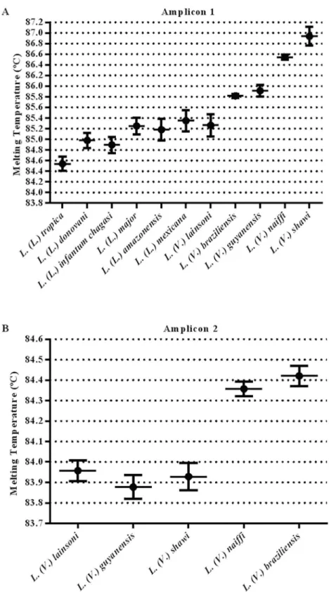
The average and standard deviation of the melting temperature (Tm) of each amplicon was determined in duplicate from three independent experiments using 50 ng of DNA as a tem-plate from each reference species. The melting profiles and obtained Tm values ofhsp70 amplicon 1 for all species studied are presented in Figs2and3andTable 1. For a reliable dis-crimination, we calculated the dispersion of Tm values and only considered differences in Tm values exceeding 0.3°C (Fig 2).
Fig 2. Tm values obtained with the HRM assay.Representative dispersion graph of individual Tm values for each studiedLeishmaniaspecies using 50 ng of genomic DNA as a template forhsp70amplicon 1 and amplicon 2. The plots show the average and standard deviation of the Tm values. Each species was tested in duplicate in three independent experiments.
doi:10.1371/journal.pntd.0004485.g002
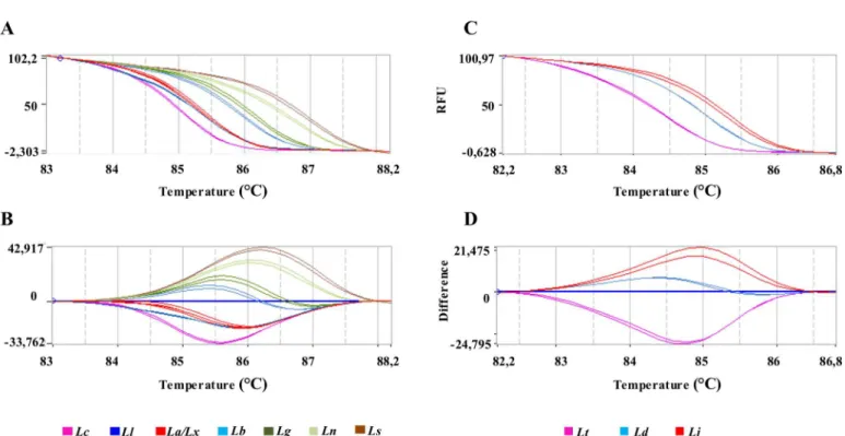
Fig 3. HRM plots ofhsp70amplicon 1.Representative melting profiles ofhsp70amplicon 1 obtained with DNA fromLeishmaniaspecies present in Brazil (A, B) or DNA fromLeishmaniaspecies of Eurasia and Africa (C, D). (A, C): Normalized melting curves; (C, D): normalized difference curves. (Lt):L.(L.) tropica;(Ld):L.(L.) donovani; (Lc):L.(L.) infantum chagasi; (Lj):L.(L.) major; (La):L.(L.) amazonensis;(Lx):L.(L.) mexicana; (Ll):L.(V.) lainsoni; (Lb):L.(V.) braziliensis; (Lg):L.(V.) guyanensis; (Ln):L.(V.) naiffiand (Ls):L.(V.) shawi. Each sample was tested in duplicate.
doi:10.1371/journal.pntd.0004485.g003
reference DNA (FMUSP-IOF-2016), and the call identification agreed 100% with the reference samples, even in samples where the call efficiency was approximately 75% (Table 2).
To test if the initial amount of target DNA caused a variation in the Tm, serial dilutions con-taining 50 ng to 50 fg (DNA amount corresponding to 5.0 x 10⁵to 0.5 of parasite) of Leish-maniaDNA from reference strains were used as a template to produce bothhsp70amplicon 1 (Fig 4A) andhsp70amplicon 2 (Fig 4B). The Tm variation obtained for both amplicons in each species showed that some species presented a fluctuation of Tm values that overlapped with other species.
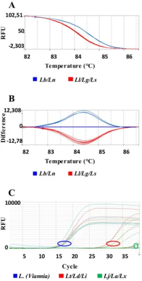
In the case of overlapping Tm values for amplicon 1, a sequential discrimination can be per-formed by HRM analysis of amplicon 2. This amplicon is specific for theL.(Viannia)subgenus species, allowing the segregation of two patterns that groupL.(V.) guyanensis,L.(V.) lainsoni andL.(V.) shawiwith Tm = 83.92 ± 0.04°C orL.(V.) naiffiandL.(V.) braziliensiswith Tm = 84.39 ± 0.04°C (Figs4and5).
The Ct values obtained in the amplification curves of amplicon 2, using DNA of all Leish-maniastudied indicated that the reactions were at least 5 orders of magnitude more specific to Leishmania (Viannia)species than for theL.(Leishmania)species (Figs5CandS2), confirming that amplicon 2 can be used to discriminateL.(Viannia)from theL.(Leishmania)species.
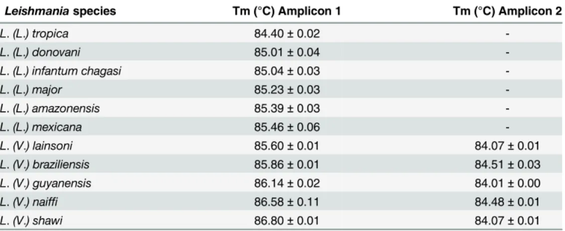
Moreover, using the information on the geographical origin of the samples associated with the HRM analysis ofhsp70amplicon 2 allowed for the discrimination betweenL.(L.) donovani Table 1. Tm values obtained in the HRM analysis targeting thehsp70gene of differentLeishmania species.Fifty ng of genomic DNA from each species was tested in duplicate in three independent experiments.
Leishmaniaspecies Tm (°C) Amplicon 1 Tm (°C) Amplicon 2
L.(L.) tropica 84.40±0.02
-L.(L.) donovani 85.01±0.04
-L.(L.) infantum chagasi 85.04±0.03
-L.(L.) major 85.23±0.03
-L.(L.) amazonensis 85.39±0.03
-L.(L.) mexicana 85.46±0.06
-L.(V.) lainsoni 85.60±0.01 84.07±0.01
L.(V.) braziliensis 85.86±0.01 84.51±0.03
L.(V.) guyanensis 86.14±0.02 84.01±0.00
L.(V.) naiffi 86.58±0.11 84.48±0.01
L.(V.) shawi 86.80±0.01 84.07±0.01
doi:10.1371/journal.pntd.0004485.t001
Table 2. Call efficiency obtained in the HRM analysis targeting thehsp70gene of differentLeishmaniaspecies in the presence of two different amounts of human reference (FMUSP-IOF-2016) DNA.The“call”column shows the identification name given by the software to the unknown samples based on names given to the reference samples; (CHA):L.(L.) infantum chagasi; (AMA):L.(L.) amazonensis; (LAI):L.(V.) lainsoni; (BRA):L.(V.) braziliensis; (GUY):L.(V.) guyanensis; (NAI):L.(V.) naiffiand (SHA):L.(V.) shawi. Each condition was tested in duplicate.
Leishmaniaspecies DNA proportion 1:1 DNA proportion 1:100
Call Efficiency (%) Call Efficiency (%)
L.(L.) infantum chagasi CHA 92.55 CHA 79.95
L.(L.) amazonensis AMA 89.90 AMA 75.80
L.(V.) lainsoni LAI 89.20 LAI 74.35
L.(V.) braziliensis BRA 89.85 BRA 78.10
L.(V.) guyanensis GUY 91.90 GUY 79.40
L.(V.) naiffi NAI 95.05 NAI 81.95
Fig 4. Effect of the amount of target DNA on the Tm values ofhsp70amplicons.Representative dispersion graph of individual Tm values for each studiedLeishmaniaspecies for amplicon 1 (A) and for amplicon 2 (B). Each point corresponds to the average and standard deviation of the variation in the Tms obtained within a range of 50 ng to 50 fg of genomic DNA used as a template. Each concentration point was measured in duplicate.
doi:10.1371/journal.pntd.0004485.g004
andL.(L.) infantum chagasi; amongL.(L.) major,L.(L.) amazonensis,L.(L.) mexicanaand L.(V.) lainsoni.
DNA from uninfected mouse, human, orTrypanosoma cruziandT.bruceiwere used as templates and compared to the standardized positive range of Tm values for the tested Leish-maniaspecies. No cross-reactivity was detected. For these controls, characteristic Tm values (T.cruzi: 83.08 ± 0.07°C andT.brucei: 83.91 ± 0.06°C) or no amplification was observed (mouse and human) (S4 Fig).
Validation of the HRM Protocol with other Leishmania strains and with
experimental, clinical and field samples
The HRM analysis ofhsp70amplicon 1 obtained with DNA from otherLeishmaniaisolates also used as reference strains resulted in a 100% correlation with the Tm values of the reference
Fig 5. HRM plots ofhsp70amplicon 2.Representative melting profiles ofhsp70amplicon 2. Panels (A) and (B) show the melting profiles of AmericanL.(Viannia)species with data organized in normalized melting curves and normalized difference curves, respectively. (Lb):L.(V.) braziliensis;(Ln):L.(V.) naiffi; (Ll):L.(V.) lainsoni; (Lg):L.(V.) guyanensisand (Ls):L.(V.) shawi. Panel (C) shows the amplification curves in relation to the Ct, using DNA of the same species as A and B plus(Lt.):L.(L.) tropica; (Ld):L.(L.) donovani; (Li):L.(L.) infantum; (La):L.(L.) amazonensis; (Lx):L.(L.) mexicanaand(Lj):L.(L.) major. Each sample was tested in duplicate.
species used in this study (Table 3). Some of those strains represent isolates obtained from dif-ferent geographical regions in Brazil, and experimentally corroborated the identification through the HRM protocol for possible polymorphisms.
The intra-specific variability was further assessed by thein silicoanalysis of polymorphism of 186hsp70entries fromL.(L.) tropica,L.(L.) donovani,L.(L.) infantum,L.(L.) major,L.(L.) amazonensis,L.(L.) mexicana,L.(V.) lainsoni,L.(V.) braziliensis,L.(V.) guyanensis,L.(V.) naiffi,L.(V.) shawi,L.(V.) peruviana,L.(V.) panamensis,L.(L.) aethiopica,L.(L.) martini-quensisandL.siamensis. All the sequences were aligned to include the regions of amplicons 1 and 2. The aligned sequences were then examined for polymorphisms among species as well as among strains of the same species. We then calculated the percentage of similarity and esti-mated the theoretical Tm value of both amplicons (S1 Table). If we assume that the nucleotide differences that we detected are real polymorphisms and not sequencing errors then we can see fromS1 Tablethat the differences in the theoretical Tm values of each species results in the same discriminatory pattern. Of the 186 strains analyzed, only two strains ofL.infantum, MCAN/IR/96/LON-49 and LEM75/zymodeme1, presented a theoretical Tm value whose dif-ference was higher than 0.3°C. We cannot rule out the possibilities that this difdif-ference is in fact a real one, due to sequencing errors or reflects different taxa.
In the absence ofbona fidesamples we also determined the theoretical Tm of amplicons 1 and 2 (S1 Table) of twoLeishmaniaspecies found in America,L.(V.) peruvianaandL.(V.) panamensis, that occur outside Brazil. The obtained data indicated that these two species could be differentiated from the othersL.(Viannia)species by the coupled HRM analysis of the two amplicons.
The theoretical Tm value of the AfricanL.(L.) aethiopica, potentially allowed the discrimi-nation fromL.(L.) donovani,L.(L.) infantum and L.(L.) major, but not fromL.(L.) tropica(S1 Table). Theenriettiicomplex membersL.(L.) martiniquensisandL.siamensispresented iden-tical theoreiden-tical Tm values.
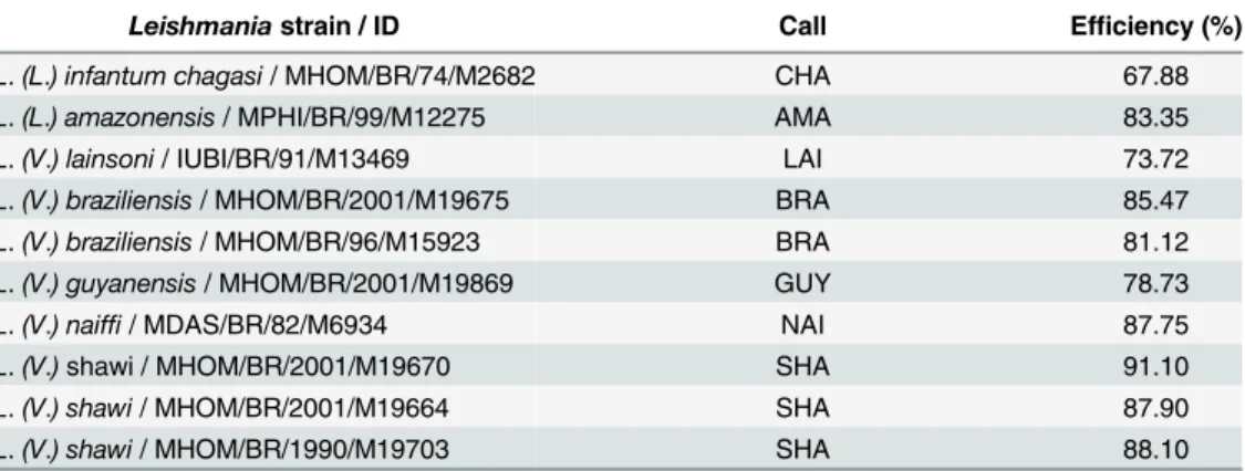
To validate the HRM protocol for different types of sample preparations, sixteen DNA obtained from real biological samples, like fresh tissue from hamster inoculated with infected sample from human or dog cases; cell culture of the human isolated strain; human fresh tissue; human paraffin-embedded tissue; tissues from experimentally infected BALB/c mice and natu-rally infected phlebotomines, that had been previously tested in our laboratory by sequencing of SSU rDNA [20] or by discriminatory PCR targetingg6pd[21], were submitted to HRM Table 3. Call efficiency obtained in the HRM analysis targeting thehsp70gene of differentLeishmania strainsThe“call”column shows the names given by the software to the unknown samples based on names given to the reference samples:L.(L.) infantum chagasi(CHA),L.(L.) amazonensis(AMA),L.(V.) lainsoni
(LAI),L.(V.) braziliensis(BRA),L.(V.) guyanensis(GUY),L.(V.) naiffi(NAI) andL.(V.) shawi(SHA). Each con-dition was tested in duplicate in three independent experiments.
Leishmaniastrain / ID Call Efficiency (%)
L.(L.) infantum chagasi/ MHOM/BR/74/M2682 CHA 67.88
L.(L.) amazonensis/ MPHI/BR/99/M12275 AMA 83.35
L.(V.) lainsoni/ IUBI/BR/91/M13469 LAI 73.72
L.(V.) braziliensis/ MHOM/BR/2001/M19675 BRA 85.47
L.(V.) braziliensis/ MHOM/BR/96/M15923 BRA 81.12
L.(V.) guyanensis/ MHOM/BR/2001/M19869 GUY 78.73
L.(V.) naiffi/ MDAS/BR/82/M6934 NAI 87.75
L.(V.)shawi / MHOM/BR/2001/M19670 SHA 91.10
L.(V.) shawi/ MHOM/BR/2001/M19664 SHA 87.90
L.(V.) shawi/ MHOM/BR/1990/M19703 SHA 88.10
doi:10.1371/journal.pntd.0004485.t003
analysis. The results obtained presented a correlation with the results obtained with the other targets (Table 4).
Discussion
The establishment of optimized protocols for the detection and identification of the aetiological agents of Leishmaniases are extremely useful tools in a clinical context. Identifying the species can lead to species-specific treatment protocols to promote a better efficacy of treatment, assessing the need for patient follow up as well as the development and understanding of the mode of action of potential new drugs.
Several methodologies targeting different genomic or mitochondrial DNA have been described in the past 20 years, and PCR is currently the preferred method in studies involving the detection and identification ofLeishmania. These methodologies have been developed by designing primers that exploit species-specific sequence polymorphisms in different targets, such as kDNA [22], the SSU rDNA gene [20,23], the glucose-6-phosphate dehydrogenase gene (g6pd) [21,24], rDNA internal transcribed spacers (ITSs) [25],hsp70[13–17] and cysteine proteinase B gene (cpb) [7,26]. However, none of these methods represents a gold standard because the targeted polymorphisms were unsuitable for simple and direct identification proto-cols. These PCR analyses involved the use of multiple targets requiring a combination of sev-eral primers creating the need of running more than one reaction to identify a single sample. The multiplex PCR that uses several pair of primers in one reaction and restriction fragment length polymorphism analysis (RFLP) of PCR products both need of a subsequent DNA frac-tionation by gel electrophoresis. These procedures require experienced operators to interpret the results, besides the risk of laboratory contamination with amplicons, due to the manipula-tion of PCR product.
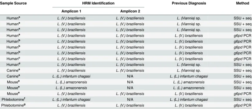
Table 4. Identification ofLeishmaniain clinical and experimentally infected and field samples by the HRM analysis targetinghsp70gene.The
hsp70amplicons 1 or 2 of DNA from each sample were submitted to the HRM analysis. The results were compared with a previous identification performed by SSU rDNA sequencing [17] org6pdPCR [18].(a)fresh tissue from hamster inoculated with infected sample;(b)cell culture of the human isolated strain;(c) human fresh tissue;(d)human paraffin-embedded tissue;(e)experimentally infected BALB/c mice;(f)naturally infectedLutzomyia (Lutzomyia) longipalpis; and (g)
naturally infectedLu.(Nyssomyia) whitmani; (N/A): not applicable.
Sample Source HRM Identification Previous Diagnosis Method
Amplicon 1 Amplicon 2
Humana L.(V.) braziliensis L.(V.) braziliensis L.(Viannia)sp. SSU + seq
Humana L.(V.) braziliensis L.(V.) braziliensis L.(Viannia)sp. SSU + seq
Humana L.(V.) braziliensis L.(V.) braziliensis L.(Viannia)sp. SSU + seq
Humana L.(V.) braziliensis L.(V.) braziliensis L.(V.) braziliensis g6pdPCR
Humanb L.(V.) braziliensis L.(V.) braziliensis L.(V.) braziliensis g6pdPCR
Humanb L.(V.) braziliensis L.(V.) braziliensis L.(V.) braziliensis g6pdPCR
Humanb L.(V.) braziliensis L.(V.) braziliensis L.(V.) braziliensis g6pdPCR
Humanc L.(V.) braziliensis L.(V.) braziliensis L.(V.) braziliensis g6pdPCR
Humand L.(V.) braziliensis L.(V.) braziliensis L.(Viannia)sp. SSU + seq
Caninea L.(V.) braziliensis L.(V.) braziliensis L.(Viannia)sp. SSU + seq
Caninea L.(L.) infantum chagasi N/A L.(L.) infantum chagasi SSU + seq
Mousee L.(L.) amazonensis N/A L.(L.) amazonensis SSU + seq
Mousee L.(L.) amazonensis N/A L.(L.) amazonensis SSU + seq
Mousee L.(V.) braziliensis L.(V.) braziliensis L.(V.) braziliensis g6pdPCR
Phlebotominef L.(L.) infantum chagasi N/A L.(L.) infantum chagasi SSU + seq
Phlebotomineg L.(V.) braziliensis L.(V.) braziliensis L.(V.) braziliensis g6pdPCR
Another way to exploit DNA polymorphisms is the determination of the C+G composition of PCR products from conserved regions by calculating the Tm of the amplicon in a melting curve. HRM methodology has been successfully used forLeishmaniaidentification using dif-ferent targets, such as the 7SL RNA gene that discriminatedL.tropica,L.majorand species that cause visceralLeishmaniases in clinical samples [5,6]. Additionally, using the same target, researchers determined that rodentCtenodactylus gundiis a potential host ofL.tropicain Tunisia [5]. Polymorphisms onhaspb(Hydrophilic Acylated Surface Protein B gene) analyzed by HRM allowed the differentiation of strains ofL.(L.) donovanifrom distinct regions of East Africa [7]. In Southeastern Iran, the rRNA ITS sequence incriminatedPhebotomus sergentias a natural vector ofL.(L.) tropica[10], or the discrimination betweenL.(L.) tropicaandL.(L.) infantumin Turkey [9]. HRM analysis of the ITS-1 rRNA region discriminatedL.(L.) major, L.(.L) tropica,L.(L.) aethiopicaandL.(L.) infantumin samples from Middle East, Asia, Africa and Europe [8]. The combination of two targets,hsp70and the rRNA ITS1 sequence, using the absolute HRM values allowed for the discrimination of six AmericanLeishmaniaspecies [11] and MPI/6PGD-FRET PCR distinguishedL.(V.) braziliensisfromL.(V.) peruviana[12].
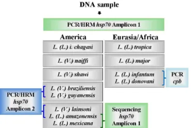
Here, we described an algorithm using HRM methodology for the rapid detection and dis-crimination ofLeishmaniaspecies circulating in Brazil and Eurasia/Africa (Fig 6). We used the sequence coding forhsp70, but in order to obtain a discriminatory PCR product, we designed the primers to encompass a region that was no larger than 144 bp and that had relevant poly-morphisms for HRM analysis, that is, shifts of AT base pairs to CG or vice-versa. Moreover, to be effective, the total amount of polymorphisms was taken into account, and compensatory changes were avoided. Using these criteria, we obtained two PCR products: amplicon 1 and amplicon 2. Using the algorithm described inFig 6, the analysis of the produced melting pro-files of amplicon 1 for the Brazilian species allowed for the discrimination ofL.(L.) i.chagasi, L.(L.) amazonensis/L.(L.) mexicana/L.(V.) lainsoni,L.(V.) braziliensis/L (V.) guyanensis,L. (V.) naiffiandL.(V.) shawiusing differences in the Tm of at least 0.3°C. For Eurasian samples, amplicon 1 produced values with the same 0.3°C interval to discriminateL.(L.) tropicafromL.
Fig 6. Schematic algorithm representation forLeishmaniaidentification in Brazilian or Eurasian/ African samples.Purified DNA was submitted tohsp70PCR to produce amplicon 1. The melting analysis of
hsp70amplicon 1 for American samples discriminatesL.(L.) infantum chagasi,L.(V.) naiffiandL.(V.) shawi. The grouping ofL.(V.) braziliensis/L.(V.) guyanensiscan be discriminated by producinghsp70 PCRamplicon 2. Amplicon 2 also resolve the groupingL.(L.) amazonensis/L.(L.) mexicana/L.(V.) lainsoni, which is positive forL.(V.) lainsoni, or by sequencinghsp70PCR amplicon 1 to discriminate betweenL.(L.) amazonensis and L.(L.) mexicana. For Eurasian/African samples, the melting analysis ofhsp70amplicon 1 discriminatesL.(L.) tropica,L.(L.) majorand theL.(L.) donovani/L (L.) infantumgroup, which can be solved bycpbPCR [7].
doi:10.1371/journal.pntd.0004485.g006
(L.) majorand fromL.(L.) donovani/L (L.) infantum, but these two species cannot be discrimi-nated from each other (Fig 2).
The occurrence of an overlap in the Tm value for the Brazilian speciesL.(L.) amazonensis andL.(L.) lainsoniafter a positive reaction of amplicon 1 can be solved by a positive reaction of amplicon 2. This amplicon sequence is specific forLeishmania (Viannia)species, soL.(L.) amazonensiswill not be amplified andL.(V.) lainsoniwill present the corresponding Tm value (Fig 5). The occurrence of an overlap in the Tm value for the American speciesL.(L.) amazo-nensisandL.(L.) mexicanacan be solved by amplicon 1 sequencing because this amplicon is not identical, but there are two mismatches (position 82 A to G and position 100 G to T inL. (L.) amazonensisandL.(L.) mexicana, respectively (Fig 1), that are compensatory in the melt-ing profile. It is interestmelt-ing to note that these two species are very closely related. Uliana et al. [23] distinguishedL.(L.) amazonensisfromL.(L.) mexicanaby SSU rDNA, but Castilho et al. [21] also failed to distinguish these species byg6pdbecause the region of theg6pdsequence that was used is identical in the two species. It is also interesting that Hernandez et al. [11], using a larger amplicon (337 bp) ofhsp70, succeeded in differentiatingL.(L.) mexicanafromL. (L.) amazonensis; however, Fraga et al. [13] failed to distinguish these two species using RFLP in another region ofhsp70. However, when the complete nucleotide sequence of thehsp70PCR fragment of 1268 bp is used, the discrimination between the two species can be achieved [27]. These problems once again emphasize that one gene or a particular sequence of a gene is not reliable to define a species or plot its phylogeny. Recently, Real et al. [28] showed thatL.(L.) mexicanaandL.(L.) majorhad, respectively, 5 and 7 species-specific orthologous gene fami-lies, whileL.(L.) amazonensishad 23 different gene families. Moreover, the geographical parameter can also be used; Uliana et al. used SSU rDNA polymorphism to show that these species present a characteristic distribution in Latin America that correlates to monoclonal antibody profiles [29].
The Tm overlap for Eurasian species occurred forL.(L.) donovaniandL.(L.) infantum, which presented identical sequences for amplicon 1. Again, the geographical origin of the sam-ple can be used becauseL.(L.) donovaniis more frequently found in India and East Africa and presents anthroponotic behavior.L.(L.) infantumis found in Africa, China and the Mediterra-nean and shows zoonotic behavior [30]. However, the two species can be discriminated by multilocus enzyme electrophoresis (MLEE) or multilocus microsatellite typing (MLMT) [30]. Recently, thehaspbcoding region was initially used in a classical PCR coupled to RFLP [31], while the gene coding forcpbwas used as a target in conventional PCR [7]. We propose to use the latter in case of doubt between the two species (Fig 6).
Thein silicoanalysis of amplicon 1 and 2 from otherLeishmaniaspecies from America or from Eurasia/Africa, also indicated the potentiality of thehsp70HRM protocol to discriminate L.(V.) peruviana,L.(V.) panamensisandL.(L.) aethiopica/ L.(L.) martiniquensis/L.siamensis fromL.(L.) donovaniandL.(L.) majorbut not fromL.(L.) tropica. It is interesting to note that the ITS-HRM analysis applied toL.(L.) tropica L.(L.) aethiopica,L.(L.) infantum,L.(L.) majorandL.(L.) donovani[8] presented exactly the same degree of resolution of thehsp70 HRM described here.
In fact, when we applied the protocol described here to otherLeishmaniaisolates, the obtained“call”(the identification of the problem sample in relation to the reference samples) presented a 100% correlation with the reference strains (Table 3).
The test of sixteen samples consisting of fresh hamster tissue from animals injected with human or dog biopsy macerates, fresh or paraffin embedded human biopsies, tissues of experi-mentally infected BALB/c mice or even naturally infected phebotominae, produced identifica-tion“calls”comparable to the identification results using SSU rDNA sequencing org6pdPCR (Table 4), showing that the source of the sample as well as its conservation do not interfere in the HRM protocol. Moreover, the use of HRM protocol is easier than the use of SSU rDNA and/org6pdPCR, since those methods require either sequencing of the product or three or more distinct PCRs followed by gel electrophoresis analysis.
Overall, thehsp70HRM protocol described herein accurately and sensitively identified Leishmaniaspecies that are important in the majority of cases of Leishmaniases in the Brazil and Eurasia. The test is simple and rapid, and its use in the clinic or in research samples has many advantages, such as a lower total cost for the identification of a sample and other charac-teristics that facilitate its application. There is no need for sequencing or gel fractionation to analyze the product, thus avoiding laboratory contamination with PCR products because these products are discarded without being manipulated. It also reduces the need for trained person-nel to analyze the fractionation profile of an electrophoretic gel or sequencing data to provide a result. Also the HRM assay presents a possibility of quantifying parasites present in samples because it is a real-time PCR-based technique. Moreover, the whole process can be automated because the analyzer software will produce the“call”result by comparing the tested samples to the reference sample identities, which must always be included in the reactions.
In conclusion, the protocol described herein is a low cost, reliable, easy to apply, potentially automated procedure that is a good alternative for the detection, quantification and identifica-tion ofLeishmaniaspecies in biological and clinical samples.
Supporting Information
S1 Fig. Nucleotide sequence ofhsp70amplicon 2 and primer localization.The underlined sequences indicate the position of the primers used in real-time PCR assays; the grey boxed nucleotides represent the mutation points found among reference strains ofLeishmania. The black boxed nucleotides represent mismatches that prevent the annealing of oligonucleotide hsp70F1 to theL.(Leishmania)spp. complementary sequence.
(TIFF)
S2 Fig. Electrophoretic profile of conventional PCR products of polymorphic regions of thehsp70gene from referenceLeishmaniaspecies.hsp70amplicons 1 (A) andhsp70amplicons 2 (B) were fractioned by electrophoresis in 1.5% agarose gel and stained with ethidium bromide. DNA from reference strains ofLeishmaniaare named as follows: (Lt):L.(L.) tropica; (Ld):L.(L.) donovani; (Lc):L.(L.) infantum chagasi; (Lj):L.(L.) major; (La):L.(L.) amazonensis;(Lx):L.(L.) mexicana;(Ll):L.(V.) lainsoni; (Lb):L.(V.) braziliensis; (Lg):L.(V.) guyanensis; (Ln):L.(V.) naiffiand (Ls):L.(V.) shawi. (L): 100 bp DNA ladder and (ntc): no template control, without DNA.
(TIFF)
S3 Fig. Efficiency ofhsp70amplicon 1 real-time PCR for DNA from distinctLeishmania species.Standard curves were constructed with recombinant plasmids containing amplicon 1 sequence fromL.(L.) amazonensis(A),L.(L.) infantum chagasi(B),L.(V.) guyanensis(C) andL. (V.) lainsoni(D). The assays used as a template underwent a 10-fold serial dilution representing
1 x 10⁶to 1 x 101plasmid copies per reaction and were performed in duplicate. (TIFF)
S4 Fig. HRM plots ofhsp70amplicon 1 fromTrypanosoma.Representative melting profiles ofhsp70amplicon 1 obtained from the genomic DNA ofT.cruziandT.brucei. (A): Normal-ized melting curves; (B): normalNormal-ized difference curves and (C): dispersion graph of individual plots fromT.cruzi(Tc) andT.brucei(Tb) compared toL.(L.) tropica(Lt).
(TIFF)
S1 Table. Polymorphisms detection byin silicoanalysis ofLeishmania hsp70sequences. The hsp70 regions compassing amplicon 1 or amplicon 2 were retrieved from GenBank Data-base [32] using the sentence“heat shock protein 70 kDa”as descriptor words in“search”field. The obtained sequences were formatted as FASTA files and aligned on BioEdit Sequence Alignment Editor v.7.1.8 [33]. The identity indexes were obtained by pairwise alignments on BioEdit software. Only sequences encompassing the whole amplicon were analyzed. Theoreti-cal melting temperatures of hypothetic amplicons were Theoreti-calculated using OligoCalc oligonucleo-tide properties on-line calculator [34].
(DOCX)
Author Contributions
Conceived and designed the experiments: RAZ MFLS JJS LMFW. Performed the experiments: RAZ SMM ACSdL. Analyzed the data: RAZ MFLS SMM ACSdL JJS LMFW. Contributed reagents/materials/analysis tools: LMFW. Wrote the paper: RAZ MFLS SMM JJS LMFW.
References
1. Alvar J, Velez ID, Bern C, Herrero M, Desjeux P, Cano J, et al. Leishmaniasis worldwide and global esti-mates of its incidence. PLoS One. 2012; 7(5):e35671. doi:10.1371/journal.pone.0035671PMID: 22693548
2. World Health O. Control of the leishmaniases. World Health Organization technical report series. 2010 (949: ):xii-xiii, 1–186, back cover. PMID:21485694
3. Desjeux P. Leishmaniasis: current situation and new perspectives. Comparative immunology, microbi-ology and infectious diseases. 2004; 27(5):305–18. PMID:15225981
4. Reithinger R, Dujardin JC. Molecular diagnosis of leishmaniasis: Current status and future applications. J Clin Microbiol. 2007; 45(1):21–5. PMID:17093038
5. Bousslimi N, Ben-Ayed S, Ben-Abda I, Aoun K, Bouratbine A. Natural Infection of North African Gundi (Ctenodactylus gundi) byLeishmania tropicain the Focus of Cutaneous Leishmaniasis, Southeast Tunisia. American Journal of Tropical Medicine and Hygiene. 2012; 86(6):962–5. doi:10.4269/ajtmh. 2012.11-0572PMID:22665601
6. Nasereddin A, Jaffe CL. Rapid Diagnosis of Old World Leishmaniasis by High-Resolution Melting Anal-ysis of the 7SL RNA Gene. J Clin Microbiol. 2010; 48(6):2240–2. doi:10.1128/JCM.00553-10PMID: 20392923
7. Zackay A, Nasereddin A, Takele Y, Tadesse D, Hailu W, Hurissa Z, et al. Polymorphism in the HASPB Repeat Region of East AfricanLeishmania donovaniStrains. PLoS neglected tropical diseases. 2013; 7(1).
8. Talmi-Frank D, Nasereddin A, Schnur LF, Schonian G, Toz SO, Jaffe CL, et al. Detection and Identifica-tion of Old WorldLeishmaniaby High Resolution Melt Analysis. PLoS neglected tropical diseases. 2010; 4(1).
9. Toz SO, Culha G, Zeyrek FY, Ertabaklar H, Alkan MZ, Vardarli AT, et al. A real-time ITS1-PCR based method in the diagnosis and species identification ofLeishmaniaparasite from human and dog clinical samples in Turkey. PLoS neglected tropical diseases. 2013; 7(5):e2205. doi:10.1371/journal.pntd. 0002205PMID:23675543
method in South-eastern Iran. Asian Pacific journal of tropical medicine. 2014; 7(2):93–6. doi:10.1016/ S1995-7645(14)60002-XPMID:24461520
11. Hernandez C, Alvarez C, Gonzalez C, Ayala MS, Leon CM, Ramirez JD. Identification of Six New WorldLeishmaniaspecies through the implementation of a High-Resolution Melting (HRM) genotyping assay. Parasites & vectors. 2014; 7:501.
12. Tsukayama P, Nunez JH, De Los Santos M, Soberon V, Lucas CM, Matlashewski G, et al. A FRET-based real-time PCR assay to identify the main causal agents of New World tegumentary leishmania-sis. PLoS neglected tropical diseases. 2013; 7(1):e1956. doi:10.1371/journal.pntd.0001956PMID: 23301111
13. Fraga J, Montalvo AM, De Doncker S, Dujardin JC, Van der Auwera G. Phylogeny ofLeishmania spe-cies based on the heat-shock protein 70 gene. Infection, genetics and evolution: journal of molecular epidemiology and evolutionary genetics in infectious diseases. 2010; 10(2):238–45. doi:10.1016/j. meegid.2009.11.007PMID:19913110
14. Montalvo AM, Fraga J, Monzote L, Montano I, De Doncker S, Dujardin JC, et al. Heat-shock protein 70 PCR-RFLP: a universal simple tool forLeishmaniaspecies discrimination in the New and Old World. Parasitology. 2010; 137(8):1159–68. doi:10.1017/S0031182010000089PMID:20441679
15. Garcia L, Kindt A, Bermudez H, Llanos-Cuentas A, De Doncker S, Arevalo J, et al. Culture-independent species typing of neotropicalLeishmaniafor clinical validation of a PCR-based assay targeting heat shock protein 70 genes. J Clin Microbiol. 2004; 42(5):2294–7. PMID:15131217
16. Montalvo AM, Fraga J, Maes I, Dujardin JC, Van der Auwera G. Three new sensitive and specific heat-shock protein 70 PCRs for globalLeishmaniaspecies identification. European journal of clinical micro-biology & infectious diseases: official publication of the European Society of Clinical Micromicro-biology. 2012; 31(7):1453–61.
17. Graça GC, Volpini AC, Romero GA, Oliveira Neto MP, Hueb M, Porrozzi R, et al. Development and vali-dation of PCR-based assays for diagnosis of American cutaneous leishmaniasis and identification of the parasite species. Mem Inst Oswaldo Cruz. 2012; 107(5):664–74. PMID:22850958
18. Miller SA, Dykes DD, Polesky HF. A simple salting out procedure for extracting DNA from human nucle-ated cells. Nucleic acids research. 1988; 16(3):1215. PMID:3344216
19. de Lima AC, Zampieri RA, Tomokane TY, Laurenti MD, Silveira FT, Corbett CE, et al.Leishmania sp. identification by PCR associated with sequencing of target SSU rDNA in paraffin-embedded skin sam-ples stored for more than 30 years. Parasitology research. 2011; 108(6):1525–31. doi:10.1007/ s00436-010-2208-0PMID:21161272
20. Uliana SR, Nelson K, Beverley SM, Camargo EP, Floeter-Winter LM. Discrimination amongst Leish-maniaby polymerase chain reaction and hybridization with small subunit ribosomal DNA derived oligo-nucleotides. The Journal of eukaryotic microbiology. 1994; 41(4):324–30. PMID:8087103
21. Castilho TM, Shaw JJ, Floeter-Winter LM. New PCR assay using glucose-6-phosphate dehydrogenase for identification ofLeishmaniaspecies. J Clin Microbiol. 2003; 41(2):540–6. PMID:12574243
22. Lopez M, Inga R, Cangalaya M, Echevarria J, Llanos-Cuentas A, Orrego C, et al. Diagnosis of Leish-maniausing the polymerase chain reaction: a simplified procedure for field work. The American journal of tropical medicine and hygiene. 1993; 49(3):348–56. PMID:8396860
23. Uliana SR, Affonso MH, Camargo EP, Floeter-Winter LM.Leishmania: genus identification based on a specific sequence of the 18S ribosomal RNA sequence. Experimental parasitology. 1991; 72(2):157–
63. PMID:2009920
24. Castilho TM, Camargo LM, McMahon-Pratt D, Shaw JJ, Floeter-Winter LM. A real-time polymerase chain reaction assay for the identification and quantification of AmericanLeishmaniaspecies on the basis of glucose-6-phosphate dehydrogenase. The American journal of tropical medicine and hygiene. 2008; 78(1):122–32. PMID:18187795
25. Cupolillo E, Grimaldi Junior G, Momen H, Beverley SM. Intergenic region typing (IRT): a rapid molecu-lar approach to the characterization and evolution ofLeishmania. Molecular and biochemical parasitol-ogy. 1995; 73(1–2):145–55. PMID:8577322
26. Quispe Tintaya KW, Ying X, Dedet JP, Rijal S, De Bolle X, Dujardin JC. Antigen genes for molecular epidemiology of leishmaniasis: polymorphism of cysteine proteinase B and surface metalloprotease glycoprotein 63 in theLeishmania donovanicomplex. The Journal of infectious diseases. 2004; 189 (6):1035–43. PMID:14999607
27. Van der Auwera G, Ravel C, Verweij JJ, Bart A, Schonian G, Felger I. Evaluation of four single-locus markers forLeishmaniaspecies discrimination by sequencing. J Clin Microbiol. 2014; 52(4):1098–104. doi:10.1128/JCM.02936-13PMID:24452158
28. Real F, Vidal RO, Carazzolle MF, Mondego JM, Costa GG, Herai RH, et al. The genome sequence of
Leishmania (Leishmania) amazonensis: functional annotation and extended analysis of gene models.
DNA research: an international journal for rapid publication of reports on genes and genomes. 2013; 20 (6):567–81.
29. Uliana SR, Ishikawa E, Stempliuk VA, de Souza A, Shaw JJ, Floeter-Winter LM. Geographical distribu-tion of neotropicalLeishmaniaof the subgenusLeishmaniaanalysed by ribosomal oligonucleotide probes. Trans R Soc Trop Med Hyg. 2000; 94(3):261–4. PMID:10974994
30. Pratlong F, Lami P, Ravel C, Balard Y, Dereure J, Serres G, et al. Geographical distribution and epide-miological features of Old WorldLeishmania infantumandLeishmania donovanifoci, based on the iso-enzyme analysis of 2277 strains. Parasitology. 2013; 140(4):423–34. doi:10.1017/
S0031182012001825PMID:23146283
31. Haralambous C, Antoniou M, Pratlong F, Dedet JP, Soteriadou K. Development of a molecular assay specific for theLeishmania donovanicomplex that discriminatesL.donovani/Leishmania infantum
zymodemes: a useful tool for typing MON-1. Diagnostic microbiology and infectious disease. 2008; 60 (1):33–42. PMID:17889482
32. Benson DA, Cavanaugh M, Clark K, Karsch-Mizrachi I, Lipman DJ, Ostell J, Sayers EW. GenBank. Nucleic Acids Res. 2013 Jan; 41 (Database issue):D36–42. doi:10.1093/nar/gks1195PMID: 23193287
33. Hall TA 1999. BioEdit: a user-friendly biological sequence alignment editor and analysis program for Windows 95/98/NT. Nucl. Acids. Symp. Ser. 41:95–98.