online | memorias.ioc.fiocruz.br Schistosoma japonicum is an important trematode
that causes human schistosomiasis in Asia, including the People’s Republic of China, the Philippines and some ar-eas in Indonesia (Zhou et al. 2010). The re-emergence and active transmission of S. japonicum in endemic areas, with a recent infection of approximately 700,000 people, have been reported (Zhou et al. 2007). The S. japonicum life cycle involves a snail intermediate host and a defini-tive host. Adult schistosomes lay eggs in the mesenteric vein capillaries, which drain the small intestine in the definitive host. The eggs embedded in the intestinal sub-mucosa and sub-mucosa break into the lumen of the bowel and appear in the faeces. In water, the eggs give rise to miracidia, the infective stage of the snail intermediate host. Miracidia penetrate into and migrate through snail tissues to develop into sporocysts, which give rise to cer-cariae. Humans become infected by coming in contact with water contaminated with cercariae, which actively penetrate into the skin. Parasite transmission requires three factors: surface water contamination with fae-cal excreta, the presence of specific fresh water snails as the first intermediate host and human water contact
(Gryseels et al. 2006). In definitive mammalian hosts, S. japonicum cause feverish syndrome, intestinal diseases and hepatosplenic inflammation in the acute stage of the infection and liver fibrosis in the chronic stage (Gryseels et al. 2006). Human infection has been associated with anaemia (King et al. 2005), hepatosplenomegaly and liver fibrosis, as well as liver and colon cancers (Qiu et al. 2005). Egg detection by stool examination remains the standard method to diagnosis human schistosomia-sis japonica. However, it is difficult to differentiate be-tween S. japonicum and Schistosoma mekongi eggs by stool examination; it is not feasible during the prepatent period and it is not sensitive enough to detect mild infec-tions (Doenhoff et al. 2004). Parasite-specific antibody detection by enzyme-linked immunosorbent assay has a high sensitivity and is useful for mass screening, but it has the limitation of a relatively low specificity due to cross-reactivity, and it does not always correlate with an active infection (Doenhoff et al. 2004). The detec-tion of parasite DNA in host-derived material is direct evidence of an actual infection, which is why molecular-based approaches to detect S. japonicum DNA, such as conventional polymerase chain reaction (c-PCR) and loop-mediated isothermal amplification, have been de-veloped (Xia et al. 2009, Kato-Hayashi et al. 2010, Xu et al. 2010). Recently, a SYBR® green-based real-time PCR (Lier et al. 2006, 2008), a minor groove-binding probe-based real-time PCR (Lier et al. 2009), and a TaqMan probe-based real-time PCR (Hung & Remais 2008) were published as sensitive and fast methods that could quan-tify specific S. japonicum DNA in biological samples and in environmental water. In contrast with previous
Financial support: NSTDA (Discovery Based Development Grant), Higher Education Research Promotion, National Research University Project of Thailand, OHEC, Thailand
+ Corresponding author: wanch_ma@kku.ac.th Received 15 March 2011
Accepted 6 September 2011
Molecular detection of
Schistosoma japonicum
in infected snails and mouse faeces
using a real-time PCR assay with FRET hybridisation probes
Tongjit Thanchomnang1,2, Pewpan Intapan1,3, Pusadee Sri-Aroon4, Viraphong Lulitanond1,5, Penchome Janwan1,3, Oranuch Sanpool1,3, Wanchai Maleewong1,3/+
1Research and Diagnostic Center for Emerging Infectious Diseases 3Department of Parasitology 5Department of Microbiology,
Faculty of Medicine, Khon Kaen University, 40002 Khon Kaen, Thailand 2Faculty of Medicine, Mahasarakham University,
Mahasarakham, Thailand 4Applied Malacology Center, Department of Social and Environmental Medicine,
Faculty of Tropical Medicine, Mahidol University, Bangkok, Thailand
A real-time polymerase chain reaction (PCR) assay with fluorescence resonance energy transfer (FRET) hy-bridisation probes combined with melting curve analysis was developed to detect Schistosoma japonicum in experi-mentally infected snails and in faecal samples of infected mice. This procedure is based on melting curve analysis of a hybrid between an amplicon from the S. japonicum internal transcribed spacer region 2 sequence, which is a 192-bp S. japonicum-specific sequence, and fluorophore-labelled specific probes. Real-time FRET PCR could detect as little as a single cercaria artificially introduced into a pool of 10 non-infected snails and a single egg inoculated in 100 mg of non-infected mouse faeces. All S. japonicum-infected snails and all faecal samples from infected mice were positive. Non-infected snails, non-infected mouse faeces and genomic DNA from other parasites were negative. This assay is rapid and has potential for epidemiological S. japonicum surveys in snails, intermediate hosts and faecal samples of final hosts.
reports, S. japonicum DNA detection based on the fast, specific and reliable real-time fluorescence resonance energy transfer (FRET) PCR combined with melting curve analysis is promising for detecting an array of par-asites from biological specimens(Intapan et al. 2008a, b, 2009a, b). Here, we report the possible applicability of this real-time FRET PCR combined with melting curve analysis to detect S. japonicum larvae in snails and eggs in faecal samples of infected mice.
SUBJECTS, MATERIALS AND METHODS
Parasite and DNA materials - S. japonicum (Japanese Yamanashi strain) - Experimentally infected Oncomela-nia nosophora snails and mice, as well as S. japonicum adults and cercariae, were obtained from the Applied Malacology Center, Department of Social and Environ-mental Medicine, Faculty of Tropical Medicine, Mahidol University, Thailand. All animal experiments were han-dled according to the Guidelines for Animal Experimen-tation of the National Research Council of Thailand.
S. japonicum (Philippine strain) adults and S. japoni-cum experimentally infected Oncomelania quadrasi snails, as well as S. mekongi (Loatian strain) adults and S. mekongi infected mice, were also provided ob-tained from the Faculty of Tropical Medicine, Mahidol University. To evaluate the analytical specificity of the real-time FRET PCR, other parasite DNA was extracted from the adult worms of Opisthorchis viverrini, Hap-lorchis taichui, Centrocestus spp, Echinostoma malaya-num, Fasciola gigantica, Paragonimus heterotremus and Stellantchasmus spp, as well as from metacercariae of Haplorchoides spp, cercariae of animal schistosomes, healthy human faeces, human leukocytes and human fae-cesinfectedwith Strongyloides stercoralis, Taenia spp, Echinostoma spp and intestinal lecithodendriid flukes. The DNA samples were kept in a DNA bank at -70ºC in the Department of Parasitology, Faculty of Medicine, Khon Kaen University until use.
Specimen preparation for real-time FRET PCR - DNA samples were extracted from all non-infected (n = 30) and experimentally infected (n = 30) snails, includ-ing their shells, and from snail tissue samples artificially inoculated with S. japonicum cercariae. Each specimen was homogenised with disposable polypropylene pestles (Bellco Glass Inc Vineland, NJ, USA) and extracted us-ing the Nucleospin Tissue kit (Macherey-Nagel GmbH & Co, Duren, Germany). For DNA extraction from mouse faeces, 100 mg of each S. japonicum-infected mouse fae-ces,non-infected mouse faeces and non-infected mouse faeces artificially inoculated with S. japonicum eggs were thoroughly mixed with 200 µL of normal saline solution (0.85% NaCl in distilled water) and centrifuged at 8,000 g for 5 min. The supernatant was then discarded and the faecal pellet was frozen at -20ºC for 30 min. The frozen pellets were homogenised with disposable poly-propylene pestles (Bellco Glass Inc) and extracted using the QIAamp® DNA stool mini kit (Qiagen, Hilden, Ger-many). The number of S. japonicum eggs in the faeces of infected mice (n = 10) was presented as eggs per gram (EPG) of faeces (ranging from 100-1,100 EPG;
geomet-ric mean = 302 EPG). The DNA was eluted in 100 μL of distilled water, 5 μL of which was used for the real-time
PCR reaction.
Limit of detection - To determine the limit of detec-tion of the real-time FRET PCR, tissues from non-in-fected snails were separately ground. Next, individual aliquots of one, five and 10 non-infected snail samples were separately inoculated with one, five, 10 and 30 S. japonicum cercariae. Similarly, 100 mg faecal aliquots of non-infected mice were separately inoculated with a serial dilution of one, two, four and eight S. japonicum eggs. These samples were also used for genomic DNA extraction as described above. The resultant DNA sam-ples were then used in the real-time FRET PCR.
To evaluate the analytical specificity of the method, genomic DNA from the S. mekongi, O. viverrini, Cen-trocestus spp, H. taichui, F. gigantica, E. malayanum, P. heterotremus, Haplorchoides spp, Stellantchasmus spp and animal schistosomes, as well as DNA extract-ed from human leukocytes, negative human faeces and human faeces infected with S. stercoralis, Taenia spp, intestinal lecithodendriid flukes and Echinostoma, were purified as mentioned above.
65ºC, incubated at 65ºC for 3 s and then slowly heated at 0.1ºC/s-80ºC. The fluorescence intensity change was measured throughout the slow heating phase. To evalu-ate the analytical specificity of the oligonucleotide hy-bridisation FRET-based technique, DNA extracted from samples other than S. japonicum-infected snail and mouse faecal samples were separately analysed. Each run contained at least one negative control consisting of 5 µL distilled water. For improved visualisation of the melting temperatures (Tm), melting curves were derived as previously described (Thanchomnang et al. 2008). Melting curves were used to determine the specific PCR products, which were confirmed by conventional gel electrophoresis. The cycle number (Cn) was presented as the number of PCR cycles needed for the amplicon fluo-rescence signal to exceed the detected threshold value.
S. japonicum-positive control plasmid - A positive control plasmid was constructed by cloning a PCR prod-uct of the S. japonicum ITS2 into the pGEM-T Easy vec-tor (Promega, Madison, WI) according to the protocol of the manufacturer. The PCR products were obtained by c-PCR using the SJ-F and SJ-R primers. The plasmid was propagated in Escherichiacoli,and the nucleotide sequence of the inserted gene was sequenced in both di-rections. The cloned ITS2 nucleotide sequence was iden-tical to the S. japonicum genomic sequence (GenBank accession U22167).
Data analysis - The correlation between the worm loads and the Cn was analysed by the Pearson’s cor-relation test.
RESULTS
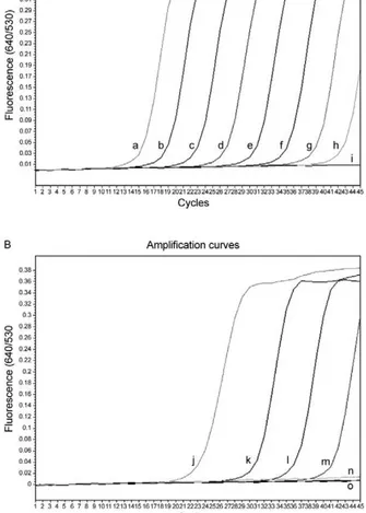
Real-time PCR standardisation - The analytical sen-sitivity of the real-time FRET PCR was evaluated using 5 µL of serial dilutions (1.42 × 10-1.42 × 108 copies) of S. japonicum positive control plasmid in distilled water. The limit of detection of the ITS2 gene target DNA sequence was 1.42 × 10² copies of positive control plasmid (Fig. 2A) or as little as 10-2 ng S. japonicum genomic DNA (Fig. 2B) when considering 35 cycles as the cut-off detection limit. No fluorescence signal was obtained when purified DNA from the following was tested: non-infected O. nosophora
snails,non-infected mouse faeces, O. viverrini, Centro-cestus spp, H. taichui, F. gigantica, E. malayanum, P. heterotremus, Haplorchoides spp, Stellantchasmus spp, animal schistosomes, S. mekongi and S. mekongi-infected mouse faeces, human leukocytes, negative human faeces and human faeces infected with S. stercoralis, Taenia spp, Echinostoma and intestinal lecithodendriid flukes.
With regards to the limit of detection, as little as a sin-gle egg could be detected in 100 mg non-infected mouse faeces based on the 192-bp band in the ethidium bromide-stained gel (Fig. 3A, Lane 2). DNA samples from each aliquot of one, five and 10 non-infected O. nosophora snail tissues artificially inoculated with one S. japonicum cercaria were amplified (Fig. 3B, Lanes 7-9).
Real-time FRET PCR for the detection of S. japonicum in infected O. nosophora snails and mice -A total of 30 S. japonicum-infected,30 non-infected O. nosophora snails,
Fig. 1: schematic illustration of the polymerase chain reaction prim-ers (SJ-F and SJ-R primprim-ers), anchor and detection probes hybridized with the Schistosoma japonicum internal transcribed spacer region 2 (ITS2) (GenBank accession U22167). The probe SJFL530 was labelled with 530 fluorescein at the 3’ end and served as anchor probe for the sensor SJLC640 probe. The sensor probe was labelled with Light-Cycler Red 640 fluorophore (LC red 640) at the 5’ end. The left and right dashed line indicate the 5.8S ribosomal RNA gene (5.8S rRNA) and 28S ribosomal RNA gene (28S rRNA), respectively, whereas the middle line indicates the ITS2. Single circle: 530 fluorescein; double circle: LC red 640.
Fig. 2: amplification plot of fluorescence (y-axis) vs. cycle numbers (x-axis) shows the analytical sensitivity of the real-time polymerase chain reaction for detecting serial dilutions of Schistosoma japonicum
plasmid DNA copies (A) and genomic DNA (B). a-h: 10-fold dilutions concentrations of S. japonicum plasmids from 1.42 × 10⁸-14.2 copies
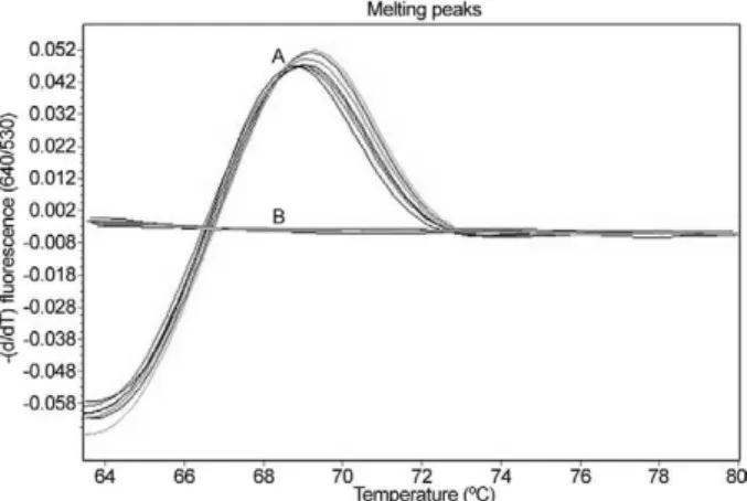
10 S. japonicum-infectedand 10 non-infected mouse fae-cal samples were analysed separately. The melting curve analyses are shown in Fig. 4. When using S. japonicum -specific primers and probes, the mean ± standard devia-tion (SD), range and the median of the Tm values of the S. japonicum-infected O. nosophora snails were 68.98 ± 0.05, 68.93-69.06 and 68.99, respectively, and those of the S. japonicum-infected mouse faecal samples were 68.64 ± 0.29, 68.12-69.00 and 68.68, respectively. A total of 30 S. japonicum-infected O. nosophora snails (Cn range = 9.61-15.07; mean ± SD = 10.97 ± 2.33; median = 9.84) and 10 S. japonicum-infected mouse faecal samples (Cn range = 22.04-30.03; mean ± SD = 25.41 ± 2.5; median = 25.78) were positive by real-time FRET PCR with melt-ing curve analysis, whereas all of the specific control DNAs were negative. However, no significant correlation between the Cn and the S. japonicum eggs intensity in the faecal samples were reported (p > 0.050) (data not shown). The positive and negative rates were 100%. To ensure this procedure can detect S. japonicum DNA from different strains,DNA extracted from S. japonicum Phil-ippine strain adults and from O. quadrasi snails experi-mentally infected with the S. japonicum Philippine strain were also examined by this method. All samples were positive and the nucleotide sequences of the amplified products were identical to that of the S. japonicum Japa-nese Yamanashi strain (data not shown).
The validity of the real-time FRET PCR method used in this study was verified by the presence of a prominent 192-bp product, which was amplified from the DNAs of the S. japonicum-infected O. nosophora snails, S. japoni-cum-infected mouse faeces, S. japonicum cercariae, S.
Fig. 3: detection limit of the real-time fluorescence resonance energy transfer polymerase chain reaction for the detection ofone, two, four and eight Schistosoma japonicum eggs artificially inoculated into 100 mg non-infected mouse faeces samples (A) and one S. japoni-cum cercaria artificially inoculated into one, five and 10 Oncomela-nia nosophora snails (B). Ethidium bromide staining patterns of the amplicon products on a 1.5% agarose gel. Lane M: DNA size markers (100 bp DNA ladder from Invitrogen); N: [cycle number (Cn) > 40]; P: (Cn = 21), negative and positive control containing distilled water and
S. japonicum plasmid 1.42 × 10⁶ copies/reaction, respectively; 1:
am-plicon product from non-infected mouse faeces (Cn > 40); amam-plicon products from one (Cn = 30.03) (Lane 2), two (Cn = 28.30) (Lane 3), four (Cn = 28.13) (Lane 4) and eight (Cn = 27.99) (Lane 5) S. japoni-cum eggs artificially inoculated into 100 mg non-infected mouse fae-ces; 6: amplicon products from non-infected O. nosophora snails (Cn > 40); amplicon products from one S. japonicum cercaria inoculated into aliquots of one (Cn = 23.64) (Lane 7), five (Cn = 28.0) (Lane 8) and 10 (Cn = 35.66) (Lane 9) O. nosophora snails. Arrows indicate the 192 bp S. japonicum specific bands.
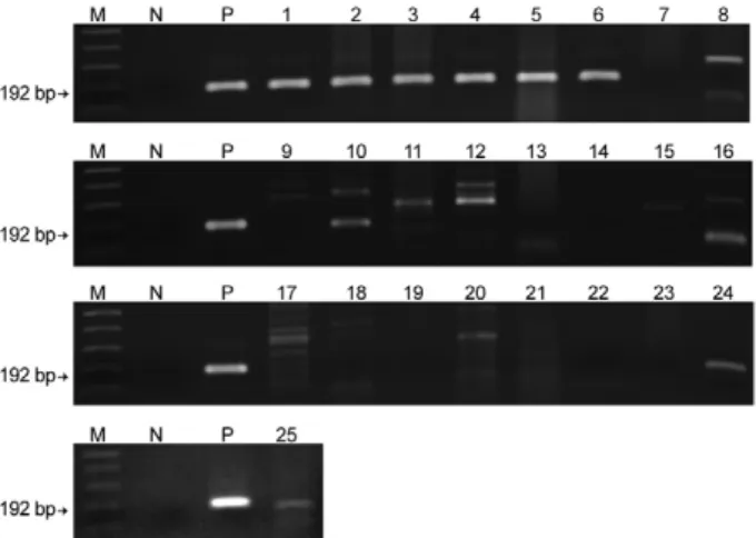
japonicum genomic DNA and the positive control plasmid (Fig. 5, Lanes 1-6, P). However, non-specific bands were also amplified using genomic DNA from some control materials. Although the genomic DNAs from non-infect-ed mouse faeces (Fig. 5, Lane 8), Centrocestus spp (Fig. 5, Lane 10), H. taichui (Fig. 5, Lane 11), F. gigantica (Fig. 5, Lane 12), Stellantchasmus spp (Fig. 5, Lane 16), animal schistosomes (Fig. 5, Lane 17), intestinal lecithodendriid flukes (Fig. 5, Lane 20), S. mekongi (Fig. 5, Lane 24) and S. mekongi-infected mouse faeces (Fig. 5, Lane 25) dem-onstrated various amplified bands, no specific fluores-cence signal was detected by the melting curve analysis.
DISCUSSION
The strategies of transmission control and the inter-ruption of schistosomiasis japonicum require sensitive and specific tests, particularly in areas where the level of endemicity decreases (Lier et al. 2009). Real-time PCR has increasingly superseded c-PCR due to its greatly improved molecular detection efficacy. Not only is this method accurate, rapid and can measure the specific DNA quantity in samples (Lyon & Wittwer 2009), it also discriminates the species or strains of several patho-genic agents by melting curve analysis. Furthermore, this method offers a high throughput and is performed in a closed system, which eliminates the risk of
poten-Fig. 4: representative melting curve analysis of two fluorophore-la-belled probes hybridized to the amplification products of internal tran-scribed spacer region 2 DNA from Schistosoma japonicum. The melt-ing temperature (Tm) of the double-stranded fragment is visualized by plotting the negative derivative of the change in fluorescence divided by the change in temperature in relation to the temperature [-(d/dT) fluorescence (640/530)]. The turning point of this converted melting curve results in a peak and permits easy identification of the fragment specific Tm. Melting curves of positive control plasmid, genomic DNA from S. japonicum adults, S. japonicum-infected Oncomelania nosophora snails, S. japonicum cercariae and S. japonicum-infected mouse faeces (A) as well as genomic DNA from non-infected O. noso-phora snails,non-infected mouse faeces, Opisthorchis viverrini, Cen-trocestus spp, Haplorchis taichui, Fasciola gigantica, Echinostoma malayanum, Paragonimus heterotremus, Haplorchoides spp, Stellant-chasmus spp, animal schistosomes, Schistosoma mekongi and S. me-kongi-infected mouse faeces, human leukocytes, healthy human fae-ces, individual human faeces infected with Strongyloides stercoralis,
tial cross-over contamination because it does not require agarose gel electrophoresis to visualise the amplicons. The real-time FRET PCR-based method has been suc-cessfully used for the detection of several parasites, in-cluding Wuchereria bancrofti (Lulitanond et al. 2004), Brugia malayi (Thanchomnang et al. 2008, 2010a), Dirofilaria immitis (Thanchomnang et al. 2010b), Toxo-plasma gondii (Brenier-Pinchart et al. 2007), Plasmodi-um falciparPlasmodi-um (Ojurongbe et al. 2007) and O. viverrini (Intapan et al. 2008a, b, 2009b).
In this report, we demonstrate that real-time FRET PCR combined with melting curve analysis using two individu-ally labelled hybridisation probes can be used for the sen-sitive and specific detection of S. japonicum infection in snails and faecal samples. This procedure could detect as little as one cercaria implanted in a pool of 10 non-infected O. nosophora snails and a single egg inoculated in 100 mg of non-infected mouse faeces. The results were positive in all S. japonicum-infectedsnails and faecal samples from infected mice equalling a 100% positive rate.
Regarding the analytical specificity of the procedure, no fluorescence was observed despite the amplification of non-specific bands when DNA from some flukes be-longing to a different species than S. japonicum was test-ed, indicating a 100% negative rate. Thus, our real-time
FRET PCR could be useful for the differentiation of S. japonicum cercariae from those of other flukes, such as S. mekongi, O. viverrini, Centrocestus spp, H. taichui, F. gigantica, E. malayanum, P. heterotremus, Haplor-choides spp, Stellantchasmus spp and animal schisto-somes. This method could also distinguish S. japonicum eggs in mouse faecal samples from DNA extracted from human leukocytes, negative human faeces and human faeces infected with S. stercoralis, Taenia spp, intestinal lecithodendriid flukes and Echinostoma.
The retime FRET PCR protocol provides an al-ternative to the classic or more modern molecular or serological methods for the detection of S. japonicum in snails, the first intermediate host and in faecal sam-ples. Because the probes and assay system can be used to detect at least two strains of S. japonicum (Japanese and Philippine strains), this method can provide stan-dardised information about S. japonicum epidemiology. The entire protocol (after the extraction of specimen DNA) can be completed within 1 h and thus is high throughput. A large number of samples can be pro-cessed simultaneously, and only small sample volumes are required. This method eliminates the need for labo-rious, time-consuming microscopic examination by ex-perienced personnel. Moreover, it is independent of the subjective bias that is sometimes present in microscopic examinations and avoids confusion with other miscel-laneous parasite eggs. Nevertheless, no significant cor-relation between Cn and the S. japonicum egg amount in the faecal samples was observed. This may be due to miracidium DNA degradation in S. japonicum eggs in the faecal samples. Further studies are needed to im-prove the quantitative efficacy of this method.
In conclusion, a specific, sensitive and fast real-time FRET PCR for the detection of S. japonicum in snail in-termediate hosts and in infected mouse faecal samples is reported here. This test is suitable for not only epidemio-logical studies and eradication programs for intermedi-ate hosts, but also for the possible diagnosis of human infection in Asian populations.
ACKNOWLEDGEMENTS
To Dr Yukifumi Nawa, for comments and suggestions, and to Dr Mark Roselieb, for his assistance in preparing the paper.
REFERENCES
Bowles J, Blair D, McManus DP 1995. A molecular phylogeny of the human schistosomes. Mol Phylogenet Evol4: 103-109.
Brenier-Pinchart MP, Morand-Bui V, Fricker-Hidalgo H, Equy V, Marlu R, Pelloux H 2007. Adapting a conventional PCR assay for Toxoplasma gondii detection to real-time quantitative PCR including a competitive internal control. Parasite14: 149-154.
Doenhoff MJ, Chiodini PL, Hamilton JV 2004. Specific and sensitive diagnosis of schistosome infection: can it be done with antibod-ies? Trends Parasitol 20: 35-39.
Gryseels B, Polman K, Clerinx J, Kestens L 2006. Human schistoso-miasis. Lancet 368: 1106-1118.
Hung YW, Remais J 2008. Quantitative detection of Schistosoma japonicum cercariae in water by real-time PCR. PLoS Negl Trop Dis 2: e337.
Fig. 5: ethidium bromide staining patterns of the polymerase chain reaction (PCR) products on a 1.5% agarose gel. The arrows indicate the 192 bp Schistosoma japonicum specific bands. Lanes M: DNA size markers (1 kb plus DNA ladder from Invitrogen); N: negative control containing no DNA; P: the PCR products obtained from the positive control plasmid 1.42 × 10⁶ copies/reaction [cycle number (Cn)
= 21.01]; 1: genomic DNA (1 ng/reaction) from S. japonicum adults (Cn = 27); 2, 3: S. japonicum-infected Oncomelania nosophora snails (Cn = 9.61-9.65); 4: five S. japonicum cercariae (Cn = 22.74); 5, 6: S. japonicum-infected mouse faeces (Cn = 22.44-26.48); 7: non-infected
Intapan PM, Thanchomnang T, Lulitanond V, Maleewong W 2009a. Rapid detection of Wuchereria bancrofti and Brugia malayi in mosquito vectors (Diptera: Culicidae) using a real-time fluo-rescence resonance energy transfer multiplex PCR and melting curve analysis. J Med Entomol 46: 158-164.
Intapan PM, Thanchomnang T, Lulitanond V, Phongsaskulchoti P, Maleewong W 2008a. Real-time fluorescence resonance energy transfer PCR with melting curve analysis for the detection of
Opisthorchis viverrini in fish intermediate hosts. Vet Parasitol 157: 65-71.
Intapan PM, Thanchomnang T, Lulitanond V, Pongsaskulchoti P, Ma-leewong W 2008b. Detection of Opisthorchis viverrini in infect-ed bithynid snails by real-time fluorescence resonance energy transfer PCR-based method and melting curve analysis. Parasitol Res 103: 649-655.
Intapan PM, Thanchomnang T, Lulitanond V, Pongsaskulchoti P, Maleewong W 2009b. Rapid molecular detection of Opisthorchis viverrini in human fecal samples by real-time polymerase chain reaction. Am J Trop Med Hyg 81: 917-920.
Kato-Hayashi N, Kirinoki M, Iwamura Y, Kanazawa T, Kitikoon V, Matsuda H, Chigusa Y 2010. Identification and differentia-tion of human schistosomes by polymerase chain reacdifferentia-tion. Exp Parasitol 124: 325-329.
King CH, Dickman K, Tisch DJ 2005. Reassessment of the cost of chronic helminthic infection: a meta-analysis of disability-relat-ed outcomes in endemic schistosomiasis. Lancet 365:1561-1569.
Lier T, Johansen MV, Hjelmevoll SO, Vennervald BJ, Simonsen GS 2008. Real-time PCR for detection of low intensity Schistosoma japonicum infections in a pig model. Acta Trop 105: 74-80.
Lier T, Simonsen GS, Haaheim H, Hjelmevoll SO, Vennervald BJ, Johansen MV 2006. Novel real-time PCR for detection of Schis-tosoma japonicum in stool. Southeast Asian J Trop Med Public Health 37:257-264.
Lier T, Simonsen GS, Wang T, Lu D, Haukland HH, Vennervald BJ, Hegstad J, Johansen MV 2009. Real-time polymerase chain reac-tion for detecreac-tion of low-intensity Schistosoma japonicum infec-tions in China. Am J Trop Med Hyg 81: 428-432.
Lulitanond V, Intapan PM, Pipitgool V, Choochote W, Maleewong W 2004. Rapid detection of Wuchereria bancrofti in mosquitoes by LightCycler polymerase chain reaction and melting curve analy-sis. Parasitol Res 94: 337-341.
Lyon E, Wittwer CT 2009. LightCycler technology in molecular diag-nostics. J Mol Diagn 11: 93-101.
Ojurongbe O, Ogungbamigbe TO, Fagbenro-Beyioku AF, Fendel R, Kremsner PG, Kun JF 2007. Rapid detection of Pfcrt and Pfmdr1 mutations in Plasmodium falciparum isolates by FRET and in vivo response to chloroquine among children from Osogbo, Ni-geria. Malar J 6: 41.
Qiu DC, Hubbard AE, Zhong B, Zhang Y, Spear RC 2005. A matched, case-control study of the association between Schistosoma japonicum and liver and colon cancers in rural China. Ann Trop Med Parasitol 99:47-52.
Thanchomnang T, Intapan PM, Chungpivat S, Lulitanond V, Malee-wong W 2010a. Differential detection of Brugia malayi and Bru-gia pahangi by real-time fluorescence resonance energy transfer PCR and its evaluation for diagnosis of B. pahangi-infected dogs.
Parasitol Res 106: 621-625.
Thanchomnang T, Intapan PM, Lulitanond V, Choochote W, Man-jai A, Prasongdee TK, Maleewong W 2008. Rapid detection of
Brugia malayi in mosquito vectors using a real-time fluorescence resonance energy transfer PCR and melting curve analysis. Am J Trop Med Hyg 78:509-513.
Thanchomnang T, Intapan PM, Lulitanond V, Sangmaneedet S, Chun-gpivat S, Taweethavonsawat P, Choochote W, Maleewong W 2010b. Rapid detection of Dirofilaria immitis in mosquito vectors and dogs using a real-time fluorescence resonance energy transfer PCR and melting curve analysis. Vet Parasitol 168: 255-260.
Xia CM, Rong R, Lu ZX, Shi CJ, Xu J, Zhang HQ, Gong W, Luo W 2009. Schistosoma japonicum: a PCR assay for the early detec-tion and evaluadetec-tion of treatment in a rabbit model. Exp Parasitol 121:175-179.
Xu J, Rong R, Zhang HQ, Shi CJ, Zhu XQ, Xia CM 2010. Sensitive and rapid detection of Schistosoma japonicum DNA by loop-mediated isothermal amplification (LAMP). Int J Parasitol 40: 327-331.
Zhou XN, Bergquist R, Leonardo L, Yang GJ, Yang K, Sudomo M, Olveda R 2010. Schistosomiasis japonica control and research needs. Adv Parasitol 72: 145-178.
Zhou XN, Guo JG, Wu XH, Jiang QW, Zheng J, Dang H, Wang XH, Xu J, Zhu HQ, Wu GL, Li YS, Xu XJ, Chen HG, Wang TP, Zhu YC, Qiu DC, Dong XQ, Zhao GM, Zhang SJ, Zhao NQ, Xia G, Wang LY, Zhang SQ, Lin DD, Chen MG, Hao Y 2007. Epidemiol-ogy of schistosomiasis in the People’s Republic of China, 2004.