1 Ankara Üniversitesi Tıp Fakültesi Hastanesi, Tıbbi Onkoloji Bilim Dalı, Ankara, Türkiye 2 Ankara Üniversitesi Tıp Fakültesi Hastanesi, İç Hastalıkları Anabilim Dalı Ankara, Türkiye
Correspondence: Ali Alkan,
Ankara Üniversitesi Tıp Fakültesi Hastanesi, Tıbbi Onkoloji Bilim Dalı, Ankara, Türkiye Email: [email protected] Received: 15.07.2015, Accepted: 19.10.2015
Copyright © JCEI / Journal of Clinical and Experimental Invesigaions 2015, All rights reserved
JCEI / 2015; 6 (4): 391-392
Journal of Clinical and Experimental Invesigaions doi: 10.5799/ahinjs.01.2015.04.0555 C A S E R E P O RT / O LG U S U N U M U
Paraneoplasic digital necrosis associated with rectum carcinoma
Rektum kanseri ilişkili paraneoplasik dijital nekroz
Ali Alkan1, Seçkin özgül2, Arzu Yaşar1, Ebru Karcı1, Elif Köksoy1, Güngör Utkan1
ÖZET
Paraneoplastik vasküler patolojiler günlük pratikte nadir-dir ve bu konuda bilgi sınırlıdır. Burada rektum kanseri ilişkili paraneoplastik dijital nekroz vakası sunulmuştur. Metastatik rektum kanseri ile izlenmekte olan hasta dijital nekroz ile başvurdu. Yapılan değerlendirmede vasküler veya romatolojik hastalığa ait bulgu saptanmadı. Lezyon-lar steroid tedavisi ile geriledi. Onkoloji pratiğinde para-neoplastik vasküler lezyonlar oldukça nadirdi. Vakamız bu nadir patolojinin klinik özellikleri, tanısı ve tedavisine dikkat çekmektedir.
Anahtar kelimeler: Rektum kanseri, dijital nekroz, para-neoplastik sendrom
ABSTRACT
Paraneoplastic vascular pathologies are rare in daily practice. There is limited data about this phenomenon. Patient with a diagnosis of metastatic rectum carcinoma presented with digital necrosis. The work up for vascular and rheumatological pathology was inconclusive. Lesions progressively improved with steroid therapy. Paraneo-plastic vascular lesions are rare in oncology practice. Our case points out important parts of a rare clinical entity. J Clin Exp Invest 2015; 6 (4): 391-392
Key words: Rectum cancer, digital necrosis, paraneo -plastic
INTRODUCTION
Paraneoplastic syndromes are common in our daily oncology practice. While endocrine and hemato
-logical pathologies are encountered, rarely vascu
-lar pathologies are seen. Paraneoplastic vasculitic syndromes are rare and dificult to diagnose. Here we present a paraneoplastic digital necrosis pre
-senting with metastatic rectum carcinoma.
CASE PRESENTATION
A 62 years old female patient presented with con
-stipation and hematochezia. Colonoscopy revealed an obstructing mass originating from rectum. Biopsy of the lesion showed an adenocarcinoma of rectum. There were no metastatic lesions in the radiological workup. She was operated with low anterior resec
-tion and lymph node dissec-tion. The histopathol
-ogy showed an adenocarcinoma, grade 2, invading through the muscularis propria into the peri-colorec
-tal tissues without lymph node metastasis. T3N0M0
rectum cancer patient was followed with adjuvant chemo-radiotherapy. The patient was treated with per oral capecitabine 2x825mg/m2/day during ra
-diotherapy and 10 cycles of FOLFOX-6 regimen (5-5-Fluorouracil, folinic acid and oxaliplatin). After 2 year follow-up, pulmonary metastatic lesion en
-sued and patient was treated with 5-FU, folinic acid and oxaliplatin regimen for palliative intent. Partial response was achieved after 6 courses of chemo
-therapy. Therapy was stopped due to incompliance of the patient and an chemo-free observation period was planned. After 3 months, the pulmonary lesions were stable and the patient was monitored with 3 months intervals.
At the 6th month in the chemo-free period, the
patient was presented with headache and somno
-lence. Progressive neurological deterioration con
Alkan A. et al. Paraneoplasic digital necrosis and rectum carcinoma
392
J Clin Exp Invest www.jceionline.org Vol 6, No 4, December 2015
therapy. During this progressive course of the dis
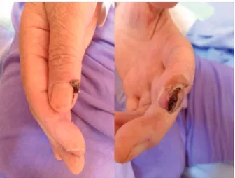
-ease, progressive necrotic lesions appeared on the tips of the ingers and toes (Figure 1). Patient pre
-sented with two weeks of history starting from the ingers and progressing to toes. The necrotic lesion was painless and arterial pulses were palpable. Cardiovascular system evaluation was inconclu
-sive. To exclude vascular pathology arterial and venous Doppler ultrasonography were performed. There was no vascular pathology. The echocardiog
-raphy was normal without any valvular pathology, vegetations of atrial septal defect.
Figure 1. Necrotic lesion on the tip of left index inger and right thumb
The connective tissue disorder workup was again inconclusive. Rheumatoid factor, ANA, Anti-DsDNA, SS-A, SS-B, SCL-70, anti-topoisomerase, C3, C4, cryoglobulin, lupus anticoagulant, p-ANCA, c-ANCA, capillaroscopy were all normal. Viral se
-rology for HBV, HCV, CMV, herpes virus was nor
-mal. The association with the cytotoxic drugs was ignored due to a 6 month of chemo-free period.
The patient was followed with the diagnosis of paraneoplastic digital necrosis and the lesions im
-proved with dexamethasone used as an anti-edema therapy in one week period without scar tissue. The lesions weren’t encountered again during follow-up.
DISCUSSION
Numerous paraneoplastic syndromes are encoun
-tered in oncology practice mostly associated with lung cancer. The cancer and digital ischemia asso
-ciation was irst reported by O’Connor in 1884 in a patient with breast cancer. Since then paraneo
-plastic vasculitic pathologies have been reported but the etiopathogenesis is still a mystery. The most
suspected mechanism for this paraneoplastic phe
-nomenon is the autoimmune reaction induced by tu
-mor antigens. Digital vasospasm provoked by sym
-pathetic hyperactivity and digital artery obstruction as a result of hyperviscosity and arteritis are other possible mechanisms [1].
Digital necrosis as a paraneoplastic syndrome has been reported in literature in different patholo
-gies. Ovarian carcinoma is the mostly related pa
-thology. Association also with lung, tonsillary, re
-nal carcinoma, leukemia, lymphoma, small bowel carcinoid have been reported [2,3]. In most of the reported cases, digital necrosis is the presenting symptom of the malignancy and addition of steroid to chemotherapeutics have provided symptomatic relieve and regression of digital lesions. Drug re
-lated vasculitis was well deined, especially with antibiotics and anti-TNF drugs [1]. Chemotherapy related vasculitic events are rarely reported. Hill and Pellegrini has been reported patients with small vessel vasculitis associated with luorouracil [2,3]. In our patient, we excluded the diagnosis of drug re
-lated vasculitis due to 6 months chemo-free period and the absence of any drugs that can be associ
-ated with the vasculitic lesions.
Our case is accepted as a paraneoplastic pa
-thology, after exclusion of possible cardiovascular and connective tissue disorders. The anti-edema therapy with 24mg dexamethasone daily provides improvement in the lesions. To the best of our knowledge our case is the irst reported digital ne
-crosis associated with colorectal carcinoma.
REFERENCES
1. Taylor L, Hauty MG, Edwards JM, Porter JM. Digital ischemia as a manifestation of malignancy. Ann Surg 1987;206:62-68.
2. Wright JR, Gudelis S. Digital necrosis associated with squamous cell carcinoma of the tonsil: Head & Neck 2002;24:1019-1021.
3. Petri M, Fye KH. Digital necrosis: a paraneoplastic syn -drome. J Rheumatol 1985;12:800-802.
4. Radic M, Kaliterna DM, Radic J. Drug-induced vascu -litis: a clinical and pathological review. Neth J Med 2012;70:12-17.
5. Hill SE, Phillips R, Francis N, Agnew K. Small-vessel vasculitis following treatment with combination 5-luo-rouracil/folinic acid and oxaliplatin. Clin Exp Dermatol 2009;34:103-105.