Original article
A synthetic peptide selectively kills only virulent
Paracoccidioides
brasiliensis
yeasts
Erika Seki Kioshima
a, Fabiana Aliperti
a, Juliana Terzi Maricato
a, Renato Arruda Mortara
a,
Eduardo Bagagli
b, Mario Mariano
a,c, Jose´ Daniel Lopes
a,*
aDepartment of Microbiology, Immunology and Parasitology, Universidade Federal de Sa˜o Paulo, Sa˜o Paulo, SP, Brazil bDepartment of Microbiology, Immunology, Institute of Biosciences of Botucatu, State University of Sa˜o Paulo, Botucatu, SP, Brazil
cInstitute of Health, Paulista University, Sa˜o Paulo, Brazil
Received 28 September 2010; accepted 26 October 2010 Available online 9 November 2010
Abstract
This work was conducted to identify virulence biomarkers for Paracoccidioides brasiliensis (Pb), the fungus responsible for
Para-coccidioidomycosis (PCM), a systemic disease endemic in Latin America. Measurement of mortality showed that all B10.A mice were killed after 250 days by the virulent Pb18 isolate while only one of the mice that received the attenuated counterpart died. Also, number of lung CFUs from virulent Pb18 inoculated mice were much higher when these isolates were compared. Phage display methodology allowed selection of three phages that specifically bound to virulent Pb18. Variability of p04 phage binding to different Pb isolates were examples of variability of
expression by the fungus of its binding molecule, strongly suggesting p04 as a biomarker of virulence.In vitro, its derived peptide pep04 killed
only virulent fungi, and confocal microscopy showed that it was internalized only by the virulent isolate. Pep04 blocked establishment of Pb infection in mice and virulent Pb18 pre-incubated with p04 showed significantly inhibited lung infection. Furthermore, infected mice treated with p04 showed highly significant reduction in lung CFUs. These findings firmly establish p04 as a biomarker of Pb virulence. Therefore, after proper peptide engineering, p04 may become a useful adjuvant for the distressing treatment of PCM.
Ó2010 Institut Pasteur. Published by Elsevier Masson SAS. All rights reserved.
Keywords: P. brasiliensis; Virulence; Biomarkers
1. Introduction
Paracoccidioides brasiliensis is the agent of Para-coccidioidomycosis (PCM) [1,2], a systemic granulomatous mycosis restricted to South American countries[3,4]. The disease develops following inhalation of infectious propagules which transform into pathogenic yeasts within the pulmonary alveolar epithelium[2,5]. The clinical forms of PCM range from asymp-tomatic patients to those that present with high mortality rates[2]. The spectrum of the disease depends on factors associated with both the host immune response and characteristics of the
infecting agent, mainly virulence [6,7]. The number of well defined virulence factors for dimorphic fungal pathogens is relatively small [8]. Several molecules were reported to be involved in the pathogenicity of the fungus[9e14]. However,
they were found in all isolates studied, and did not discrimi-nate either phenotypical differences or isolates that could be correlated with the severity of the disease.
The characterization of molecules associated with pathoge-nicity will enhance our understanding of the hosteparasite
relationship, lead to better evaluation of prognosis, and may be useful in the development of new therapeutic agents. One of the techniques used for this purpose is phage display, a technology which has already facilitated the identification of disease markers and has driven drug design based on ligand directed targeting
[15]. Using phage display, we identified three different peptide-displaying phages that bound to the surface of the high-virulent, * Corresponding author. Rua Botucatu 862, 4andar. Sa˜o Paulo, SP
04023-901, Brazil. Tel.:þ55 11 5549 6073; fax:þ55 11 55723328.
E-mail address:jdlopes@unifesp.br(J.D. Lopes).
1286-4579/$ - see front matterÓ2010 Institut Pasteur. Published by Elsevier Masson SAS. All rights reserved. doi:10.1016/j.micinf.2010.10.019
but not the attenuated, isolates of P. brasiliensis. A derived synthetic peptide from one of the phages (pep04) exhibited fungicidal activityin vitroagainstP. brasiliensisas demonstrated by inhibition of colony forming units (CFUs). Furthermore, the corresponding phage, p04, prevented thein vivolung coloniza-tion by the fungus as well as its disseminacoloniza-tion to other organs.
2. Material and Methods
2.1. Animals
Six-week-old male B10.A mice, bred under specific path-ogen-free conditions at the animal facilities of the Federal University of Sa˜o Paulo, Brazil, were used. All procedures and experiments were performed according to the regulations of the institutional Ethical Committee for animal experimentation, Federal University of Sa˜o Paulo, Brazil, by which the study was approved (CEP 0522/04, 25/03/2004). The animals were handled according to National Institutes of HealtheUSA (NIH)
guide for care and use of laboratory animals (http://oacu.od.nih. gov/ARAC/index.htm).
2.2. Fungi and growth
Six different P. brasiliensisisolates were used. Pb18, after more than 5 years in culture, was demonstrably non-virulent. Two isolates (T4B17 and T10B1) were originally obtained from armadillos[16,17]. The virulence of T10B1 and T4B17 isolates were assumed as described[17], high and intermediate virulence, respectively. Mg16 was isolated from a single dental alveolus lesion of a patient from the Laboratory of Medical Mycology, State University of Maringa´, Brazil. This restricted type of infection, with no open disease or dissemination, allows for the assumption that it was a simple infection with very low virulence. Pb339, a very well known isolate, was obtained from our department fungal culture collection. To restore the Pb18 viru-lence for the purpose of comparison to the attenuated Pb18 strain, the strain was only used after serial passages in mice, the animal model used throughout for experimental analysis as well as for reestablishing lost virulence. This concept of attenuation or loosing virulence by long term cultures came from Brummer et al. (1990) and Kashino et al. (1990)[18,19]. The attenuated Pb18 isolate had been previously maintained in culturein vitrofor 5e6
years. All isolates were sub-cultivated weekly in semisolid Fava-Netto’s medium[20]at 36
C and used on the 5th day of culture.
2.3. Pathogenicity and virulence ofP. brasiliensis
isolates
Groups of 6 mice of the B10.A strain were used to evaluate mortality, number of CFUs, and histology. Briefly, the fungal viability of five day cultures (generally over 90%) was verified by trypan blue exclusion in a Neubauer chamber. Groups of mice (6e8 weeks old) were infected i.p. with 1106viable yeast cells
in 50ml of phosphate-buffered saline (PBS). Mortality of the mice was recorded for 250 days after intra-tracheal (i.t.) infection[21]. The number of viable microorganisms in the lungs, liver, and
spleen from experimental and control mice were determined by counting CFUs. Animals from each group were sacrificed at different time intervals after infection and the number of viable organisms in the organs were estimated as described[22]. Briefly, the number of CFU was recovered in supplemented BHI plates. The weighed organs of each mouse were macerated in 5 ml of PBS, and 100ml was plated in supplemented BHI. CFU for each organ of individual mice was counted until no further growth could be detected. The results were presented as log 10 of the average valuestandard error. For histopathological analysis, the lung, spleen, and liver from each mouse were removed, fixed in 10% formalin, and embedded in paraffin. Five-micrometer sections were stained with hematoxylineeosin (H&E) and
examined microscopically.
2.4. Biopanning and rapid analysis of selective interactive ligands (BRASIL)
The phage display library was screened for virulent isolate of
P. brasiliensis(Pb18) binding ligands by the BRASIL method
[23]. The library used in the present study contained the insert CX7C (C, cysteine; X, any amino acid residue)[15]. Yeasts of attenuated P. brasiliensis isolates suspended in PBS-BSA 1% (w/v) were incubated with 109transducing units (TU) of the phage library. After 4 h the suspension was transferred to the top of a nonmiscible organic lower phase with an intermediate specific density solution and centrifuged (12,000 RPM, 15 min, 25C) [23]. The upper aqueous phase, containing phages that were unbound to the attenuated isolate, was incubated with yeasts of virulentP. brasiliensis, as described above. Following centrifu-gation, yeasts of virulent Pb18 entered the lower organic phase and settled at the bottom of the tube, carrying only specifically bound phages. The pellet was then transferred to another clean tube as described[23]. Phages bound to virulentP. brasiliensisisolate yeasts were rescued by infection of log-phaseEscherichia coli
K91kan[24]. The next day, amplified phages were separated from bacteria by polyethylene-glycol-NaCl precipitation and used for subsequent selection rounds (109TU) to further enrich phages binding specifically to yeasts of virulentP. brasiliensis. After three complete rounds of selection, the DNA corresponding to peptide inserts of randomly picked phage clones were sequenced (approximately two hundred). Enriched peptide sequences were analyzed by Clustal W, [EMBL in Heidelberg, Germany ([http:// www.ebi.ac.uk/Tools/clustalw2/index.html] Version 2) sequence alignment. To measure phage binding, the insert-less phage (fd-tet) was used as control[25]. Virulent or attenuated isolates of
P. brasiliensiswere co-incubated with selected peptide-displaying or control phages. The binding to P. brasiliensis isolates was quantified relative to fd-tet. In parallel, four isolatesP. brasiliensis
(T4B17, T10B1, Pb339, Mg16) were co-incubated with selected peptide-displaying or control phages and similarly quantified.
2.5. Peptide synthesis and purification for screening
(CGSYGFNAC), pep05 (CGLRLESTC), pep06 (CSLERLGFC), irrelevant peptide (pepIR) (CGSTSAYGC), linear pep04 (AGSYGFNAA) and its specific scrambled peptide (CSFA-GYNGC) were used to synthesize the peptides as described[26]. Briefly, the synthesis was realized by solid-phase technology using the 9-fluorenylmethoxy carbonyl strategy. Therefore, all peptides were obtained with the C-terminal carboxyl group in an amide form. The resulting peptides were analyzed by reverse-phase high-performance liquid chromatography (Shimadzu, Tokyo, Japan). The peptide quality was assessed by a matrix-assisted laser desorption ionization-time of flight instrument (Micromass, Manchester, United Kingdom) using a -cyano-4-hydroxy cinnamic acid as the matrix.
2.6. Confocal fluorescence microscopy
Labeling for fluorescence microscopy was performed as described, with modifications[27]. Briefly,P. brasiliensisyeast cells growing in aerated liquid cultures, in logarithmic phase, were collected and adjusted to a concentration of 2 106 to 3106viable cells/ml. The cells were fixed in cold methanol for 30 min in the dark. The washed cells (three times with PBS) were incubated with 3% (wt/vol) BSA in PBS (blocking buffer) for 16 h at 4C. After washing, the cells were incubated in the dark with
either 100mg/ml of pep04 or pepIR labeled with 6-Carboxy-fluorescein (6-FAM) for 1 h at room temperature. For confocal fluorescence microscopy, samples were prepared as described above. However, in order to better define the fungal surface, samples were also labeled with monoclonal antibody toP. bra-siliensisgp75, a well characterized surface protein[13]developed with Alexa-610 goat anti-Mouse IgG (Invitrogen), and 20mm DAPI (40
-6-Diamidino-2-phenylindole di-lactate, Invitrogen); specimens were imaged on a BioRad 1024UV instrument attached to a Zeiss Axiovert microscope using a 631.4 NA DIC
oil immersion lens[28]. Z-series were processed with ImageJ and renderized with VolumeJ (http://rsb.info.nih.gov/ij/).
2.7. Peptides activity onP. brasiliensis
Thein vitroantifungal activity of peptides against virulent or attenuated P. brasiliensis isolates, as demonstrated by inhibition of CFU after treatment, was measured. Briefly, yeast cells (virulent or attenuated isolates) were incubated for 24 h in the presence of peptides (1 mg/ml) in Fava-Netto’s medium. A final volume of 200ml was added to each well of 96-wells plates, at 37
C with shaking, as described[29]. Readings were performed from day 8e20 and results were expressed in CFU.
The serial concentrations of pep04 and pepIR (1000, 500, 250, 125 and 15.6mg/ml) were only tested against virulent isolates ofP. brasiliensis.
2.8. Blocking infection implantation
Yeast cells (106) were incubated with 1 mg/ml of pep04, pepIR, or PBS for 1 h. After that, the viability of fungal suspensions, determined by trypan blue exclusion in a Neu-bauer chamber, was always higher than 90%. Then, this fungal
suspension was inoculated i.v. (100 ml of PBS) in the lateral tail vein of B10.A mice (six animals per group). Control mice received only PBS. Animals were sacrificed after 3 or 45 days post-infection, and the number of viableP. brasiliensiscells in the lungs, spleen and liver were determined by counting CFUs.
2.9. Phage treatment of mice infected withP. brasiliensis
Mice, after anesthesia, were subjected to i.t.P. brasiliensis
infection as described [21]. After 24 h, fd-tet or p04 were administered i.v. (1011TU phage/100ml of PBS) in the lateral tail vein of BALB/c mice (five animals per group) every day. Treatment was given for 10 days, 7 days after which they were sacrificed and the number of viableP. brasiliensiscells in the lungs were determined by number of CFUs.
2.10. Statistical analysis
Results were expressed as the meanthe standard deviations (SD). Data was analyzed by unpaired Student’s t test with Welch’s correction (two-tailed) used for comparison of the two groups when the data met the assumptions of thettests. Analysis of variance (ANOVA) followed by the TukeyeKramer test were
sometimes applied (INSTAT software, GraphPad, San Diego, CA).Pvalues under 0.05 indicated statistical significance.
3. Results
3.1. Virulence of P. brasiliensisisolates
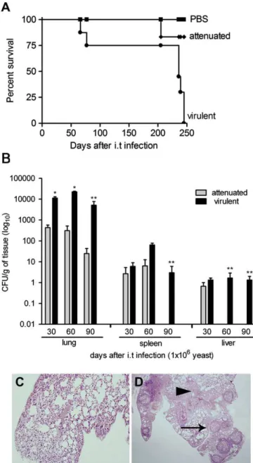
The virulence ofP. brasiliensisisolates (virulent and attenu-ated Pb18) were compared following i.t. administered infection. After 250 days of infection, virulent Pb18 killed all the animals and its attenuated counterpart killed a single mouse out of six. All animals of the control group (PBS) survived (P<0.001 relative to control group) (Fig. 1A).Fig. 1B shows the number of viable yeast cells recovered from lungs, spleen, and liver of mice after 30, 60 and 90 days post i.t. inoculation. The number of lung CFUs from virulent Pb18 inoculated mice were significantly higher than those inoculated with attenuated Pb18. Dissemination of virulent Pb18 to the spleen and liver was always observed. Histopathology reflected the CFU data as shown inFig. 1C and D. Mice infected with attenuated Pb18 showed diffuse inflammatory cells in the lungs (Fig. 1C) and a lack of defined granulomas in the liver (data not shown) at 30 days post inoculation. Mice infected with virulent Pb18 showed isolated granulomas after 30 days in both the lungs (Fig. 1D) and liver (data not shown).
3.2. Selection of peptide motifs that bind to the surface of virulent P. brasiliensisisolates
In order to begin identification of markers forP. brasiliensis
second and third rounds were selected. Two phages were considerably enriched, the CGSYGFNAC and CSLERLGFC-displaying phages, denominated p04 (three repetitions in second and third rounds) and p06 (seven repetitions in third round), respectively. However, some phages presented two repetitions in the third round and were also selected for binding assays e CYRLTGLWC (p01), CLDSASRGC (p02),
CGLRLESTC (p05), CWLFSVSAC (p03).
3.3. Validation of phage binding to yeast cells of a highly virulent isolate ofP. brasiliensis
The degree to which phages bound to virulent or attenuated Pb18 isolates, compared to control, was analyzed by the BRASIL method. As expected, binding of the p04, p05 and p06 phages recovered from virulent Pb18 was significantly higher when compared to attenuated Pb18 and control in all experimental conditions tested (P < 0.05) (Fig. 2A). These phages, with specific binding to virulent isolates, were selected for further experiments. Binding of the p04 phage to yeast cells of virulent isolates during independent experiments (106e1010 TUs) was dose-dependent as compared to control
(Fig. 2B). However, a competitive inhibition assay using synthetic peptides showed that only pep04 inhibited binding of its homologous phage. As shown in Fig. 2C, increased concentration of pep04 decreased the titer of phages recovered from the virulent isolate, while the inhibition rate of p04 binding increased gradually up to 1 mM or above the pep04 concentration. Pep06 did not inhibit binding of homologous phage (p06), but unexpectedly, such binding was increased. Regarding this result, it can only be speculated that some type of homotypic interaction occurred between the displaying phage and its derived peptide.
3.4. P04 as a possible marker of virulence
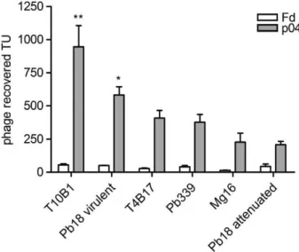
To determine whether p04 phage could be used as a novel virulence marker, binding assays to six isolates with pre-defined degrees of virulence were performed. As shown in
Fig. 3, p04 phage showed great binding variability to yeast cells of different isolates. These results suggest that the fungal binding molecule is differently expressed by each isolate, as expected, possibly leading to various degrees of virulence. No correlation with the original description of each isolate’s virulence was ever assumed here.
3.5. Pep04 binding to the virulent Pb18 isolate
Yeast cells of virulent or attenuated Pb18 isolates were incubated with pep04 or pepIR, both coupled to 6-FAM. PepIR did not bind to either isolate (data not shown). Most of the attenuated Pb18 cells showed no labeling with pep04-6FAM (Fig. 4A). Conversely, this labeled peptide was inter-nalized by the virulent isolate (Fig. 4B and F). Imaging of sequential optical sections indicated that the labeling was intracellular and frequently close or overlapping DAPI stain-ing, as shown inFig. 4F.
3.6.In vitroantifungal activity of peptides selected by phage display
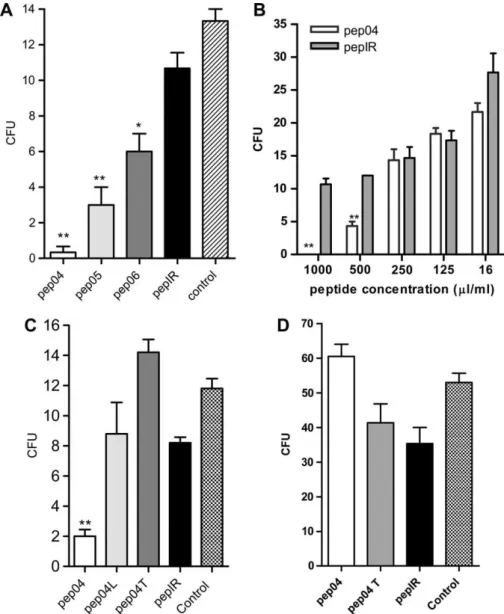
Since the selected phages bound specifically to the virulent isolate, we tested if synthetic peptides, derived from these phages, possessed similar biological activity. Fig. 5A shows that pep04 (P<0.01), pep05 (P<0.01) and pep06 (P<0.05) exertedin vitro fungicidal activity against P. brasiliensisand Fig. 1. Pb18 isolates displayed clear differences in degree of virulence. (A)
that the effects were statistically significant. Other concen-trations of pep04 were tested and results showed dose dependence (Fig. 5B). The pep04 and its vehicle (DMSO) did not show any toxicity to peritoneal macrophages at any of the concentrations tested during MTT cell viability assays (data not shown).
3.7. Thein vitroantifungal activity of pep04 is specific for virulent isolates
To confirm the specificity of in vitro pep04 antifungal activity, additional peptides similar to pep04 were engineered and synthesized, as described in Material and Methods. As shown in Fig. 5C, yeast cells of the virulent isolate were
completely inhibited by pep04. In the same concentration, none of the other peptides had any effect. These results suggest a conformational importance to pep04, dependent on the presence of two cysteines, necessary to form a disulfide bond and to make the cyclic peptide. This conformation can permit the correct interaction with the receptor or molecule important to the fungicidal activity. As shown in Fig. 5D, when pep04 was incubated with the attenuated isolate, no fungicidal activity was observed. The fact that the peptide did not interfere with the proliferation rate of the attenuated isolate suggests that this peptide is nontoxic and does not recognize the same, but attenuated, fungus isolate.
3.8. Phage p04 prevented the deployment of infection
Since p04 could be utilized to distinguish degrees of viru-lence by simply binding to molecules of P. brasiliensis, and pep04 showed in vitro antifungal activity, we decided to determine if p04 could be employed to influence or block the development of mycosis. As shown in Fig. 6A, after 3 days, the number of lung CFUs from mice infected with virulent Pb18, incubated with p04, were significantly lower than controls. This result demonstrated that p04 was able to prevent the implantation of the fungus in the lungs, its primary site of infection. This difference increased in animals sacrificed 45 days after infection (Fig. 6B). The CFUs recovered from the lung, spleen, and liver of animals infected with p04 treated Pb18 were always lower than those from animals treated with fd-tet. These data suggest that the interaction of the fungus with p04 inhibits both implantation (Fig. 6A) and deployment (Fig. 6B) of infection in mice.
3.9. Treatment of mice infected with P. brasiliensiswith p04 phage
Since p04 prevented the establishment of infection, the next step was to check whether it was capable of treating mice already infected with P. brasiliensis. Such mice treated with p04 showed a significantly reduced CFU average in the lungs as compared to controls (P<0.001) (Fig. 6C).
Fig. 2. Biopanning strategy to isolate phages binding only to highly virulent isolate yeast cells ofP. brasiliensis. (A) Cell binding assay with selected phages. Clones were incubated with highly virulent and attenuated isolates to identify three phages (p04, p05 and p06) presenting higher specificity and affinity to the former. (B) Binding of the pep04 displaying phage to yeast cells using increasing amounts of phages TU was always higher with the virulent isolate. (C) Inhibition results between the synthetic peptide (pep04 or pep06) and its homologous phage (p04 or p06); with pep04 concentrations higher than 0.001mM led to significant
inhibition. [*(P<0.05) and **(P<0.01) relative to control, PBS only].
4. Discussion
For decades, the virulence of P. brasiliensis has been a controversial issue because it embodies a great number of variables[30e33]. Being a characteristic agent that is
respon-sible for a human disease, its definition necessarily bears a correlation with the disease severity produced by any given isolate. In humans, however, virulence cannot be measured. In animal models, virulence varies from the inoculation site to the animal species used. Furthermore, besides patient’s lungs, isolates come from a variety of different sources such that the proper habitat of the fungus is, as yet, unknown[34]. In order to achieve the goal of identifying a P. brasiliensis virulence biomarker that would provide for some much needed clarity in this field of research, the use of a model in which clearly defined parameters are defined is indispensable. Among these parame-ters are the use of the same animal, the same inoculation route, and the same isolate, whose degree of virulence is clear, even if dependent on artificial procedures such as long times ofin vitro
cultivation. We closely followed these parameters and proce-dures here. None of these considerations, however, restrained the reporting of several results from different groups that, so far, left us without the identification of any reliableP. brasiliensis
virulence marker[30e32,35e37]. The lack of efficient classical
genetic techniques for silencing suspected virulence genes contributed to this failure. Characterizing such a molecule or its ligand, for virulent isolate(s), represented the main objective of this work.
Two variables were taken into account from the beginning of our studies. Firstly, long cultivation decreases virulence, a concept that came from Brummer et al. (1990) and Kashino et al. (1990), and that previously assumed virulence could be
recovered by repeated animal passage [18,19], a fact confirmed by others[38,39]. Secondly, virulence itself is an ill defined characteristic, as discussed above.
We compared isolate Pb18 with its counterpart that was maintained in culture for longer than five years and had thus, lost virulence. The virulent Pb18 isolate not only showed greater pathogenicity than its non-virulent counterpart, but also presented striking morphological differences (not shown) These characteristics could be associated with adaptation of the fungus in order to survive within the host[30,40,41]. They also demonstrate that, despite derivation from the same isolate, these strains are evidently different.
Some Pb proteins and genes have been reported as putative virulence factors by several authors [33,36,41e43], with no
marker clearly established. To find such a marker or its ligand, the BRASIL method was employed which involved a peptide approach for selecting cellular surface motifs based on phage display libraries[23]. At least one possible virulence marker for Pb18, the motif CGSYGFNAC, was characterized and validated. The phage displaying this sequence was named p04 and its homologous synthetic peptide, pep04. Using binding assays, p04 phage could be utilized for the ranking of various isolates according to their described level of virulence. Based on this methodology, we can state that there is a variable expression of the fungal molecule that binds the peptide. Nishikaku et al. (2008) evaluated the ability ofP. brasiliensis
isolated from armadillos to cause disease in mice. Higher fungal loads were recovered from mice infected with arma-dillo isolates as compared with two clinical isolates, suggest-ing that armadillo isolates were more virulent. One of these
to five years ofin vitroculture, this armadillo isolate produced high CFU counts in all the organs[39]. This virulence profile was also observed by others [17,44]. Canteros et al. (2000) ranked the isolate Pb339 as slightly virulent and Motta et al. (2002) as intermediate when compared with virulent Pb18 (after animal passage) [38,45]. In our studies, all isolates, including Mg16, gave binding results that correlated with their presumed virulence.
Since p04 could be used to distinguish levels of virulence by simply binding to molecules of P. brasiliensis, we inves-tigated whether synthetic peptide pep04 displayed fungicidal activity in vitro. Similar activity has been demonstrated by other peptides against Cryptococcus neoformans [46], P. brasiliensis [29] and Candida albicans [47]. Tested against both isolates of Pb18, virulent and non-virulent, killing
activity was clearly observed only against the virulent isolate. The conformational structure of pep04 seemed important since linear or scrambled peptides did not kill fungi as efficiently. Our efforts to clarify the mechanisms of fungicidal activity displayed by pep04 proved unsuccessful. For instance, confocal experiments showed that this peptide could only be detected after internalization by yeast. No binding to the fungal surface was observed. Different experiments designed to show surface binding were always unsuccessful, suggesting that peptide entry into the cell is a rapid process. Pep04 showed hydrophobic characteristics and so possibly acts by distinct mechanisms from those seen with other antimicrobial peptides (AMP). The general properties of AMP, such as amphipathicity and cationicity, allow them to form pores in biological membranes, resulting in microbial death [48]. Fig. 5. Thein vitroantifungal activity of selected peptides. (A) Pep04 (P<0.01), pep05 (P<0.01) and pep06 (P<0.05) peptides incubated with yeasts of virulent
Pb18. These peptides demonstrated fungicidal activityin vitroagainstP. brasiliensis. (B) The fungicidal activity of pep04in vitrowas dose-dependent (P<0.01)
as compared with pepIR. Bars indicate standard errors. (C) Fungicidal activity of pep04in vitroagainst virulentP. brasiliensisisolate depends on molecular conformation. Pep04L (linear pep04, with cysteine amino acids substituted by alanine), pep04T (scrambled peptide) and pepIR (irrelevant peptide).P<0.01, as
Moreover, pep04 displayed a marked association with DAPI labeling, suggesting that after spontaneously diffusing into the cytoplasm of fixed or unfixed cells, it might bind to the fungal DNA or to nuclear proteins with regulatory functions, thus suggesting a possible mechanism of fungal killing. Though highly speculative, these possibilities cannot be ruled out.
These findings led us to determine whether similar effects could be obtained in vivo. Phage p04, when given together with the fungus, was able to inhibit implantation as well as dissemination of the disease. To demonstrate how p04 does this, as we confirmed by repeating the experiment several times, may be an almost impossible task. No fungus or its dissemination could be found, making it impracticable to check whether the agent died, became incapable of multi-plying, or had some important adhesion molecule blocked.
These results indicate that both p04 and pep04 can be considered biomarkers of virulence in PCM by binding assays, as are similarly used nowadays some monoclonal antibodies. On the other hand,in vivoassays demonstrated that p04 phage or pep04 could interfere with the pathogenicity of this mycosis. Therefore, beyond selectively binding to yeast cells of virulentP. brasiliensis
isolates, recognition of a specific virulence marker by p04 phage and pep04 make plausible the development of an effective ther-apeutic adjuvant. This task should be pursued by a different approach since peptides, in the long run, are very unstable. Although these results are preliminary, the model of selection based on molecules preferentially expressed in virulent isolates suggests alternatives to current antifungal therapy.
Specific peptides as those herein described are the first real biomarkers of virulence. By opening the possibility of char-acterizing and purifying their target proteins, whose functions may then be specifically hindered, such peptides can reveal new approaches for the control of PCM.
Acknowledgments
We are indebt to Dr. Patricia Xander Batista and Creuza R. Oliveira for technical assistance, to Drs. Zoilo Pires de
Camargo, Terezinha Inez Estivalet Svidzinski, Vera Lu´cia Garcia Calich who kindingly providedP. brasiliensisisolates and Dr. Alexandre Basso for critically reading of the manu-script. This work was supported by Fundac¸a˜o de Ampar-oa`Pesquisa do Estado de Sa˜o Paulo (FAPESP - 04/04471-9).
References
[1] G. San-Blas, Paracoccidioidomycosis and its etiologic agent Para-coccidioides brasiliensis, J. Med. Vet. Mycol. 31 (1993) 99e113. [2] M.I. Borges-Walmsley, D. Chen, X. Shu, A.R. Walmsley, The
pathobi-ology of Paracoccidioides brasiliensis, Trends Microbiol. 10 (2002) 80e87.
[3] G. San-Blas, G. Nin˜o-Vega, T. Iturriaga,Paracoccidioides brasiliensis
and paracoccidioidomycosis: molecular approaches to morphogenesis, diagnosis, epidemiology, taxonomy and genetics, Med. Mycol. 40 (2002) 225e242.
[4] Z.F. Coutinho, D. Silva, M. Lazera, V. Petri, R.M. Oliveira, P.C. Sabroza, B. Wanke, Paracoccidioidomycosis mortality in Brazil (1980e1995), Cad. Saude. Publica. 18 (2002) 1441e1454.
[5] A. Restrepo, G. Benard, C.C. de Castro, C.A. Agudelo, A.M. Tobo´n, Pulmonary paracoccidioidomycosis, Semin. Respir. Crit. Care Med. 29 (2008) 182e197.
[6] M. Franco, Host-parasite relationships in paracoccidioidomycosis, J. Med. Vet. Mycol. 25 (1987) 5e18.
[7] M. Franco, E. Bagagli, M. Cunha, L.G. Chama, D. Fecchio, Para-coccidioides brasiliensis antigens batches from the same isolate show immunological and biochemical differences, Mycopathologia 135 (1996) 13e19.
[8] C.A. Rappleye, W.E. Goldman, Defining virulence genes in the dimor-phic fungi, Annu. Rev. Microbiol. 60 (2006) 281e303.
[9] D. Mattos Grosso, S.R. de Almeida, M. Mariano, J.D. Lopes, Charac-terization of gp70 and anti-gp70 monoclonal antibodies in Para-coccidioides brasiliensis pathogenesis, Infect. Immun. 71 (2003) 6534e6542.
[10] L. Ganiko, R. Puccia, V.S. Mariano, O.A. Sant’Anna, E. Freymu¨ller, M.C. Roque-Barreira, L.R. Travassos, Paracoccin, an N-acetyl-glucos-amine-binding lectin of Paracoccidioides brasiliensis, is involved in fungal growth, Microbes. Infect. 9 (2007) 695e703.
[11] M.S. Barbosa, S.N. Ba´o, P.F. Andreotti, F.P. de Faria, M.S. Felipe, L. dos Santos Feitosa, M.J. Mendes-Giannini, C.M. Soares, Glyceraldehyde-3-phosphate dehydrogenase of Paracoccidioides brasiliensis is a cell surface protein involved in fungal adhesion to extracellular matrix proteins and interaction with cells, Infect. Immun. 74 (2006) 382e389. Fig. 6. P04 inhibits both implantation and deployment of infection in mice. (A) Virulent Pb18 was incubated with p04, p03 (the latter unable to bind to
P. brasiliensis, as shown inFig. 2) or fd-tet (non inserted) phages for 1 h and then five B10.A mice were infected with each preparation and sacrificed 72 h after the infection. The number of lung CFUs from virulent Pb18 incubated with p04 were significantly lower than p03, fd-tet phages, or controls (P<0.01). (B) Virulent
Pb18 was incubated with p04 or fd-tet phages for 1 h after which B10.A mice were infected (5 animals). Sacrificed 45 days after the infection, these mice showed that the difference in number of lung CFUs became more pronounced (P<0.01), with no evidence of dissemination. Spleen and liver CFUs were not different. (C)
[12] A.L. Matsuo, I.I. Tersariol, S.I. Kobata, L.R. Travassos, A.K. Carmona, R. Puccia, Modulation of the exocellular serine-thiol proteinase activity ofParacoccidioides brasiliensis by neutral polysaccharides, Microbes Infect. 8 (2006) 84e91.
[13] P. Xander, A.F. Vigna, L.S. Feitosa, L. Pugliese, A.M. Baila˜o, C.M. Soares, R.A. Mortara, M. Mariano, J.D. Lopes, A surface 75-kDa protein with acid phosphatase activity recognized by monoclonal antibodies that inhibitParacoccidioides brasiliensisgrowth, Microbes Infect. 9 (2007) 1484e1492.
[14] J.T. Maricato, W.L. Batista, E.S. Kioshima, L.S. Feitosa, R.R. e Brito, G.H. Goldman, M. Mariano, R. Puccia, J.D. Lopes, The Para-coccidioides brasiliensisgp70 antigen is encoded by a putative member of the flavoproteins monooxygenase family, Fungal Genet. Biol. 47 (2010) 179e189.
[15] A. Sergeeva, M.G. Kolonin, J.J. Molldrem, R. Pasqualini, W. Arap, Display technologies: application for the discovery of drug and gene delivery agents, Adv. Drug Deliv. Rev. 58 (2006) 1622e1654. [16] E. Bagagli, A. Sano, K.I. Coelho, S. Alquati, M. Miyaji, Z.P. de
Camargo, G.M. Gomes, M. Franco, M.R. Montenegro, Isolation of
Paracoccidioides brasiliensisfrom armadillos (Dasypus noveminctus) captured in an endemic area of paracoccidioidomycosis, Am. J. Trop. Med. 58 (1998) 505e512.
[17] F. Hebeler-Barbosa, M.R. Montenegro, E. Bagagli, Virulence profiles of ten Paracoccidioides brasiliensis isolates obtained from armadillos (Dasypus novemcinctus), Med. Mycol. 41 (2003) 89e96.
[18] S.S. Kashino, L.M. Singer-Vermes, V.L. Calich, E. Burger, Alterations in the pathogenicity of one Paracoccidioides brasiliensisisolate do not correlative with its in vitro growth, Mycopathologia 111 (1990) 173e180.
[19] E. Brummer, A. Restrepo, L.H. Hanson, D.A. Stevens, Virulence of
Paracoccidioides brasiliensis: the influence of in vitro passage and storage, Mycopathologia 109 (1990) 13e17.
[20] C.F. Netto, V.S. Vegas, I.M. Scianname´a, D.B. Guarnieri, The poly-saccharidic antigen fromParacoccidioides brasiliensis. study of the time of cultivation necessary for the preparation of the antigen, Rev. Inst. Med. Trop. Sao Paulo 11 (1969) 177e181.
[21] L.E. Cano, L.M. Singer-Vermes, T.A. Costa, J.O. Mengel, C.F. Xidieh, C. Arruda, D.C. Andre´, C.A. Vaz, E. Burger, V.L. Calich, Depletion of CD8(þ) T cells in vivo impairs host defense of mice resistant and susceptible to pulmonary paracoccidioidomycosis, Infect. Immun. 68 (2000) 352e359.
[22] L.M. Singer-Vermes, M.C. Ciavaglia, S.S. Kashino, E. Burger, V.L. Calich, The source of the growth-promoting factor(s) affects the plating efficiency of Paracoccidioides brasiliensis, J. Med. Vet. Mycol. 30 (1992) 261e264.
[23] R.J. Giordano, M. Cardo´-Vila, J. Lahdenranta, R. Pasqualini, W. Arap, Biopanning and rapid analysis of selective interactive ligands, Nat. Med. 7 (2001) 1249e1253.
[24] G.P. Smith, J.K. Scott, Libraries of peptides and proteins displayed on filamentous phage, Meth. Enzymol. 217 (1993) 228e257.
[25] F.I. Staquicini, A. Tandle, S.K. Libutti, J. Sun, M. Zigler, M. Bar-Eli, F. Aliperti, E.C. Pe´rez, J.E. Gershenwald, M. Mariano, R. Pasqualini, W. Arap, J.D. Lopes, A subset of host B lymphocytes controls mela-noma metastasis through a melamela-noma cell adhesion molecule/MUC18-dependent interaction: evidence from mice and humans, Cancer Res. 68 (2008) 8419e8428.
[26] I.Y. Hirata, M.H.S. Cezari, C.R. Nakaie, P. Boschcov, A.S. Ito, M.A. Juliano, L. Juliano, Internally quenched fluorogenic protease substrates: solid-phase synthesis and fluorescence spectroscopy of peptides con-taining ortho-aminobenzoyl-dinitrophenyl groups as donor-acceptor pairs, Lett. Pept. Sci. 1 (1994) 299e308.
[27] W.L. Batista, A.L. Matsuo, L. Ganiko, T.F. Barros, T.R. Veiga, E. Freymu¨ller, R. Puccia, The PbMDJ1 gene belongs to a conserved MDJ1/ LON locus in thermodimorphic pathogenic fungi and encodes a heat shock protein that localizes to both the mitochondria and cell wall of
Paracoccidioides brasiliensis, Eukaryot. Cell 5 (2006) 379e390. [28] H.C. Barros, N.V. Verbisck, S. Da Silva, M.F. Araguth, R.A. Mortara,
Distribution of epitopes ofTrypanosoma cruziamastigotes during the
intracellular life cycle within mammalian cells, J. Eukaryot. Microbiol. 44 (1997) 332e344.
[29] L.R. Travassos, L.S. Silva, E.G. Rodrigues, S. Conti, A. Salati, W. Magliani, L. Polonelli, Therapeutic activity of a killer peptide against experimental paracoccidioidomycosis, J. Antimicrob. Chemother. 54 (2004) 956e958.
[30] T.I. Svidzinski, M.H. Miranda Neto, R.G. Santana, O. Fischman, A.L. Colombo,Paracoccidioides brasiliensesisolates obtained from patients with acute and chronic disease exhibit morphological differences after animal passage, Rev. Inst. Med. Trop. Sao Paulo 41 (1999) 279e283. [31] D. Zacharias, A. Ueda, M. Moscardi-Bacchi, M. Franco, G. San-Blas, A
comparative histopathological, immunological, and biochemical study of experimental intravenous paracoccidioidomycosis induced in mice by three Paracoccidioides brasiliensis isolates, J. Med. Vet. Mycol. 24 (1986) 445e454.
[32] E. Burger, C.C. Vaz, A. Sano, V.L. Calich, L.M. Singer-Vermes, C.F. Xidieh, S.S. Kashino, K. Nishimura, M. Miyaji,Paracoccidioides bra-siliensisinfection in nude mice: studies with isolates differing in viru-lence and definition of their T cell-dependent and T cell-independent components, Am. J. Trop. Med. Hyg. 55 (1996) 391e398.
[33] E.E. Molinari-Madlum, M.S. Felipe, C.M. Soares, Virulenceof Para-coccidioides brasiliensisisolates can be correlated to groups defined by random amplified polymorphic DNA analysis, Med. Mycol. 37 (1999) 269e276.
[34] A. Restrepo, D.J. Baumgardner, E. Bagagli, C.R. Cooper Jr., M.R. McGinnis, M.S. La´zera, F.H. Barbosa, S.M. Bosco, Z.P. Camargo, K.I. Coelho, S.T. Fortes, M. Franco, M.R. Montenegro, A. Sano, B. Wanke, Clues to the presence of pathogenic fungi in certain environments, Med. Mycol. 1 (2000) 67e77.
[35] L.M. Singer-Vermes, E. Burger, M.F. Franco, M.M. Di-Bacchi, M.J. Mendes-Giannini, V.L. Calich, Evaluation of the pathogenicity and immunogenicity of seven Paracoccidioides brasiliensis isolates in susceptible inbred mice, J. Med. Vet. Mycol. 27 (1989) 71e82. [36] K.C. Carvalho, L. Ganiko, W.L. Batista, F.V. Morais, E.R. Marques,
G.H. Goldman, M.F. Franco, R. Puccia, Virulence ofParacoccidioides brasiliensis and gp43 expression in isolates bearing known PbGP43 genotype, Microbes. Infect. 7 (2005) 55e65.
[37] E. Castaneda, E. Brummer, D. Pappagianis, D.A. Stevens, Chronic pulmonary and disseminated paracoccidioidomycosis in mice: quantitation of progression and chronicity, J. Med. Vet. Mycol. 25 (1987) 377e387. [38] T.R. Motta, C.A. Moreira-Filho, R.P. Mendes, L.R. Souza, M.F. Sugizak,
S. Baueb, V.L. Calich, C.A. Vaz, Evaluation of DNA polymorphisms amplified by arbitrary primers (RAPD) as genetically associated elements to differentiate virulent and non-virulent Paracoccidioides brasiliensis
isolates, FEMS Immunol. Med. Microbiol. 33 (2002) 151e157. [39] S.A. Macoris, M.F. Sugizaki, M.T. Perac¸oli, S.M. Bosco, F.
Hebeler-Barbosa, L.B. Simo˜es, R.C. Theodoro, L.A. Trinca, E. Bagagli, Virulence attenuation and phenotypic variation of Paracoccidioides brasiliensis
isolates obtained from armadillos and patients, Mem. Inst. Oswaldo Cruz 101 (2006) 331e334.
[40] E.E. McClelland, W.T. Perrine, W.K. Potts, A. Casadevall, Relationship of virulence factor expression to evolved virulence in mouse-passaged
Cryptococcus neoformanslines, Infect. Immun. 73 (2005) 7047e7050. [41] B.L. Ortiz, A.M. Garcia, A. Restrepo, J.G. McEwen, Immunological
char-acterization of a recombinant 27-kilodalton antigenic protein from Para-coccidioides brasiliensis, Clin. Diagn. Lab. Immunol. 3 (1996) 239e241. [42] A. Sano, J. Defaveri, R. Tanaka, K. Yokoyama, N. Kurita, M. Franco,
K.I. Coelho, E. Bagagli, M.R. Montenegro, M. Miyaji, K. Nishimura, Pathogenicities and GP43kDa gene of three Paracoccidioides brasi-liensis isolates originated from a nine-banded armadillo (Dasypus novemcinctus), Mycopathologia 144 (1999) 61e65.
[43] D.R. Matute, L.M. Quesada-Ocampo, J.T. Rauscher, J.G. McEwen, Evidence for positive selection in putative virulence factors within the
Paracoccidioides brasiliensisspecies complex, PLoS. Negl. Trop. Dis. 2 (2008) e296.
[44] M.T. Perac¸oli, M.F. Sugizaki, R.P. Mendes, R. Naiff, M.R. Montenegro,
[45] C.E. Canteros, M.A. Soria, M.C. Rivas, C. Pe´rez, M. Tous, W. Lee, L. Rodero, G. Davel, In vitroinfection by different strains of Para-coccidioides brasiliensis, Rev. Argent. Microbiol. 32 (2000) 116e122. [46] E. Cenci, F. Bistoni, A. Mencacci, S. Perito, W. Magliani, S. Conti,
L. Polonelli, A. Vecchiarelli, A synthetic peptide as a novel anti-cryptococcal agent, Cell Microbiol. 6 (2004) 953e961.
[47] M. Manfredi, E. Merigo, A. Salati, S. Conti, A. Savi, L. Polonelli, M. Bonanini, P. Vescovi,In vitrocandidacidal activity of a synthetic killer decapeptide (KP) against Candida albicans cells adhered to resin acrylic discs, J. Oral Pathol. Med. 36 (2007) 468e471. [48] K.A. Brogden, Antimicrobial peptides: pore formers or metabolic