Dysregulation of the Descending Pain System
in Temporomandibular Disorders Revealed by
Low-Frequency Sensory Transcutaneous
Electrical Nerve Stimulation: A Pupillometric
Study
Annalisa Monaco1, Ruggero Cattaneo1, Luca Mesin2, Eleonora Ortu1, Mario Giannoni1, Davide Pietropaoli1*
1University of L’Aquila, Department of Life, Health and Environmental Sciences, Building Delta 6 Dental Unit, St Salvatore Hospital—Via Vetoio 67100 L’Aquila, Italy,2Department of Electronics and
Telecommunications, Politecnico di Torino, Turin, Italy
*davide.pietropaoli@univaq.it
Abstract
Using computerized pupillometry, our previous research established that the autonomic nervous system (ANS) is dysregulated in patients suffering from temporomandibular disor-ders (TMDs), suggesting a potential role for ANS dysfunction in pain modulation and the eti-ology of TMD. However, pain modulation hypotheses for TMD are still lacking. The
periaqueductal gray (PAG) is involved in the descending modulation of defensive behavior and pain throughμ,κ, andδopioid receptors. Transcutaneous electrical nerve stimulation (TENS) has been extensively used for pain relief, as low-frequency stimulation can activate
µreceptors. Our aim was to use pupillometry to evaluate the effect of low-frequency TENS stimulation ofμreceptors on opioid descending pathways in TMD patients. In accordance with the Research Diagnostic Criteria for TMD, 18 females with myogenous TMD and 18 matched-controls were enrolled. All subjects underwent subsequent pupillometric evalua-tions under dark and light condievalua-tions before, soon after (end of stimulation) and long after (recovery period) sensorial TENS. The overall statistics derived from the darkness condition revealed no significant differences in pupil size between cases and controls; indeed, TENS stimulation significantly reduced pupil size in both groups. Controls, but not TMD patients, displayed significant differences in pupil size before compared with after TENS. Under light conditions, TMD patients presented a smaller pupil size compared with controls; the pupil size was reduced only in the controls. Pupil size differences were found before and during TENS and before and after TENS in the controls only. Pupillometry revealed that stimulating the descending opioid pathway with low-frequency sensory TENS of the fifth and seventh pairs of cranial nerves affects the peripheral target. The TMD patients exhibited a different pattern of response to TENS stimulation compared with the controls, suggesting that im-paired modulation of the descending pain system may be involved in TMD.
OPEN ACCESS
Citation:Monaco A, Cattaneo R, Mesin L, Ortu E, Giannoni M, Pietropaoli D (2015) Dysregulation of the Descending Pain System in Temporomandibular Disorders Revealed by Low-Frequency Sensory Transcutaneous Electrical Nerve Stimulation: A Pupillometric Study. PLoS ONE 10(4): e0122826. doi:10.1371/journal.pone.0122826
Academic Editor:Claudia Sommer, University of Würzburg, GERMANY
Received:October 5, 2014
Accepted:February 15, 2015
Published:April 23, 2015
Copyright:© 2015 Monaco et al. This is an open access article distributed under the terms of the Creative Commons Attribution License, which permits unrestricted use, distribution, and reproduction in any medium, provided the original author and source are credited.
Data Availability Statement:Data are available by request due to ethical restrictions. Interested researchers who meet the criteria for access to confidential data may contact Ruggero Cattaneo ( catrug@libero.it). A disclosure form by applicant Scientist is mandatory.
Funding:The authors received no specific funding for this work.
Introduction
Recent literature has suggested that TMD patients may suffer from dysfunction in the brain network that supports sensory, pain, emotional, and cognitive processes [1–8]. Some authors have focused on the dysregulation of the autonomic nervous system (ANS) in TMD patients [9–13], suggesting that TMD could be the clinical manifestation of multisystem dysregulation [14].
Among the structures that are involved in the brain network regulating the sensory, pain, emotional, and cognitive systems, the periaqueductal gray (PAG) has a key role. It receives afferent connections from cortical areas associated with cognition and motivation related to sensory and pain perception [15–17], and it then projects the connections to the centers con-trolling the peripheral afferent inputs and couples autonomic reactions in a specific manner [18,19].
The role of the PAG in descending pain control via endogenous opioids is one of the most studied pathways [20–22], and several findings suggest that chronic pain is promoted by an ab-normal modulation of the descending endogenous pain system [23,24].
Transcutaneous electric nerve stimulation (TENS) has been used for a long time to relieve pain [25–27]. Its main effect is believed to be achieved through the modulation of descending inputs from the ventral-lateral PAG to the rostroventral medial medulla (RVM) [28,29]. In particular, low-frequency TENS is suggested to activateμopioid receptors [30–32].
Bilateral low-frequency TENS of the fifth and seventh cranial nerves has been suggested to treat TMD [33–36] on the basis of the resulting pain relief [37,38] monitored by surface elec-tromyography (EMG) of masticatory muscles and kinesiography of jaw movements [39,40]. However, despite the clinical relevance of TENS in TMD, the above-cited studies focused on clinical pain or jaw muscle effects. Therefore, no information can be drawn regarding the hy-pothesis of autonomous or central dysfunction in patients with TMD.
Recent findings promote the use of pupillometry to study the effects of opioids in opioid-maintained patients and healthy subjects [41–43]. Moreover, the dysregulation of the ANS has been reported in TMD [44], obstructive sleep apnea syndrome [45–48], and other autonomous disorders [49–52] by pupillometry. For these reasons, pupillometry can be considered a non-invasive, sensitive and cost-effective tool for investigating, via pupil constriction (miosis), the ANS and opioid-related miosis [53–55].
According to the above-mentioned literature, it is possible that the effects of low-frequency TENS that are mediated by the supraspinal center and the endogenous opioid path can be dem-onstrated at the pupil level (miosis). The aim of this study was to evaluate pupillometric index-es using low-frequency TENS to tindex-est the hypothindex-esis that the opioid dindex-escending pathway is dysregulated in patients with TMD.
Materials and Methods
Subjects
This study was conducted in accordance with the Declaration of Helsinki. The Committee on Ethics in Science of the University of L’Aquila, L’Aquila, Italy, approved the study, and written informed consent was obtained from each subject and electronically stored as suggested by our institutional guidelines.
Inclusion/Exclusion Criteria
duration longer than 3 months; presence of complete permanent dentition, with the possible exception of the third molars; normal occlusion. Patients were excluded from the study if they met one or more of the following criteria: presence of systemic or metabolic diseases; eye dis-eases or visual defects; history of local or general trauma; neurological or psychiatric disorders; muscular diseases; cervical pain; bruxism, as diagnosed by the presence of parafunctional facets and/or anamnesis of parafunctional tooth clenching and/or grinding; pregnancy; assumed use of anti-inflammatory, analgesic, anti-depressant, opioid, or myorelaxant drugs; smoking; fixed or removable prostheses; fixed restorations that affected the occlusal surfaces; and either previ-ous or concurrent orthodontic or orthognathic treatment.
For comparison with previous literature [7,8,56,57], the diagnosis of myofascial-type TMD was provided after clinical examination by a trained clinician according to group 1a and 1b of the Research Diagnostic Criteria for TMD (RDC/TMD) [58], in a blinded manner (RC).
The control group consisted of 18 age- and gender-matched Caucasian subjects scheduled for a routine checkup at the University Clinic, without signs or symptoms of TMD, who ful-filled the inclusion and exclusion criteria (mean age 25.1±5.7 years).
Each enrolled subject underwent the experimental protocol described inFig 1.
Fig 1. Diagram of the Experimental Protocol.Before beginning the experimental session, each enrolled subject was prepared with TENS electrodes and connected to a turned-off TENS device. Subsequently, they underwent three experimental phases: Pre TENS stimulation (green); During TENS stimulation (red); and Post TENS stimulation (blue). Pupillometer application and its removal occurred after the acclimation period and at the end of the experimental session, respectively (green). Based on the literature, central nervous system stimulation was obtained by sensory stimulation of cranial nerves V and VII with low-frequency TENS for 21 minutes (red) [28–32]. To avoid operator interference, the pupillometer, electrodes and TENS device were removed only at the end of the experimental session.
Pupillometry
Pupillometry was performed with an infrared pupillometer (Oculus system, Inventis srl, Pa-dova, Italy), which was composed of two infrared CCD cameras (resolution of 720x576 pixels, 256 gray levels) mounted on a light helmet (1.5 kg), with a sampling frequency of 25 frames/s. To stabilize accommodation, the subjects were asked to focus their eyes on the light point in the pupillometer [42].
The assessment of pupil size was performed under light conditions, with the eyes illuminat-ed using a yellow-green LED with a 740-nanometer wavelength, as well as under dark condi-tions that were obtained using only three infrared diodes with a wavelength of 880 nanometers. Pupillometric recordings were acquired in digital form and processed using the Tarjan algo-rithm designed to evaluate strongly connected components [59] to obtain frame-by-frame measurements of the pupil area, expressed as the number of pixels covering it. A template was positioned on the computer screen to allow for the correction of the eye position to avoid errors in the alignment of the pupils, as previously described [44].
Pupillometry was performed with the subjects in a horizontal supine position on a bed. Room temperature (21°C) and relative humidity (50%) remained constant. Any external or in-ternal noise sources were excluded.
Before the pupillometric recording sessions, patients were invited to lie on the bed for clini-cal examination with their eyes open for at least 10 minutes to adapt to the temperature and humidity of the room, as well to reduce their anxiety (Fig 1). Then, the pupillometer was ap-plied and maintained until the end of the recording session.
Recording Procedure
All recording procedures are described in the protocol diagram (Fig 1). Briefly, after 2 minutes of darkness under an infrared light condition, 1 minute of recording was obtained, followed by the application of yellow-green light for 2 minutes and subsequent recording for 1 minute; this procedure was applied before, during and after sensory TENS stimulation, for a total of 6 pupil-lometric recordings of 60 seconds each (Fig 1). The recording sessions were performed by an expert operator (RC) in a blinded manner.
Based on previous literature [44], darkness and light adaptation was minimized, due to the short duration of infrared and yellow-green stimulation, and any potential, residual adaptive effect, if present, could be observed in both groups.
Stimulation Procedure
The method for sensory TENS was described previously [60]. Briefly, a J5 Myomonitor TENS Unit device (Myotronics-Noromed, Inc., Tukwila, WA, USA) with disposable electrodes (Myo-trode SG Elec(Myo-trodes, Myotronics-Noromed, Inc., Tukwila, WA, USA) was used. This low-fre-quency neurostimulator generates a repetitive synchronous and bilateral stimulus delivered at 1.5-s intervals, with an adjustable amplitude of approximately 0–24 mA, a duration of 500μs,
and a frequency of 0.66 Hz. The two TENS electrodes were placed bilaterally over the cutane-ous projection of the notch of the fifth pair of cranial nerves, which was located between the coronoid and condylar processes and was retrieved by manual palpation of the zone anterior to the tragus; a third grounding electrode was placed in the center of the back of the neck.
attention was paid to avoid reaching the threshold of motor stimulation; if any movement of the investigated muscles was observed, the patient was excluded from the study. One patient with TMD and 1 control subject were excluded because the amplitude reached the motor threshold of the seventh cranial nerve, as shown by the contraction of the masseter and temporal muscles.
The same operator (RC) applied the pupillometer and delivered the TENS according to the manufacturer’s guidelines.
Statistical Analysis
Statistical analysis was performed using STATA 10 (StataCorp LP, College Station, TX, USA) on average pupil sizes, computed on 60 seconds of recordings, as previously described [44]. Preliminary analysis of pupil size showed, as expected, a high correlation between the left and right pupil sizes, allowing the use of the left-right mean value to simplify the statistics. The ratio between pupil size in the presence of yellow-green light and that in darkness (herein re-ferred to as the L/D ratio) was calculated. The Shapiro-Wilk test revealed a normal distribution of data. Within-group differences in the pupil size and L/D ratio were analyzed using a paired t test, while differences in pupil size between groups were analyzed using an unpaired t test. The level of significance was set at p = 0.05 for all tests. The results are expressed as the mean and standard deviation (SD).
Results
The size of the pupil in darkness (infrared light) was significantly reduced during sensory low-frequency TENS both in the control (overall mean of 7886.51 before TENS vs. 7434.33 during TENS; p = 0.02) and TMD (7599.88 vs. 7148.94; p = 0.001) groups (Table 1). After TENS, the reduction of pupil size remained significant in the control group (7886.51 vs. 7427.11; p = 0.003) but not in the TMD group (7599.88 vs. 7490.61; p = 0.082) because of the return of pupil size to that prior to TENS.
The unpaired t test did not reveal any significant difference in pupil size between the control and TMD groups before TENS (7886.51 vs. 7599.88; p = 0.24), during TENS (7434.33 vs. 7184.94; p = 0.11), or after TENS (7427.11 vs. 7490.61; p = 0.47). In the TENS condition, it is possible that the higher value of data dispersion in the TMD group (SD 1983.31) was responsi-ble for the lack of significance.
Table 2shows the statistics of pupil size under yellow-green light. The size of the pupils in the control group was significantly reduced during TENS compared with the size prior to
Table 1. Comparison between the Pupil Size in the Control and TMD Groups in Infrared Light.
Infrared pupil size Infrared pupil size
Control group TMD group
Before TENS 7886.51 (1231.0) 7599.88 (1254.6)
During TENS 7434.33 (1464.0)* 7148.94 (1983.3)*
After TENS 7427.11 (1168.6)** 7490.61 (1477.6)
The data are expressed as the mean and standard deviation (in parentheses). Asterisks indicate statistically significant differences.
*paired t test within groups between the pre-TENS and TENS conditions: p = 0.02 in the control group and p = 0.001 in the TMD group.
**paired t test within groups between the pre-TENS and post-TENS conditions in the control group: p = 0.003.
TENS (3934.98 vs. 3523.55; p = 0.005), and the pupil size continued to decrease post-TENS (3934.98 vs. 3294.16; p = 0.005). The TMD group did not show significant differences in pupil size between the pre-TENS and TENS conditions (2911.51 vs. 2836.38: p = 0.21) or between the pre-TENS and post-TENS conditions (2911.51 vs. 2973.83: p = 0.34). The unpaired t test between groups showed that the pupil size in the TMD group was significantly lower than that in the controls in the pre-TENS (2911.51 vs. 3934.38: p = 0.001) and TENS (2836.38 vs. 3523.55: p = 0.02) conditions, but no significant differences between groups were found in the post-TENS condition (2973.83 vs. 3294.16: p = 0.14). It is possible that, as in the post-TENS condition in darkness, the higher dispersion of data in the TMD group was responsible for the lack of significance. Note that the dispersion of data in the control group was lower than that in the TMD group, and, in the post-TENS condition, it was approximately one-third of that in the TMD group (sd: 617.32 vs. 1978.37). The absolute value of size differed by 10% (control 3294.16 vs. TMD 2973.83), suggesting more homogeneous pupil behavior among the control subjects.
Results of the L/D ratio within and between groups are shown inTable 3in the pre-TENS, TENS, and post-TENS conditions. The ratio decreased significantly in the control group
Table 2. Comparison between Pupil Size in the Control and TMD Groups in the Yellow-Green Light Condition.
Yellow-Green pupil size Yellow-Green pupil size
Control group TMD group
Before TENS 3934.38 (821.35) 2911.51 (1041.97)°
TENS 3523.55 (867.38)* 2836.38 (1184.54)°°
After TENS 3294.16 (617.32)** 2973.83 (1978.37)
The data are expressed as means and standard deviations (in parentheses). Asterisks and circles show the statistically significant differences.
*paired t test within groups between the pre-TENS and TENS conditions in the control group: p = 0.003.
**paired t test within groups between the pre-TENS and post-TENS conditions in the control group: p = 0.0005.
°unpaired t test between the groups before TENS: p = 0.001. °°unpaired t test between the groups during TENS: p = 0.02.
doi:10.1371/journal.pone.0122826.t002
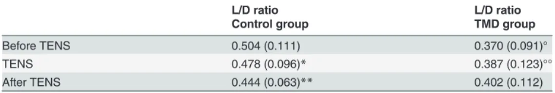
Table 3. Comparison of the Pupil Size and Yellow-Green/Infrared Light (L/D) Ratio between the Con-trol and TMD Groups.
L/D ratio L/D ratio
Control group TMD group
Before TENS 0.504 (0.111) 0.370 (0.091)°
TENS 0.478 (0.096)* 0.387 (0.123)°°
After TENS 0.444 (0.063)** 0.402 (0.112)
The data are expressed as means and standard deviations (in parentheses). Asterisks and circles show the statistically significant differences.
*paired t test within groups between the pre-TENS and TENS conditions in the control group: p = 0.03.
**paired t test within groups between the pre-TENS and post-TENS conditions in the control group: p = 0.01.
°unpaired t test between the groups before TENS: p = 0.0002. °°unpaired t test between the groups during TENS: p = 0.01.
pre-TENS compared with TENS (0.504 vs. 0.478: p = 0.03) and pre-TENS compared with post-TENS (0.504 vs. 0.444: p = 0.01). The TMD group did not show statistically significant differences in this ratio during the within-group comparisons.
The L/D ratio was significantly higher in the control group compared with the TMD group, both pre-TENS (0.504 vs. 0.370: p = 0.0002) and during TENS (0.478 vs. 0.387: p = 0.01). Post-TENS, the L/D ratio remained higher in the control group (0.444 vs. 0.402), but the difference between the two groups was not significant (p = 0.08). Note that the ratio in the TMD group trended toward the value of the control group, with a value less than that in the TENS and pre-TENS conditions.
Discussion
The data in our study can be summarized as follows:
1. No significant difference was found in pupil size in darkness between the control and TMD groups.
2. Significant within-group differences in pupil size in the darkness condition before TENS compared with during TENS in both the control and TMD groups were found; specifically, the pupil size was reduced during TENS.
3. Significant differences in pupil size in the darkness condition were found pre- compared with post-TENS in the control group but not in the TMD group. In the control group, the pupil size was reduced post-TENS, with the same reduction obtained during TENS; in the TMD group, the size of the pupil post-TENS returned close to the pre-TENS value.
4. Significant differences in pupil size in the light condition were found between the control and TMD groups; specifically, the TMD group showed a significantly smaller pupil size.
5. Significant within-group differences in pupil size in the light condition were found pre-TENS compared with during pre-TENS and pre-pre-TENS compared with post-pre-TENS in the con-trol group but not in the TMD group; pupil size in the light condition was reduced only in the control group.
Pupil size is a peripheral effect of a central network that controls the balance between sym-pathetic and parasymsym-pathetic outflow. The ambient light and the physiological and pathologi-cal causes of sympathetic excitation and/or parasympathetic inhibition evoke pupil dilation, and, vice versa, causes inducing sympathetic inhibition and/or parasympathetic excitation evoke pupil constriction. For this reason, pupil dynamics have been studied in physiological conditions that are suspected to impair autonomic balance and enhance arousal and sympa-thetic drive [61,62], enabling the assessment of the relationship between pupil dilation and nor-adrenergic activity [63,64]. Some authors have focused on the locus coeruleus-noradrenergic neuromodulatory system [65,66], which is considered to be one of the main centers regulating arousal, vigilance, alertness [67–70], pain, and sensory afferents, and it is likely involved in a variety of disorders, such as chronic pain and TMD, characterized by dysfunction of the norad-renergic arousal system [71–73].
Low-frequency TENS induces a release of endorphins from the ventral-lateral PAG and the activation of the opioid descending path to the RVM. Simultaneously, PAG fibers extend to the pericoeruleus region, in which opioids are released in the dendritic system from the locus coe-ruleus neurons [80]. For this reason, it is possible that low-frequency TENS induces a reduction
Fig 2. The Edinger-Westphal Nucleus (EWN) Exerts a Tonic Activity on the Pupil Sphincter.Larson [55] proposed that in anesthetized patients, opioid action can be exerted, blocking the efferents of inhibitory neurons to the EWN. Translating this hypothesis to non-anesthetized patients, it could be suggested that opioids reduce the inhibition of interneurons, increasing the effects of excitatory afferents of the EWN. The final effect was opioid miosis. In the figure, excitatory components of the model are shown in red, while inhibitory components are shown in black.
in the firing frequency in the locus coeruleus, simultaneously reducing the state of arousal, the perception of pain, and the diameter (size) of the pupil.
According to the above data, in our study, the pupil size decreased with low-frequency sen-sory TENS in darkness, both in the control and TMD groups. Five minutes after the end of stimulation, the effect was still present in the control group, but not in the TMD group.
The dilation of the pupil in darkness is mediated by the adrenergic sympathetic branch of the ANS and is supplied by a nerve originating from Budge’s cilio-spinal center that excites the dilator muscles of the pupil. The action of the dilator muscles is counterbalanced by the cholin-ergic parasympathetic branch, which originates from the Edinger-Westphal nucleus and inhib-its the dilation. Moreover, pupil dilation obtained by sphincter inhibition can be equal to 1/3 of the maximum physiological dilation [81], and during mental or physical efforts, the pupil dila-tion may occur via central inhibidila-tion of the parasympathetic center [82].
It is possible that in the darkness condition in TMD patients, the endorphin effect due to TENS is sufficient to stimulate the parasympathetic inhibition of the dilator muscle; however, after the stimulation, if the baseline condition is characterized by impaired parasympathetic control, the stimulation provided by TENS rapidly completes its action on the pupillary muscles.
In the light condition, the parasympathetic branch responds to light that activates the sphincter muscle of the iris, causing miosis. At the same time, the sphincter receives beta-ad-renergic innervation that is sufficient to reduce and/or counterbalance the contraction [83,84].
In our study, the subjects in the control and TMD groups responded to light as evidenced by a reduction in pupil size, demonstrating the proper activation of the central parasympathetic pathway that controls this basic function. Compared with the control group, the TMD patients showed a significantly higher reduction of pupil size, as indicated by both the absolute values and the ratio between the pupil size in light and in darkness. This finding might reflect im-paired counterbalance of adrenergic inhibition or hyperexcitation of parasympathetic effectors in the TMD group. The result that TENS in the light condition reduces the size of the pupil in the control group but has no effect in the TMD group could suggest the impairment of the ad-renergic counterbalance system, given that the opioid path works as an inhibitor of the para-sympathetic branch of pupil control [55].
Taken together, the data from the present study show that TMD patients suffer from a type of impairment of the descending pathway that controls the sensory, pain, and autonomic out-put. It is possible that one of the mechanisms involved in this impairment can be addressed in the opioid descending pathway, considering that TMD patients have a weaker response to low-frequency sensory TENS than controls, with respect to modulating the inhibition of physiologi-cal parasympathetic excitation. This study focused on adrenergic dysfunction in TMD and confirmed previous studies in which TMD patients showed impairment in the recruitment of adrenergic activity in a weakly stressful task of muscle activation, such as forced voluntarily clenching [44] and other cognitive or emotional tasks [8].
It is probable that the impairments could be due to changes in the connectivity among the structures that belong to the distributed network of pain processing, such as the PAG, which is a node between cortical descending control and midbrain/spinal effectors [56,57,85,86] (Figs2
and3).
remaining 20–25% showed signs of irritation after TENS, namely, an increase in EMG values of the jaw elevator muscles at rest and a lack of interocclusal freeway space. This result dis-agrees with the activation of the opioid descending system and directs attention to the activa-tion of the sympathetic pathway, perhaps due to the stimulaactiva-tion of the dorsolateral PAG root.
As in a previous study [88], we used low-frequency TENS of the fifth and seventh pairs of cranial nerves to stimulate the descending control system and to examine the effect on the pe-ripheral target. Beyond the absolute values of pupil size and the L/D ratio, it is noteworthy to mention that the data from TMD subjects are characterized by a higher standard deviation, an
Fig 3. Mechanism of Endogenous Opioid Action Induced by LF TENS in Non-Anesthetized Subjects with Pupil Miosis.The locus coeruleus-noradrenergic system (LC) has been suggested to be a key structure in arousal, and its firing frequency tone has been correlated with wakefulness, vigilance, and sensory and pain perception. The size of the pupil correlates with the activity of LC, and light increases the frequency of firing. The action of the LC excites the excitation pathway that directly induces pupil constriction and excites inhibitory interneurons in the inhibitory pathway, exciting the inhibitor of the inhibitor; the net result is a reduction of inhibition and, indirectly, an agonist effect of the excitatory pathway of pupil constriction. Opioids act on the inhibitory pathway, and TENS has been suggested to be activated via the ventral lateral PAG and the opioid pathway. According to Valentino [74] and Curtis [75], opioids reduce the activation of the LC, and corticotropin-releasing hormone (CRH) increases the firing rate. It is possible that low-frequency sensory TENS inhibits LC inhibitory interneurons, increasing the tone of the pupillary constrictor of the LC. TMD patients could suffer from an impairment of the inhibitory pathway stemming from the ventral lateral PAG. Consequently, pupil constriction in response to light is not counterbalanced by inhibition (smaller pupil size under yellow-green light), and low-frequency sensory TENS is unable to further induce pupil constriction. Red, excitation pathway; black, inhibitory pathway.
index of the dispersion of the values, compared with the controls. This higher dispersion of TMD data was particularly seen during and after TENS, while the dispersion was reduced in the control subjects. We speculate that low-frequency sensory TENS could aid in differentiat-ing TMD patients with a higher impairment of the descenddifferentiat-ing pain modulation pathway from those with a less impaired system. Unfortunately, we did not assess the response to TENS for the prediction of therapy. Thus, we were unable to determine whether different degrees of im-pairment, such as a lower light-darkness ratio or greater TENS reduction of the pupil size in the light condition, could predict the therapy outcome. Another limitation of our study is that we did not assess whether TENS induced a reduction in pain perception in the examined sub-jects; therefore, we could not directly assess the efficacy of our stimulation protocol on experi-mental pain. However, our goal did not concern the symptom“pain”and the efficacy of low-frequency TENS used in dentistry for its relief, and thus we did not take into account a specific study design to address the response to experimental pain. These topics are beyond the scope of the present work and are suggested for future studies.
Compared with the previous work [44], TMD patients in this study exhibited a smaller pupil size in darkness. The average patient age in the present study was 5.32 years older than that in the previous investigation. It is possible that this age discrepancy is responsible for this difference; in fact, it is known that pupil size decreases with age [49,50]. Moreover, it is possible that the duration of the disorder influences the degree of dysregulation, which shifts from a high sympathetic tone (greater pupil size in darkness) in the early stage of TMD to the failure of sympathetic counterbalance (lower pupil size in darkness) with the progression of the disor-der. We did not assess the TMD duration in our sample; however, to assess the possible effect of this variable, the duration of the disorder must be investigated in the future through de-signed studies.
Conclusion
The stimulation of the descending opioid pathway with low-frequency sensory TENS of the fifth and seventh pairs of cranial nerves and the evaluation of the effect on the peripheral target with a simple pupillometric parameter (pupil size) lead to the conclusion that TMD patients suffer from an impairment in the modulation of the descending pain system. Further studies are necessary to confirm this hypothesis and to relate the impairment of the descending pain system to the therapeutic outcome.
Author Contributions
Conceived and designed the experiments: AM RC DP. Performed the experiments: EO DP RC. Analyzed the data: LM RC. Contributed reagents/materials/analysis tools: AM MG. Wrote the paper: AM RC DP LM.
References
1. Maixner W, Fillingim R, Booker D, Sigurdsson A. Sensitivity of patients with painful temporomandibular disorders to experimentally evoked pain. Pain. 1995; 63: 341–351. PMID:8719535
2. Maixner W, Fillingim R, Sigurdsson A, Kincaid S, Silva S. Sensitivity of patients with painful temporo-mandibular disorders to experimentally evoked pain: evidence for altered temporal summation of pain. Pain. 1998; 76: 71–81. PMID:9696460
3. Svensson P, List T, Hector G. Analysis of stimulus-evoked pain in patients with myofascial temporo-mandibular pain disorders. Pain. 2001; 92: 399–409. PMID:11376913
5. Ayesh EE, Jensen TS, Svensson P. Somatosensory function following painful repetitive electrical stim-ulation of the human temporomandibular joint and skin. Exp Brain Res. 2007; 179: 415–425. PMID:
17146645
6. Schmidt JE, Carlson CR. A controlled comparison of emotional reactivity and physiological response in masticatory muscle pain patients. J Orofac Pain. 2009; 23: 230–242. PMID:19639103
7. Nebel MB, Folger S, Tommerdahl M, Hollins M, McGlone F, Essick G. Temporomandibular disorder modifies cortical response to tactile stimulation. J Pain. 2010; 11: 1083–1094. doi:10.1016/j.jpain. 2010.02.021PMID:20462805
8. Weissman-Fogel I, Moayedi M, Tenenbaum HC, Goldberg MB, Freeman BV, Davis KD. Abnormal cor-tical activity in patients with temporomandibular disorder evoked by cognitive and emotional tasks. Pain. 2011; 152: 384–396. doi:10.1016/j.pain.2010.10.046PMID:21167644
9. Korszun A, Young EA, Singer K, Carlson NE, Brown MB, Crofford L. Basal circadian cortisol secretion in women with temporomandibular disorders. J Dent Res. 2002; 81: 279–283. PMID:12097314 10. Nilsson AM, Dahlström L. Perceived symptoms of psychological distress and salivary cortisol levels in
young women with muscular or disk-related temporomandibular disorders. Acta Odontol Scand. 2010; 68: 284–288. doi:10.3109/00016357.2010.494620PMID:20500119
11. Maixner W, Greenspan JD, Dubner R, Bair E, Mulkey F, Miller V, et al. Potential autonomic risk factors for chronic TMD: descriptive data and empirically identified domains from OPPERA case-contrl study. J Pain. 2011: 12(11 Suppl): T75–T91. doi:10.1016/j.jpain.2011.09.002PMID:22074754
12. Light KC, Bragdon EE, Grewen KM, Brownley KA, Girdler SS, Maixner W. Adrenergic dysregulation and pain with and without acute beta-blockade in women with fibromyalgia and temporomandibular dis-order. J Pain. 2009; 10: 542–552. doi:10.1016/j.jpain.2008.12.006PMID:19411061
13. Eze-Nliam CM, Quartana PJ, Quain AM, Smith MT. Nocturnal heart rate variability is lower in temporo-mandibular disorder patients than in healthy, pain-free individuals. J Orofac Pain. 2011; 25: 232–239. PMID:21837290
14. Chen H, Nackley A, Miller V, Diatchenko L, Maixner W. Multisystem dysregulation in painful temporo-mandibular disorders. J Pain. 2013; 14: 983–996. doi:10.1016/j.jpain.2013.03.011PMID:23721875 15. Lumb BM. Inescapable and escapable pain is represented in distinct hypothalamic-midbrain circuits:
specific roles for Adelta- and C-nociceptors. Exp Physiol. 2002; 87: 281–286. PMID:11856975 16. Ossipov MH. The perception and endogenous modulation of pain. Scientifica. 2012; 2012: 561761. doi:
10.6064/2012/561761PMID:24278716
17. Kobayashi S. Organization of neural systems for aversive information processing: pain, error, and pun-ishment. Front Neurosci. 2012; 6: 136. doi:10.3389/fnins.2012.00136PMID:23049496
18. Bandler R, Keay KA, Floyd N, Price J. Central circuits mediating patterned autonomic activity during ac-tive vs. passive emotional coping. Brain Res Bull. 2000; 53: 95–104. PMID:11033213
19. Keay KA, Clement CI, Depaulis A, Bandler R. Different representations of inescapable noxious stimuli in the periaqueductal gray and upper cervical spinal cord of freely moving rats. Neurosci Lett. 2001; 313: 17–20. PMID:11684329
20. Heinricher MM, Tavares I, Leith JL, Lumb BM. Descending control of nociception: specificity, recruit-ment and plasticity. Brain Res Rev. 2009; 60: 214–225. doi:10.1016/j.brainresrev.2008.12.009PMID:
19146877
21. Bruehl S, Burns JW, Chung OY, Chont M. Pain-related effects of trait anger expression: neural sub-strates and the role of endogenous opioid mechanisms. Neurosci Biobehav Rev. 2009; 33: 475–491. doi:10.1016/j.neubiorev.2008.12.003PMID:19146872
22. Graeff FG. New perspective on the pathophysiology of panic: merging serotonin and opioids in the peri-aqueductal gray. Braz J Med Biol Res. 2012; 45: 366–375. PMID:22437485
23. Staud R. Abnormal endogenous pain modulation is a shared characteristic of many chronic pain condi-tions. Expert Rev Neurother. 2012; 12: 577–585. doi:10.1586/ern.12.41PMID:22550986
24. Linnman C, Moulton EA, Barmettler G, Becerra L, Borsook D. Neuroimaging of the periaqueductal gray: state of the field. Neuroimage. 2012; 60: 505–522. doi:10.1016/j.neuroimage.2011.11.095PMID:
22197740
25. DeSantana JM, Walsh DM, Vance C, Rakel BA, Sluka KA. Effectiveness of transcutaneous electrical nerve stimulation for treatment of hyperalgesia and pain. Curr Rheumatol Rep. 2008; 10: 492–499. PMID:19007541
26. Johnson M, Martinson M. Efficacy of electrical nerve stimulation for chronic musculoskeletal pain: a meta-analysis of randomized controlled trials. Pain. 2007; 130: 157–165. PMID:17383095
28. Ainsworth L, Budelier K, Clinesmith M, Fiedler A, Landstrom R, Leeper BJ, et al. Transcutaneous elec-trical nerve stimulation (TENS) reduces chronic hyperalgesia induced by muscle inflammation. Pain. 2006; 120: 182–187. PMID:16360266
29. DeSantana JM, Da Silva LF, De Resende MA, Sluka KA. Transcutaneous electrical nerve stimulation at both high and low frequencies activates ventrolateral periaqueductal grey to decrease mechanical hyperalgesia in arthritic rats. Neuroscience. 2009; 163: 1233–1241. doi:10.1016/j.neuroscience.2009. 06.056PMID:19576962
30. Sluka KA, Deacon M, Stibal A, Strissel S, Terpstra A. Spinal blockade of opioid receptors prevents the analgesia produced by TENS in arthritic rats. J Pharmacol Exp Ther. 1999; 289: 840–846. PMID:
10215661
31. Kalra A, Urban MO, Sluka KA. Blockade of opioid receptors in rostral ventral medulla prevents antihy-peralgesia produced by transcutaneous electrical nerve stimulation (TENS). J Pharmacol Exp Ther. 2001; 298: 257–263. PMID:11408550
32. Radhakrishnan R, King EW, Dickman JK, Herold CA, Johnston NF, Spurgin ML, et al. Spinal 5-HT(2) and 5-HT(3) receptors mediate low, but not high, frequency TENS-induced antihyperalgesia in rats. Pain. 2003; 105: 205–213. PMID:14499437
33. Jankelson B. Electronic control of muscle contraction—a new clinical era in occlusion and prosthodon-tics. Sci Educ Bull. 1969; 2: 29–31. PMID:4949886
34. Katch EM. Application of transcutaneous electrical nerve stimulation in dentistry. Anesth Prog. 1986; 33: 156–160. PMID:3488697
35. Cooper BC. International College of Cranio-Mandibular Orthopedics (ICCMO). Temporomandibular disorders: A position paper of the International College of Cranio-Mandibular Orthopedics (ICCMO). Cranio. 2011; 29: 237–244. PMID:22586834
36. Chipaila N, Sgolastra F, Spadaro A, Pietropaoli D, Masci C, Cattaneo R, et al. The effects of ULF-TENS stimulation on gnathology: the state of the art. Cranio. 2014; 32: 118–130. PMID:24839723
37. Rodrigues D, Siriani AO, Bérzin F. Effect of conventional TENS on pain and electromyographic activity of masticatory muscles in TMD patients. Braz Oral Res. 2004; 18: 290–295. PMID:16089258 38. Cooper BC, Kleinberg I. Establishment of a temporomandibular physiological state with neuromuscular
orthosis treatment affects reduction of TMD symptoms in 313 patients. Cranio. 2008; 26: 104–117. PMID:18468270
39. Monaco A, Sgolastra F, Ciarrocchi I, Cattaneo R. Effects of transcutaneous electrical nervous stimula-tion on electromyographic and kinesiographic activity of patients with temporomandibular disorders: a placebo-controlled study. J Electromyogr Kinesiol. 2012; 22: 463–468. doi:10.1016/j.jelekin.2011.12. 008PMID:22245620
40. Kamyszek G, Ketcham R, Garcia R, Radke J. Electromyographic evidence of reduced muscle activity when ULF-TENS is applied to the Vth and VIIth cranial nerves. Cranio. 2001; 19: 162–168. PMID:
11482827
41. Murillo R, Crucilla C, Schmittner J, Hotchkiss E, Pickworth WB. Pupillometry in the detection of concom-itant drug use in opioid-maintained patients. Methods Find Exp Clin Pharmacol. 2004; 26: 271–275. PMID:15319805
42. Fliegert F, Kurth B, Göhler K. The effects of tramadol on static and dynamic pupillometry in healthy sub-jects—the relationship between pharmacodynamics, pharmacokinetics and CYP2D6 metaboliser sta-tus. Eur J Clin Pharmacol. 2005; 61: 257–266. PMID:15906019
43. Matouskova O, Slanar O, Chytil L, Perlik F. Pupillometry in healthy volunteers as a biomarker of trama-dol efficacy. J Clin Pharm Ther. 2011; 36: 513–517. doi:10.1111/j.1365-2710.2010.01203.xPMID:
21729116
44. Monaco A, Cattaneo R, Mesin L, Ciarrocchi I, Sgolastra F, Pietropaoli D. Dysregulation of the autono-mous nervous system in patients with temporomandibular disorder: a pupillometric study. PLOS ONE. 2012; 7: e45424. doi:10.1371/journal.pone.0045424PMID:23028999
45. Monaco A, Cattaneo R, Mesin L, Fiorucci E, Pietropaoli D. Evaluation of autonomic nervous system in sleep apnea patients using pupillometry under occlusal stress: a pilot study. Cranio. 2014; 32: 139– 147. PMID:24839725
46. Wilhelm B, Giedke H, Lüdtke H, Bittner E, Hofmann A, Wilhelm H. Daytime variations in central nervous system activation measured by a pupillographic sleepiness test. J Sleep Res. 2001; 10: 1–7. PMID:
11285049
48. Yamamoto K, Kobayashi F, Hori R, Arita A, Sasanabe R, Shiomi T. Association between pupillometric sleepiness measures and sleep latency derived by MSLT in clinically sleepy patients. Environ Health Prev Med. 2013; 18: 361–367. doi:10.1007/s12199-013-0331-0PMID:23420264
49. Bär KJ, Schulz S, Koschke M, Harzendorf C, Gayde S, Berg W, et al. Correlations between the auto-nomic modulation of heart rate, blood pressure and the pupillary light reflex in healthy subjects. J Neurol Sci. 2009; 279: 9–13. doi:10.1016/j.jns.2009.01.010PMID:19195664
50. Keivanidou A, Fotiou D, Arnaoutoglou C, Arnaoutoglou M, Fotiou F, Karlovasitou A. Evaluation of auto-nomic imbalance in patients with heart failure: a preliminary study of pupillomotor function. Cardiol J. 2010; 17: 65–72. PMID:20104459
51. Jain S, Siegle GJ, Gu C, Moore CG, Ivanco LS, Jennings JR, et al. Autonomic insufficiency in pupillary and cardiovascular systems in Parkinson’s disease. Parkinsonism Relat Disord. 2011; 17: 119–122. doi:10.1016/j.parkreldis.2010.11.005PMID:21115264
52. Muppidi S, Adams-Huet B, Tajzoy E, Scribner M, Blazek P, Spaeth EB, et al. Dynamic pupillometry as an autonomic testing tool. Clin Auton Res. 2013; 23: 297–303. doi:10.1007/s10286-013-0209-7PMID:
23880969
53. Gutstein HB, Akil H. Opioid analgesics. In: Brunton LL, Lazo JS, Parker KL, editors. Goodman and Gil-man’s the pharmacological basis of therapeutics. 11th ed. New York: McGraw-Hill; 2006. pp. 547– 590.
54. Verster JC, Veldhuijzen DS, Volkerts ER. Effects of an opioid (oxycodone/paracetamol) and an NSAID (bromfenac) on driving ability, memory functioning, psychomotor performance, pupil size, and mood. Clin J Pain. 2006; 22: 499–504. PMID:16772806
55. Larson MD. Mechanism of opioid-induced pupillary effects. Clin Neurophysiol. 2008; 119: 1358–1364. doi:10.1016/j.clinph.2008.01.106PMID:18397839
56. Ichesco E, Quintero A, Clauw DJ, Peltier S, Sundgren PM, Gerstner GE, et al. Altered functional con-nectivity between the insula and the cingulate cortex in patients with temporomandibular disorder: a pilot study. Headache. 2012; 52: 441–454. doi:10.1111/j.1526-4610.2011.01998.xPMID:21929661 57. Younger JW, Shen YF, Goddard G, Mackey SC. Chronic myofascial temporomandibular pain is
associ-ated with neural abnormalities in the trigeminal and limbic systems. Pain. 2010; 149: 222–228. doi:10. 1016/j.pain.2010.01.006PMID:20236763
58. Dworkin SF, LeResche L. Research diagnostic criteria for temporomandibular disorders: review, crite-ria, examinations and specifications, critique. J Craniomandib Disord. 1992; 6: 301–355. PMID:
1298767
59. Tarjan R. Depth-first search and linear graph algorithms. SIAM J Comput. 1972; 1: 146–160.
60. Monaco A, Sgolastra F, Pietropaoli D, Giannoni M, Cattaneo R. Comparison between sensory and motor transcutaneous electrical nervous stimulation on electromyographic and kinesiographic activity of patients with temporomandibular disorder: a controlled clinical trial. BMC Musculoskelet Disord. 2013; 14: 168. doi:10.1186/1471-2474-14-168PMID:23672400
61. Hayashi N, Someya N, Fukuba Y. Effect of intensity of dynamic exercise on pupil diameter in humans. J Physiol Anthropol. 2010; 29: 119–122. PMID:20558970
62. Ishigaki H, Miyao M, Ishihara S. Change of pupil size as a function of exercise. J Hum Ergol. 1991; 20: 61–66. PMID:1820381
63. Preuschoff K,‘t Hart BM, Einhäuser W. Pupil dilation signals surprise: evidence for noradrenaline’s role in decision making. Front Neurosci. 2011; 5: 115. doi:10.3389/fnins.2011.00115PMID:21994487 64. Gabay S, Pertzov Y, Henik A. Orienting of attention, pupil size, and the norepinephrine system. Atten
Percept Psychophys. 2011; 73: 123–129. doi:10.3758/s13414-010-0015-4PMID:21258914 65. Murphy PR, O’Connell RG, O’Sullivan M, Robertson IH, Balsters JH. Pupil diameter covaries with
BOLD activity in human locus coeruleus. Hum Brain Mapp. 2014; 35: 4140–4154. doi:10.1002/hbm. 22466PMID:24510607
66. Alnaes D, Sneve MH, Espeseth T, Endestad T, van de Pavert SHP, Laeng B. Pupil size signals mental effort deployed during multiple object tracking and predicts brain activity in the dorsal attention network and the locus coeruleus which represents one of the most important center of arousal. J. Vision. 2014; 14: 1–20.
67. Aston-Jones G. Brain structures and receptors involved in alertness. Sleep Med. 2005; 6(Suppl 1): S3– S7. PMID:16140243
68. Aston-Jones G, Cohen JD. Adaptive gain and the role of the locus coeruleus-norepinephrine system in optimal performance. J Comp Neurol. 2005; 493: 99–110. PMID:16254995
70. Berridge CW, Schmeichel BE, España RA. Noradrenergic modulation of wakefulness/arousal. Sleep Med Rev. 2012; 162: 187–197.
71. Berridge CW, Waterhouse BD. The locus coeruleus-noradrenergic system: modulation of behavioral state and state-dependent cognitive processes. Brain Res Brain Res Rev. 2003; 42: 33–84. PMID:
12668290
72. Szabady E. Modulation of physiological reflexes by pain: role of the locus coeruleus. Front Inegr Neu-rosci. 2012; 6: 94.
73. Brightwell JJ, Taylor BK. Noradrenergic neurons in the locus coeruleus contribute to neuropathic pain. Neuroscience. 2009; 160: 174–185. doi:10.1016/j.neuroscience.2009.02.023PMID:19223010 74. Valentino RJ, Van Bockstaele E. Opposing regulation of the locus coeruleus by corticotropin-releasing
factor and opioids. Potential for reciprocal interactions between stress and opioid sensitivity. Psycho-pharmacology. 2001; 158: 331–342. PMID:11797054
75. Curtis AL, Bello NT, Valentino RJ. Evidence for functional release of endogenous opioids in the locus ceruleus during stress termination. J Neurosci. 2001; 21: RC152. PMID:11406637
76. Mazei-Robinson MS, Nestler EJ. Opiate-induced molecular and cellular plasticity of ventral tegmental area and locus coeruleus catecholamine neurons. Cold Spring Harb Perspect Med. 2012; 2: a012070. doi:10.1101/cshperspect.a012070PMID:22762025
77. Valentino RJ, Van Bockstaele E. Convergent regulation of locus coeruleus activity as an adaptive re-sponse to stress. Eur J Pharmacol. 2008; 583: 194–203. doi:10.1016/j.ejphar.2007.11.062PMID:
18255055
78. Hou RH, Freeman C, Langley RW, Szabadi E, Bradshaw CM. Does modafinil activate the locus coeru-leus in man? Comparison of modafinil and clonidine on arousal and autonomic functions in human vol-unteers. Psychopharmacology. 2005; 181: 537–549. PMID:15983798
79. Nikolaou A, Schiza SE, Giakoumaki SG, Roussos P, Siafakas N, Bitsios P. The 5-min pupillary alert-ness test is sensitive to modafinil: a placebo controlled study in patients with sleep apnea. Psychophar-macology. 2008; 196: 167–175. PMID:17899016
80. Ishimatsu M, Williams JT. Synchronous activity in locus coeruleus results from dendritic interactions in pericoerulear regions. J Neurosci. 1996; 16: 5196–5204. PMID:8756448
81. Tachado SD, Akhtar RA, Abdel-Latif AA. Activation of beta-adrenergic receptors causes stimulation of cyclic AMP, inhibition of inositol trisphosphate, and relaxation of bovine iris sphincter smooth muscle. Biochemical and functional interactions between the cyclic AMP and calcium signalling systems. Invest Ophthalmol Vis Sci. 1989; 30: 2232–2239. PMID:2551839
82. Steinhauer SR, Siegle GJ, Condray R, Pless M. Sympathetic and parasympathetic innervation of pupil-lary dilation during sustained processing. Int J Psychophysiol. 2004; 52: 77–86. PMID:15003374 83. Topalkara A, Karadas B, Toker MI, Kaya T, Durmus N, Turgut B. Relaxant effects of beta-adrenoceptor
agonist formoterol and BRL 37344 on bovine iris sphincter and ciliary muscle. Eur J Pharmacol. 2006; 548: 144–149. PMID:16973159
84. Patil PN, Weber PA. In vivo functional implications of isoproterenol-mediated relaxation of isolated human iris sphincter. J Ocul Pharmacol. 1991; 7: 297–300. PMID:1809789
85. Ploner M, Lee MC, Wiech K, Bingel U, Tracey I. Flexible cerebral connectivity patterns subserve con-textual modulations of pain. Cereb Cortex. 2011; 21: 719–726. doi:10.1093/cercor/bhq146PMID:
20713505
86. Ploner M, Lee MC, Wiech K, Bingel U, Tracey I. Prestimulus functional connectivity determines pain perception in humans. Proc Natl Acad Sci U S A. 2010; 107: 355–360. doi:10.1073/pnas.0906186106
PMID:19948949
87. Suvinen TI, Reade PC, Kemppainen P, Könönen M, Dworkin SF. Review of aetiological concepts of temporomandibular pain disorders: towards a biopsychosocial model for integration of physical disor-der factors with psychological and psychosocial illness impact factors. Eur J Pain. 2005; 9: 613–633. PMID:15978854