The article was published by Academy of Chemistry of Globe Publications www.acgpubs.org/RNP © Published 05/01/2014 EISSN:1307-6167
Rec. Nat. Prod
. 8:3 (2014) 217-227
Characterization and Antioxidant Properties of the Condensed
Tannins from Alaska Cedar Inner Bark
Martha Rosales-Castro
1*, Rubén F. González-Laredo
2, Young-Soo Bae
3,
Jin Kgu Kim
3, Jeff Morre
4, and Joseph J. Karchesy
51
Biotechnology Group, CIIDIR Durango, Instituto Politécnico Nacional, Sigma 119 Fracc. 20 de Noviembre, 34220 Durango, Dgo., México
2
Departamento de Ingenierías Química y Bioquímica, Instituto Tecnológico de Durango, Durango, Dgo., México,
3
Department of Wood Science and Engineering, Kangwon National University, Chuncheon, Korea,
4
Department of Chemistry, Oregon State University, Corvallis OR, USA
5
Department of Wood Science and Engineering, Oregon State University, Corvallis OR, USA
(Received September 8, 2013; Revised October 24, 2013; Accepted January 24, 2014)
Abstract: The structure and antioxidant activity of condensed tannins isolated from Alaska Cedar inner bark have been investigated. Oligomers of flavan-3-ol were purified by column chromatography (Sephadex LH-20) and analyzed by 13CNMR and MALDI-TOF MS spectrometers. Their antioxidant activities were measured using 1,1’-diphenyl-2-picrylhydrazyl (DPPH), 2,2-azino-bis-3-ethylbenzothiazoline-6-sulfonic acid (ABTS) radicals scavenging, ferric reducing/antioxidant power (FRAP), and β-carotene-linoleic acid model system (β-CLAMS) assays. Results showed that the condensed tannins consents of both homogeneous and heterogeneous oligomers of procyanidins (catechin/epicatechin) and prodelphinidins (gallocatechin/ epigallocatechin) flavan-3-ol units; and oligomers from trimmers to heptamers with dominant interflavan linkages B-type as it is most common in proanthocyanidins. Condensed tannins showed significant antioxidant activity as the median inhibition capacity IC50 is comparable to the catechin control response. Alaska Cedar inner bark oligomers show high antioxidant
capacity, evaluated by both methods based on electron transfer mechanisms and hydrogen atom transfer reactions. This bark may be considered as a new source of natural antioxidants for nutraceutical ingredients.
Keywords: Chamaecyparis nootkatensis; MALDI-TOF; Proanthocyanidins; ABTS; DPPH; FRAP. © 2014 ACG Publications. All rights reserved.
1. Introduction
Alaska Cedar (Chamaecyparis nootkatensis), also known as yellow cedar or Nootka cypress, is an important timber and ecological species of the coastal Pacific Northwest of Canada and the United States. Indigenous peoples have valued and used this tree for centuries as an important material resource. The strong, fine grained wood was used to carve items such as bows, canoe paddles and chests, while the inner bark was highly prized for making fibrous materials such as baskets and clothing items [1, 2]. Because of the durability and commercial value of the heartwood, there have been many chemical studies over the years [3]. Most recently, these studies have concerned terpenes and bioactivity towards arthropods of public health concern [4–7].
*
The bark, typically an underutilized forest by-product, has received in contrast relatively little attention. The outer bark extract was shown to have activity against M. tuberculosis due to the diterpene (+)–totarol [8]. The inner bark to the best of our knowledge has not been studied. In this paper, we wish to report on the characterization by MALDI-TOF MS and 13C NMR as well as the antioxidant properties of the purified condensed tannins.
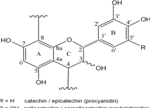
Condensed tannins (also called proanthocyanidins) are oligomers of flavan-3-ol monomer units commonly linked C8→C4 or C6→C4 in what are called B-type interflavan linkages (Figure 1). Some proanthocyanidins also have A-type linkages which additionally have an ether linkage between the C-2 position of an upper unit and the hydroxyl group at either C-5 or C-7 of the lower unit. Structural diversity is additionally added to this family of compounds because of the variability of hydroxylation patterns of the aromatic A and B rings and different stereochemistry at chiral centers at C-2 and C-3 of the C ring. Five distinct families of proanthocyanidins based on the hydroxylation patterns found in their A and B rings are commonly found in nature. These are the procyanidins, prodelphinidins, propelargonidins, profisetinidins, and prorobinetinidins. Perhaps the two more common types of proanthocyanidins are the procyanidins (PC), which are composed of catechin and epicatechin monomer flavan-3-ol units and the prodelphinidins (PD), which are composed of gallocatechin and epigallocatechin monomer flavan-3-ol units. Such oligomers can occur as pure procyanidin chains, pure prodelphinidin chains, and in mixed procyanidin – prodelphinidin oligomers. MALDI-TOF MS has been found to be a highly effective tool for analysis of such polydisperse and heterogeneous proanthocyanidins compounds, especially when combined with 13C NMR data to give a condensed tannin profile, which includes size and monomer composition of individual oligomer chains [9, 10]. Such structural information is important when considering chemical and biological functions such as antioxidant activity.
Proanthocyanidins are of great interest from the nutritional and medical perspective because of their strong antioxidant capacity and related protective effects on human health. The biological, pharmacological, and medicinal properties of tannins have been related to their free radical scavenging and antioxidant activities. Polyphenol oligomers have shown notable functions, such as anti-allergic, vasodilator, anti-carcinogenic, anti-inflammatory, antibacterial, antiviral, and cardioprotective activities.
Figure 1. Basic proanthocyanidin units in Alaska Cedar inner bark
2. Materials and Methods
2.1. Plant Material
An Alaska Cedar tree was obtained from the Hungry Mountain area in the Sol Duc drainage of the Olympia National Forest, Washington State (Oregon State University Herbarium voucher specimen #188046).
2.2 Extraction and Isolation
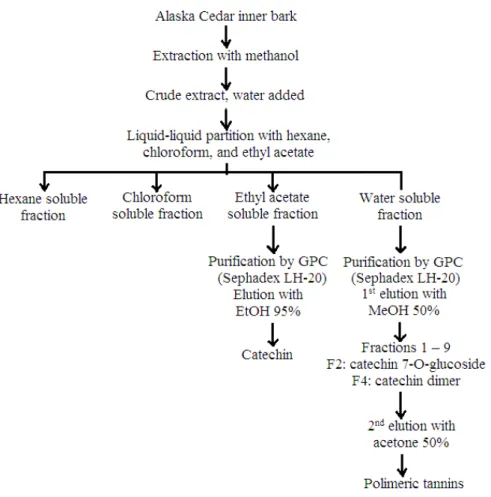
temperature with methanol. The methanol extract was filtered and concentrated on a rotary evaporator under reduced pressure to give a crude extract, which was diluted with water and the resulting aqueous solution was successively partitioned with hexane, chloroform, and ethyl acetate. The ethyl acetate fraction gave 20g and the water fraction 108 g of solid material after drying. The ethyl acetate fraction was chromatographed on Sephadex LH-20 to give (+)-catechin. 40 g of the water soluble fraction was applied to a Sephadex LH-20 column and then washed with 50 % aqueous methanol until washings were almost colorless. Catechin-7-0-glucoside and catechin – (4→8) –catechin were isolated from this elution. The condensed tannin fraction was then eluted from the column with 50% aqueous acetone to give 10g of purified condensed tannin material after freeze drying.
Figure 2. Chromatographic purification of Alaska cedar inner bark.
2.3 MALDI-TOF MS and
13C NMR
MALDI-TOF-MS (matrix-assisted laser desorption/ionization time-of-flight mass spectrometry) was performed using an ABSciEX 4700 tandem time of flight/time-of-flight (TOF/TOF) mass spectrometer run in reflectron positive ion mode. The scan range was from m/z 800-4000. Samples were mixed with 3-indole acrylic acid (t-IAA) matrix and acetonitrile as described by Taylor et al. [12]. Peak assignments for the [M + Na⁺] adduct ion for each proanthocyanidin oligomer molecular weight and monomer composition was based on the formula given by Monagas et al. [9]. 13
C NMR spectra were obtained on a Bruker model AM 100 MHz with d4-MeOH as the solvent.
Determination of Antioxidant Capacity
2.4. Chemicals
The 1,1’-diphenyl-2-picrylhydrazyl radical (DPPH), 2,2’-azinobis(3-ethylbenzothiazoline-6-sulfonic acid) (ABTS), TPTZ (2,4,6-tripyridyl-s-triazine), linoleic acid, β-carotene, ascorbic acid and catechin were purchased from Aldrich from Sigma Chemical Co.
2.5 DPPH Radical Scavenging Activity
The free radical scavenging activity on the DPPH (1,1-diphenyl-2-picrylhydrazyl) radical was determined according to the method described by Brad-Williams et al. [13]. 50 µL of sample of catechin as standard at different concentrations in methanol (50, 100, 150 and 200 µg/mL) was added to 1950 µL of a methanolic solution of DPPH (6.1×10⁻⁵ M). An equal amount of methanol and DPPH served as control. After incubation by 30 min at room temperature, the decrease in absorbance was measured at 515 nm. Lower absorbance of the reaction mixture indicated higher free radical scavenging activity. The antioxidant capacity, defined as the concentration of antioxidant necessary to scavenge the initial DPPH free radicals, was calculated using the following equation:
DPPH scavenging effect (%) = {(A1 – A2) / A1} × 100
Where A1 is the absorbance of the reaction control; A2 is the absorbance in the presence of the sample. Catechin was used as standard. And the EC50 calculated as the median effective concentration.
2.6 ABTS Radical Scavenging Activity
The ABTS antioxidant capacity assay was determined according to the method described by Re et al. [14], and modified by Shu et al. [15]. The blue-green ABTS• radical cation (2,2’-azinobis(3-ethylbenzothiazoline-6-sulfonic acid)) was generated by reaction of ABTS (7 mM) and potassium persulfate (2.42 mM), after incubation at room temperature in dark for 16 h until reaching a stable oxidative state. On the day of analysis, the ABTS• solution was diluted with 96% ethanol to an absorbance of 0.700 ± 0.05 at 734 nm. 50 µL of extract or catechin standard (20, 40, 50, 60, 80, 100 and 200 µg/mL) dissolved in 80% ethanol was added to 1950 µL of ABTS• solution and mixed thoroughly. The reactive mixture was allowed to stand at room temperature for 6 min and the absorbance was recorded at 734 nm. The results were expressed the same as the DPPH assay described above, with ABTS• inhibition and IC50 value.
2.7.
β
-carotene-linoleic acid model system (
β
-CLAMS) assay
2.8. Ferric-reducing/antioxidant power (FRAP) potential assay
The FRAP method measures the ability of antioxidants to reduce ferric-2,4,6-tripyridyl-s-triazine (Fe3+-TPTZ) to a ferrous form (Fe2+), which absorbs light at 593 nm. The ferro- and ferric-iron ions form complexes with TPTZ reagent and are the main products of this reaction [18]. To prepare the FRAP reagent, a mixture of 0.3 mol/L acetate buffer (pH 3.6), 10 mmol/L TPTZ dissolved in 40 mmol/L hydrochloric acid, and 20 mmol/L ferric chloride (10:1:1 v:v:v) was made. 100 µL sample or standard at concentrations 25, 50, 75 and 100 µg/mL was added to 3 mL of FRAP reagent and incubated at 25°C for 10 min. The absorbance was measured at 593 nm. The blank consisted of methanol into sample. The FRAP level was calculated by plotting a standard curve of absorbance against concentration of ascorbic acid standard solution (100 to 600 µM).
3. Results and Discussion
3.1. Tannin characterization
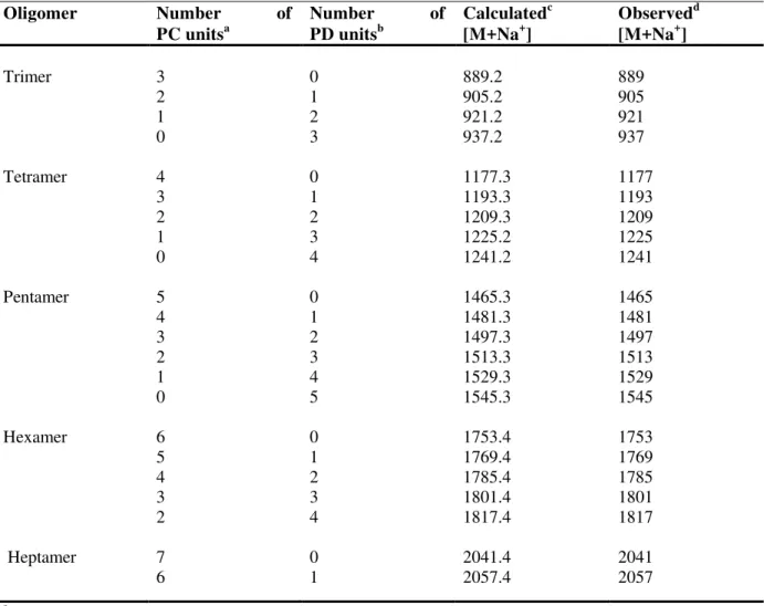
MALDI-TOF MS and 13C NMR spectra of the purified condensed tannin showed that it consents of both homogeneous and heterogeneous oligomers of procyanidins (catechin/epicatechin flavan-3-ol units) and prodelphinidins (gallocatechin/epigallocatechin flavan-3-ol units) as shown in figure 1. MALDI-TOF MS showed the existence of oligomer clusters from trimmers to heptamers as shown in Table 1 and Figure 3. Figure 4 shows an expanded view of the tetramer oligomers. Procyanidin monomer units clearly dominate the oligomer composition in these clusters. Each oligomer size, trimmer through heptamer, shows two dominant [M + Na⁺] adduct peaks. The first is a homogeneous procyanidin oligomer and the second one a 16 Da higher due to one more hydroxyl group on the B ring of a gallocatechin/epigallocatechin flavan-3-ol unit. These second peaks are heterogeneous oligomers, which consist of catechin/epicatechin monomer units plus on gallocatechin/epigallocatechin monomer unit. The rest of the oligomers with higher prodelphinidin content are significantly less abundant. The heptamer ions are barely seen above the S/N ratio.
method estimates 86% procyanidin/ 14% prodelphinidin. The rest of the signals in Table 2 are in full agreement with reported values for flavan-3-ol signals with a B-type interflavan linkage.
Table 1. MALDI-TOF MS of condensed tannins from Alaska Cedar inner bark.
Oligomer Number of
PC unitsa
Number of
PD unitsb
Calculatedc [M+Na+]
Observedd [M+Na+]
Trimer 3 0 889.2 889
2 1 905.2 905
1 2 921.2 921
0 3 937.2 937
Tetramer 4 0 1177.3 1177
3 1 1193.3 1193
2 2 1209.3 1209
1 3 1225.2 1225
0 4 1241.2 1241
Pentamer 5 0 1465.3 1465
4 1 1481.3 1481
3 2 1497.3 1497
2 3 1513.3 1513
1 4 1529.3 1529
0 5 1545.3 1545
Hexamer 6 0 1753.4 1753
5 1 1769.4 1769
4 2 1785.4 1785
3 3 1801.4 1801
2 4 1817.4 1817
Heptamer
7 6
0 1
2041.4 2057.4
2041 2057
a
procyanidin (catechin/epicatechin),
b
prodelphinidin (gallocatechin/epigallocatechin),
c
calculation based on Monagas et al. [9]
d
Figure 3. MALDI-TOF MS of condensed tannins from Alaska Cedar inner bark.
Table 2. 13C NMR data (δ ppm) of condensed tannins from Alaska Cedar inner bark (d4 –MeOH).
C number (as Fig. 1) δ ppm
C-2 76.1 cis, 82.6 trans
C-3 67.4 t, 72 ext
C-4 30 – 31 t, 38 – 39.5 ext
C-4a 100 – 102
C-5, 7, 8a 154.2 – 157.6
C-6,8 (unsubstituted)
C-6,8 (substituted), C-2’, 6’(PD)
96.5 – 98.2 106.4-107.1
C-1’, 4’ (PD) 131.2 – 131.6
C-2’(PC), 5’(PC) 115.6 –116.4
C-3’, 4’(PC), C-3’, 5’(PD)
144.8 –145.2 146
C-6’(PC) 119
t = terminal unit of oligomer,
ext = extending unit of oligomer, PC = procyanidin unit,
PD = prodelphinidin unit
Figure 4. MALDI-TOF MS of condensed tannins from Alaska Cedar inner bark. Expanded view of tetramer [M+ Na+] peaks
C= catechin/epicatechin flavan-3-ol monomer units
G= gallocatechin/epigallocatechin flavan-3-ol monomer units
3.2 Antioxidant assays
Several methods have been developed to determine the antioxidant potential of extracts and plant products. The trolox equivalent antioxidant capacity (TEAC) using ABTS (2,2-azino-bis-3-ethylbenzothiazoline-6-sulfonic acid) as an oxidant, the ferric reducing antioxidant power (FRAP), the DPPH (2,2’-diphenyl-1-picrylhydrazyl) and the β-carotene-linoleic acid model system (β-CLAMS) assays. Depending upon the reactions involved, these tests can roughly be classified into two types: assays based on electron transfer (ET) and assays based on hydrogen atom transfer (HAT) reactions [25].
ET-based assays measure the capacity of an antioxidant in the reduction of an oxidant, which changes color when reduced. The degree of color change is correlated with the antioxidant concentration of the samples. These tests include the total phenolic content using the Folin-Ciocalteu reagent (FCR), the ABTS, the ferric ion reducing antioxidant power (FRAP), and the DPPH assays.
The majority of HAT-based assays imply a competitive reaction scheme, in which antioxidant and substrate compete for thermally generated peroxyl radicals through the decomposition of azo compounds. These tests include the β-carotene-linoleic acid model system (β-CLAMS) assay, the inhibition of induced low-density lipoprotein autoxidation, and others [26]. To measure the antioxidant potential of condensed tannin from Alaska cedar inner bark, we have chosen the ABTS, FRAP and DPPH methods, which utilize the same single ET- mechanism, and the β-CLAMS assay, based on HAT reactions.
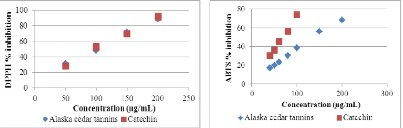
concentration, IC50 values (the concentration with scavenging activity of 50%), are shown is Table 3. A lower value of IC50 indicates greater antioxidant capacity. The results for DPPH (100.5 ± 0.5 µg/mL) were similar to the catechin standard (97.0 ± 3.4 µg/mL). Results of IC50 for Alaska cedar inner bark tannins were about 10% higher (less effective) than those reported for Delonix regia bark (90.0 ± 2.0 µg/mL) [19], as well for Acacia confusa stem bark (87.85 ± 0.52 µg/mL), and root bark (89.03 ± 0.50 µg/mL) [27].
The results for ABTS were different to DPPH assays, in this case the inhibition by condensed tannins from Alaska cedar inner bark (IC50 = 138.5 ± 2.5 µg/mL) was significantly inferior than the catechin standard (IC50 = 69.5 ± 1.5 µg/mL). The difference between ABTS and DPPH assays might be due to color interferences, the more color present in a sample, the smaller the absorbance decrease and lower the corresponding antioxidant activity measured [28].
Figure 5. DPPH radical inhibition by condensed tannins from Alaska Cedar inner bark
Figure 6. ABTS radical inhibition by condensed tannins from Alaska Cedar inner bark
Table 3. Antioxidant activity of condensed tannin from Alaska Cedar inner bark using the DPPH and ABTS radicals scavenging assays
Antioxidant activity
Sample IC50 DPPH (µg/mL) IC50 ABTS (µg/mL)
Condensed tannins from Alaska Cedar inner bark 100.5 ± 0.5 138.5 ± 2.5
Catechin 97.0 ± 3.4 69.5 ± 1.5
LSD < 0.05, = 3
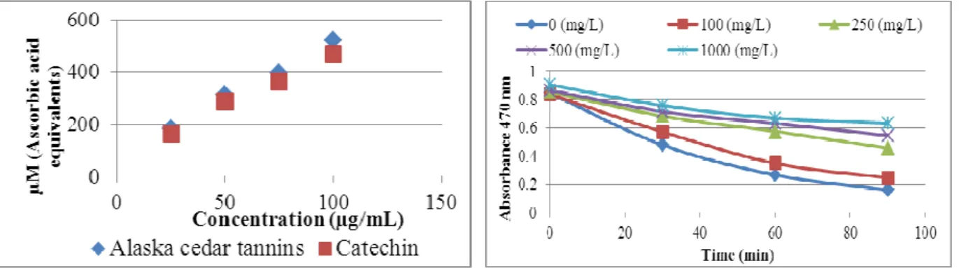
The antioxidant activity of condensed tannins measured by the FRAP assay, expressed in µM of ascorbic acid equivalents/g dried tannin, is shown in Figure 7. A higher absorbance corresponds to a higher ferric reducing power. In this assay, the higher activity shown by condensed tannins (522.4 ± 1.4 µM) was similar to the catechin standard (469.7 ± 4.2 µM). In the FRAP assay, the reducing ability of extracts is evident through the conversion of ions Fe3+ to Fe2+. This reaction is nonspecific and under the assay conditions, any reaction having lower redox potential than the ferric-ferrous half reaction, will contribute to the ferrous ion formation. The change in absorbance is therefore directly related to the total reducing power of the electron-donating antioxidants present in the samples of the reaction mixture [29]. An oligomeric phenolic fraction, consisting of proanthocyanidins trimmers and tetramers from Quercus sideroxyla bark presented similar results [30]. The reported outcome was 499
µM ascorbic acid equivalents per gram of sample at 100 µg/mL.
different concentrations of condensed tannins from Alaska cedar inner bark, with the oxidation of β -carotene and linoleic acid is shown in Figure 8.
The antioxidant capacity observed was 12.8% at 100 mg/L, 43.2% at 250 mg/L, 54.7% at 500 mg/L, and 64.6% at 1000 mg/L of tannin concentrations. The higher relative increase was observed at tannin concentrations between 100 and 250 mg/L, even superior to the 250 – 1000 mg/L range. The time needed by the blank to reduce the absorbance by a 50% factor was 29 min. However, when adding tannin (100 mg/L) it takes 42 min and even more up to 90 min at higher concentration (250 mg/L) as shown in Figure 8.
The Alaska Cedar inner bark shows significant antioxidant capacity, evaluated by both methods based on single ET- mechanism and HAT reactions. This bark may be considered as a new source of natural antioxidants for nutraceutical products.
Figure 7. Antioxidant activity of condensed tannins from Alaska Cedar inner bark by the FRAP assay.
Figure 8. Antioxidant activity of condensed tannins from Alaska Cedar inner bark by the β–carotene bleaching assay.
References
[1] S.F. Arno and R.P. Hammerly (2007). Northwest Trees. The Mountaineers Books, Seattle, pp. 125-129. [2] N.J. Turner (1998). Plant Technology of First Peoples in British Columbia. University of British
Columbia Press, pp.66-68.
[3] G. M. Barton (1976). A review of yellow cedar (Chamaecyparis nootkatensis [D. Don Spach) extractives and their importance to utilization, Wood Fiber Sci. 8, 172-176.
[4] N.A. Panella, M.C. Dolan, J.J. Karchesy, Y. Xiong, J. Peralta-Cruz, M. Khasawneh, J.A. Montenieri and G.O. Maupin (2005). Use of novel compounds for pest control: Insecticidal and acaricidal activity of essential oil components from heartwood of Alaska yellow cedar (Chamaecyparis nootkatensis), J. Med. Entomol. 42, 352-358.
[5] G. Dietrich, M.C. Dolan, J. Peralta-Cruz, J.Schmidt, J Piesman, R.J. Eisen, J.J. Karchesy (2006). Repellent activity of fractionated compounds from Chamaecyparis nootkatensis essential oil against
Nymphal Ixodes scapularis (Acari: Ixodidae), J. Med. Entomol. 43, 957-961.
[6] M.A. Khasawneh and J.J. Karchesy (2011). Terpenoids of the heartwood of Chamaecyparis nootkatensis, Am. J. Org. Chem. 1, 1-5.
[7] M.A. Khasawneh, Y. Xiong, J. Peralta-Cruz and J. J. Karchesy (2011). Biologically important eremophilane sesquiterpenes from Alaska cedar heartwood essential oil and their semi-synthetic derivatives, Molecules. 16, 4775-4785.
[8] G.H. Constantine, J.J. Karchesy, S.G. Franzblau and L.F. LaFleur (2001). (+)-Totarol from
Chamaecyparis nootkatensis and activity against Mycobacterium tuberculosis. Fitoterapia, 72, 572-574. [9] M. Monagas, J.E. Quintanilla-López, C. Gómez-Cordovés, B. Bartolomé and R. Lebrón-Aguilar (2010).
MALDI-TOF MS analysis of plant proanthocyanidins, J. Pharm. Biomed. Anal. 51, 358–372
[10] A. Behrens, N. Maie, H. Knicker and I. Kogel-Knabner (2003). MALDI-TOF mass spectrometry and PSD fragmentation as means for the analysis of condensed tannins in plant leaves and needles,
Phytochem. 62, 1159–1170.
[12] A.W. Taylor, E. Barofsky , J. A. Kennedy and M. L. Deinzer (2003). Hop (Humulus lupulus L.) proanthocyanidins characterized by mass spectrometry, acid catalysis, and gel permeation chromatography, J. Agric. Food Chem. 51, 4101-4110.
[13] W. Brand-Williams, M.E. Cuvelier and C. Berset (1995). Use of free radical method to evaluate antioxidant activity, Lebensmittel-Wissenschalt und Technology. 28, 25-30.
[14] R. Re, N. Pellegrini, A. Proteggente, A. Pannala, M. Yang and C. Rice-Evans (1999). Antioxidant activity applying and improved ABTS radical cation decolorization assay, Free Radical Biology & Medicine. 26, 1231-1237.
[15] W. Shu-Dong, C. Rui-Yan, L. Meng-Meng, T. Nian-Wan, Z. Hai-Chao and L. Yi-Ming (2011). Antioxidant condensed tannins from Machilus pauhoi leaves, J. Med. Plants Res. 5, 796-804.
[16] M. Rosales‒Castro, R.F. González‒Laredo, N.E. Rocha‒Guzmán, J.A. Gallegos‒Infante, J. Peralta‒Cruz,
J. Morré and J.J. Karchesy (2011). Chromatographic analysis of bioactive proanthocyanidins from
Quercus durifolia and Quercus eduardii barks, Acta Chromatographica. 23, 521–529.
[17] Y.S. Velioglu, G. Mazza, L. Gao and B.D. Oomah (1998). Antioxidants activity and total phenolics in selected fruits vegetables and grain products, J. Agric. Food Chem. 46, 4113-4117.
[18] I.F.F. Benzie and J.J. Strain (1996). The ferric reducing ability of plasma (FRAP) as a measure of “antioxidant power”: The FRAP assay, Analytical Biochemistry. 239, 80-76.
[19] C. Wei-Ming, S. Yan, F. Hui-Ling, Q. Ling, Z. Hai-Chao, D. Zi-Wei, Y. Chong-Ling and C. Qing-Xi (2012). NMR, HPLC-ESI-MS, and MALDI-TOF MS analysis of condensed tannins from Delonix regia
(Bojer ex Hook.) Raf. and their bioactivities, J. Agric. Food Chem. 60, 5013−5022.
[20] C. S. Ku and S. P. Mun (2007). Characterization of proanthocyanidin in hot water extract isolated from
Pinus radiata bark, Wood Sci. Technol. 41, 235–247.
[21] Z. Czochanska , L.Y. Foo, R.H. Newman and L.J. Porter (1980). Polymeric proanthocyanidins: stereochemistry, structural units and molecular weight, J. Chem. Soc. Perkin Trans. 1, 2278–2286.
[22] D. Roesch, C. Muegge, V. Fogliano, and L. Kroh (2004). Lothar W. Antioxidant oligomeric proanthocyanidins from sea buckthorn (Hippophae rhamnoides) pomace, J. Agric.Food Chem. 52, 6712-6718.
[23] P. Spencer, S. Sivakumaran, K. Fraser, L.Y. Foo, G.A. Lane, P. J. Edwards and L.P. Meagher (2007). Isolation and characterization of procyanidins from Rumex obtusifolius,Phytochem. Anal. 18, 193–203. [24] L.Y. Foo, Y. Lu, A. B. Howell and N. Vorsa (2000). The structure of cranberry proanthocyanidins which
inhibit adherence of uropathogenic P-fimbriated Escherichia coliin vitro,Phytochem. 54, 173-181. [25] D. Huang, B. Ou and R. Pryor (2005). The chemistry behind antioxidant capacity assays, J. Agric. Food
Chem. 53, 1841–1856.
[26] M. Ozgen, R. N. Reese, A.Z. Tulio, J.C. Scheerens and A.R. Miller (2006). Modified 2,2-Azino-bis-3-ethylbenzothiazoline-6-sulfonic acid (ABTS) method to measure antioxidant capacity of selected small fruits and comparison to ferric reducing antioxidant power (FRAP) and 2,2’-Diphenyl-1-picrylhydrazyl (DPPH) methods, J. Agric Food Chem. 54, 1151-1157.
[27] W. Shu-Dong, Z. Hai-Chao, L. Yi-Ming, L. Meng-Meng and C. Wei-Ming (2010). MALDI-TOF MS analysis of condensed tannins with potent antioxidant activity from the leaf, stem bark and root bark of
Acacia confusa, Molecules, 15, 4369-4381.
[28] M.B. Arnao (2000). Some methodological problems in the determination of antioxidant activity using chromogen radicals: a practical case, Trends in Food Sci. & Technol. 11, 419-421.
[29] F.F. Benzie and J.J. Strain (1999). Ferric reducing/antioxidant power assay: direct measure of total antioxidant activity of biological fluids and modified version for simultaneous measurement of total antioxidant power and ascorbic acid concentration, Methods Enzymol. 299, 15 – 23.

![Figure 4. MALDI-TOF MS of condensed tannins from Alaska Cedar inner bark. Expanded view of tetramer [M+ Na + ] peaks](https://thumb-eu.123doks.com/thumbv2/123dok_br/16412706.194571/8.892.257.650.111.497/figure-maldi-condensed-tannins-alaska-cedar-expanded-tetramer.webp)