O
h
r
c
i
r
g
a
in
e
a
s
l
R
e
Ahmet Tütüncü1, Taner Kuştarcı2, Can Özbek3, Ali Aydinlar4, Dilek Yesilbursa4 1Yüksek İhtisas Eğitim Araştırma Hastanesi, Kardiyoloji Bölümü, Bursa, 2Sungurlu Devlet Hastanesi, Kardiyoloji Bölümü, Çorum,
3İnegöl Devlet Hastanesi, Kardiyoloji Bölümü, Bursa, 4Uludağ Üniversitesi Tıp Fakültesi, Kardiyoloji Bölümü, Bursa, Türkiye
Fetuin-A and Aortic Stenosis
The Efects of Fetuin-A Levels on Aortic Stenosis
Fetuin-A Düzeylerinin Aort Darlığı Üzerine Etkisi
DOI: 10.4328/JCAM.2906 Received: 08.10.2014 Accepted: 25.11.2014 Printed: 01.07.2016 J Clin Anal Med 2016;7(4): 445-8
Corresponding Author: Ahmet Tütüncü, Kardiyoloji Kliniği, Bursa Yüksek İhtisas Eğitim ve Araştırma Hastanesi, Yıldırım, Bursa, Turkey. T.: +90 2243605050 F.: +90 2243605055 E-Mail: [email protected]
Özet
Amaç: Çalışmamızda böbrek fonksiyonları normal olan non-diyabetik kalsi-fik aort darlığı hastalarında, aort darlığı ile serum fetuin-A arasındaki ilişki-yi değerlendirmeilişki-yi amaçladık. Gereç ve Yöntem: Çalışmaya polikliniğinimiz-de aort darlığı ile takip edilen 26 hasta ve aort darlığı olmayan kontrol grubu-nu oluşturan 25 gönüllü olgu alınmıştır. Hastalarımızın venöz kan örneklerin-den Fetuin-A düzeyi çalışıldı. Tüm olguların ekokardiyografik olarak aort ka-pak alanları ve sol ventrikül parametreleri ölçüldü. Bulgular: Dejeneratif aort darlığı olan grubun yaş ortalaması istatistiksel anlamlı olarak daha yüksek olarak saptandı. Aort kapakla ilgili parametreler doğal olarak dejeneratif aort kapak olan olgularda yüksekti. Fetuin-A değerleri açısından iki grup arasın-da anlamlı farklılık saptanmadı. Ayrıca serum fetuin-A düzeyinin aort arasın- darlı-ğının ciddiyetine göre dağılımında da anlamlı fark gözlenmedi. Tartışma: So-nuç olarak fetuin-A başlıca sistemik kalsifikasyonun inhibisyonunda ve valvü-ler kalsifikasyonda rol alan multifonksiyonel özelliğe sahip bir glikoproteindir. Aktif bir süreç olan aort darlığının progresyonunun ve prognozunun değerlen-dirmesine yönelik fetuin-A ile ilgili daha geniş hasta gruplarıyla yapılmış ça-lışmalara ihtiyaç vardır.
Anahtar Kelimeler
Aort Darlığı; Fetuin-A; Ekokardiyografi
Abstract
Aim: We aimed to investigate the relation between fetuin-A and calciic aor-tic stenosis in non diabeaor-tic patients whose renal function were normal. Mate-rial and Method: 26 patients followed for aortic stenosis by our cardiology clinic for outpatients and 25 voluntary healthy subjects were included in the study. The fetuin–A levels were measured from the venous blood samples of the study population. All patients underwent transthorasic echocardiography, the aortic valvular area and let ventricular parameters of the patients were measured. Results: The average age of the patients in degenerative aortic stenosis group was signiicantly higher than the control group. The param-eters related to aortic valve were naturally higher in patients with dejenera-tive aortic valve. There was no signiicant diference between two groups about fetuin-A levels. Further more there was no signiicant relation between fetuin-a levels and aortic stenosis severity. Discussion: In conclusion fetuin-A is a multifunctional glycoprotein that plays important role in systemic calcii-cation inhibition and valvular calciicalcii-cation. Finally aortic stenosis is an active process and larger studies that investigate the relation between fetuin-a and the progression and prognosis of aortic stenosis are needed.
Keywords
Aortic Stenosis; Fetuin-A; Echocardiography
2012 yılında düzenlenen 28. Ulusal Kardiyoloji Kongresi’nde poster olarak sunulmuştur.
Fetuin-A ve Aort Darlığı / Fetuin-A and Aortic Stenosis
Introduction
The most frequent reason of aortic stenosis in adults is
de-generative aortic stenosis due to aging [1]. Dede-generative aortic
stenosis is seen frequently through elderly patients, it can
oc-cur with exertional dyspnea, angina, syncope, heart failure and
sudden cardiac death and if not treated it decreases the life
quality of the patients [2]. The prevalence of aortic stenosis
increases with age, clinically signiicant aortic stenosis occurs
in 2% among people over 65 years old and 5.5% over the age
85 [3-5], whereas aortosclerosis occurring due to aortic
calcii-cation and stifness can be seen in 50% of people between the
age 75-80 and %75 over the age 85 [6]. Serum fetuin-A is a
negative acute phase reactant glycoprotein that is synthesized
in hepatocytes, it is an indicator of acute inlammation [7,8].
Besides it is an important systemic calciication inhibitor.
Se-rum fetuin-A forms colloid formations with calcium phosphate
remains and helps it to resolve [9].
In this study we investigated the relation between aortic
steno-sis and serum fetuin-A in patients with calciic aortic stenosteno-sis
whose renal functions were normal and had no diabetes.
Material and Method
Our study population was composed of totally 51 people whose
approvals determined by the ethic committee were taken. The
patient group contained 26 patients who had been followed by
our cardiology clinic for outpatients with the diagnosis of aortic
stenosis (13 women, 13 men; mean age 67,2 ± 5,4) and the
con-trol group contained 25 voluntary healhty people (17 women,
8 men; mean age 60,5 ± 6,8). The exclusion criteria for aortic
stenosis were as follows: age under 18 or over 80, malignancy,
hipercalsemia, diabetes mellitus and renal failure. The systolic
and diastolic blood pressures of the patients were measured.
Then the venous blood samples were gained fort the
follow-ing laboratory measurements ater a night of fastfollow-ing; complet
blood counting, glucose, urea, creatinine, potasium, calcium,
phosphorus, liver function test, total chlosterol, HDL-C, LDL-C
and triglycerids. Fetuin-A analyses were made using
enzyme-linked immunosorbent assay (Biovendor Laboratory Medicine
İns). The GFR of all patients were estimated by using
modiica-tion diet of renal disease (MDRD) formula.
Echocardiographic Evaluation: All patients underwent
trans-thorasic echocardiographic evaluation in lateral decubitus
po-sition by GE-Vingmed Vivid 3 system (GE-Vingmed Ultrasound
AS, Horten, Norway) using multiHz probe transducer. The
mea-surements were made according to American Heart
Associa-tion criteria [10]. Let ventricular ejecAssocia-tion fracAssocia-tion (LVEF), Let
ventricular end diastolic diameter (LVEDD), Let ventricular
end systolic diameter (LVESD), interventricular septum
thick-ness (IVSD) ve posterior wall thickthick-ness (PWD) were measured.
Transaortic gradient was estimated with Bernoulli formula
(4v
2). Furthermore the area of aortic valve was estimated using
continuity equation. Valve area > 1.5cm2 was deined as mild
aortic stenosis, valve area between 1-1.5cm2 was deined as
moderate aortic stenosis and valve area < 1cm2 was deined
as severe aortic stenosis. Devereux Formula was used to
esti-mate let ventricular mass (gram) [11]. Let ventricular mass
index (LVMI) was estimated by dividing let ventricular mass to
body surface area.
Let ventricular mass (g) = 0,8 x 1,04 x [ (LVEDD + IVSD + PWT)3
– LVEDD3] + 0,6
Statistical Analyses
Statistical analyses were made using SPSS 13.00 package
pro-gramme. All data were deined as mean ± standart deviation.
Shapiro- Wilk test was used to investigate whether the data
shows normal distribution or not. T-test was used to compare
normally distributed data, Mann – Whitney U test was used to
compare data not normally distributed and Kruskal Wallis test
was used to compare three or more groups. The relations
be-tween variables were investigated with Pearson and Spearman
correlation coeicients. p values <0.05 were accepted as
sta-tistically signiicant.
Results
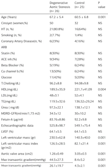
The demographic characteristics of groups involved are shown
in table 1. Gender, hypertension, obesity, smoking, diabetes
mellitus, presence of coronary artery disease, medications and
blood analyses were similar in both groups. The average age in
degenerative aortic stenosis group was higher and this
difer-ence was statistically signiicant (p<0,001). On the other hand
Table 1. The demographic and echocardiographic characteristiscs of the
pa-tients
Degenerative Aortic Stenosis
(n= 26)
Control
(n= 25) P value
Age (Years) 67.2 ± 5.4 60.5 ± 6.8 0.001
Cinsiyet (women,%) 50 32 NS
HT (n, %) 21(80.8%) 16(64%) NS
Smoking (n, %) 2(7.7%) 1(4%) NS
Coronary Artery Disease(n, %) 6(23%) 4(16%) NS
ARB
Statin (%) 8(30%) 8(30%) NS
ACE inh.(%) 9(34%) 7(28%) NS
Beta Blocker (%) 5(19%) 6(24%) NS
Ca channel b.(%) 13(50%) 6(24%) NS
Glucose 11(42%) 5(20%) NS
T.Chol(mg/dL) 96.2±8.8 94.08±9.8 NS
HDL(mg/dL) 189,5±35,9 221,7±41,09 0.004
LDL(mg/dL) 48±3.1 52±4.1 NS
TG(mg/dL) 119.5±32.6 136,52±29,24 NS
Ürea ( mg/dl) 97,5±22.1 138,1±12.1 NS
MDRD-GFR(ml/min/1,73 m2) 34.5±12 30±10.2 NS
Fetuin-A (µg/ml) 83.76±8.86 92.2±9.8 NS
Echocardiographic data 525.8±98.7 549.1±75.4 NS
LVEF (%) 64.1±5.5 64.1±5.5 NS
Let ventricular mass (gr) 230.5±62.8 140.3±40.0 0.001
Let ventricular mass index (gr/m2)
126.5±28.5 82.1±21.4 0.001
Aortic valve area (cm2) 1.26±0.49 3.05±0.3 0.001
Max transaortic gradient(mmHg) 44.5±27.3 8.4±5.2 0.001
Mean transaortic gradient(mmHg) 26.1±19.7 4.3±2.1 0.001
DM: Diabetes Mellitus, HT: Hypertension, T.Chol: Total Cholesterol, HDL: High Density Lipoprotein LDL: Low Density Lipoprotein, TG: Triglycerids, ACE: An-giotensin Converting Enzyme , ARB: AnAn-giotensin receptor blocker, Ca channel B: Calcium channel blocker, MDRD-GFR: Modiication diet of renal disease-Glomerüler iltration rate, LVEF: Let ventricular ejection fraction, NS: Not sig-niicant
| Journal of Clinical and Analytical Medicine
446
Fetuin-A ve Aort Darlığı / Fetuin-A and Aortic Stenosis
total cholesterol levels in this group were signiicantly lower
(p=0.004). The parameters associated to aortic valve were
nat-urally higher in patients with degenerative valves. But there was
no diference between the two groups about fetuinin-A levels.
Moreover serum fetuin-A levels did not difer signiicantly
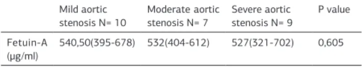
ac-cording to aortic stenosis severity (Table 2).
Further more there was no signiicant relation between fetuin-A
both aortic valve stenosis and let ventricular mass index. (Table
3).
Discussion
Serum fetuin-A is the most important systemic calciication
inhibitor. It does not efect the bone mineralization while
pre-venting ectopic calciication [12]. It can be thought to be a
mul-tifunctional protein. In this study we investigated the relation
between calciic aortic stenosis which is an active process and
serum fetuin-A which is the most important systemic
calciica-tion inhibitor showing multifunccalciica-tional properties. Furthermore
we investigated the relation between fetuin-A and the severity
of aortic stenosis diferently from other studies.
Wang et al showed that fetuin-A levels were signiicantly low
in patients with valvular calciication, atherosclerosis,
inlam-mation and malnutrition in their study on patients undergoing
periton dialysis [13]. In another study it was showed that low
fetuin-A levels were associated to cardiovascular death and
all cause mortality on patients undergoing dialysis [14]. In our
study we excluded the patients with renal failure to eliminate
the efects of renal failure on fetuin-A levels.
Ix et al [15] found negative correlation between mitral annular
calciication and fetuin-A on patients with coronary artery
dis-ease who have mitral annular calciication and aortic stenosis
but no serious renal disease. Also a negative correlation was
detected between serum fetuin-A levels and aortic stenosis in
non diabetic patients. However, in diabetic patients no such
sig-niicant correlation was established. In our study to avoid the
inluence of diabetes mellitus, diabetic patients were excluded.
In another study on patients with renal failure but no need
for dialysis, the relation between fetuin-A and progression of
aortic calciication was investigated and the calcium scores of
patients with low fetuin-A levels were found to be higher [16].
Kaden et al. investigated systemic and local fetuin-A levels in
patients with severe aortic stenosis. They found that serum
fetuin-A levels were low in patients with severe aortic stenosis
[17].
In our study there was no relation between fetuin-A levels and
calciic aortic stenosis. Further more there was no relation
be-tween fetuin-A and aortic stenosis severity. The control group
was younger than the patient group. This was possibly caused
by small number of patients meeting the inclusion criteria and
the small number of patients who do not have additional
valvu-lar diseases simultaneously. The total cholesterol levels were
found to be higher in control group. The patients with aortic
stenosis are followed more frequently and they are given
medi-cal treatment if needed. Thus, this causes the lower levels of
total cholesterol in patient group. The let ventricular mass was
found higher in patient group due to the compensatory
mecha-nisms of the let ventricle.
In conclusion fetuin-A is a multifunctional glycoprotein that
plays important role in systemic calciication inhibition, acute
inlammation, insulin resistance, metabolic syndrome, vascular
and valvular calciication. Because it is inluenced by multiple
independent factors, the patient groups should be determined
carefully. Finally aortic stenosis is an active process and larger
studies that investigate the relation between fetuin-A and the
progression and prognosis of aortic stenosis are needed.
Competing interests
The authors declare that they have no competing interests.
References
1. Roberts WC, Ko JM. Frequency by decades of unicuspid, bicuspid, and tricuspid aortic valves in adults having isolated aortic valve replacement for aortic stenosis, with or without associated aortic regurgitation. Circulation 2005;111(7):920-5. 2. Supino PG, Borer JS, Preibisz J, Bornstein A. The epidemiology of valvular heart disease: a growing public health problem. Heart Fail Clin 2006;2(4):379-93. 3. Iung B, Baron G, Butchart EG, Delahaye F, Gohlke-Bärwolf C, Levang OW et al. A prospective survey of patients with valvular heart disease in Europe: The Euro Heart Survey on Valvular Heart Disease. Eur Heart J 2003;24(13):1231-43. 4. Rajamannan NM. Calciic aortic stenosis: medical and surgical management in the elderly. Curr Treat Options Cardiovasc Med 2005;7(6):437-42.
5. Stewart BF, Siscovick D, Lind BK, Gardin JM, Gottdiener JS, Smith VE et al. Clini-cal factors associated with Clini-calciic aortic valve disease. Cardiovascular Health Study. J Am Coll Cardiol 1997;29(3):630-4.
6. Lindroos M, Kupari M, Heikkilä J, Tilvis R. Prevalence of aortic valve abnormali-ties in the elderly: an echocardiographic study of a random population sample. J Am Coll Cardiol 1993;21(5):1220-5.
7. Denecke B, Gräber S, Schäfer C, Heiss A, Wöltje M, Jahnen-Dechent W. Tissue
distribution and activity testing suggest a similar but not identical function of
fetuin-B and fetuin-A. Biochem J 2003;376(Pt 1):135-45.
8. Kalabay L, Cseh K, Jakab L, Pozsonyi T, Jakab L, Benedek S et al. Diagnos-tic value of the determination of serum alpha2-HS-glycoprotein. Orv Hetil 1992;133(25):1559-60.
9. Heiss A, DuChesne A, Denecke B, Grötzinger J, Yamamoto K, Renné T et al. Structural basis of calciication inhibition by alpha 2-HS glycoprotein/fetuin-A. Formation of colloidal calciprotein particles. J Biol Chem 2003;278(15):13333-41. 10. Schiller NB, Shah PM, Crawford M, DeMaria A, Devereux R, Feigenbaum H et al. Recommendations for quantitation of the let ventricle by two-dimensional echocardiography. American Society of Echocardiography Committee on Stan-dards, Subcommittee on Quantitation of Two-Dimensional Echocardiograms. J Am Soc Echocardiogr 1989;2(5):358-67.
11. Devereux RB, Alonso DR, Lutas EM, Gottlieb GJ, Campo E, Sachs I et al. Echo-cardiographic assessment of let ventricular hypertrophy: comparison to necropsy indings. Am J Cardiol 1986;57(6):450-8.
12. Mathews ST, Singh GP, Ranalletta M, Cintron VJ, Qiang X, Goustin AS et al. Improved insulin sensitivity and resistance to weight gain in mice null for the Ahsg gene. Diabetes 2002;51(8):2450-8.
13. Wang AY, Woo J, Lam CW, Wang M, Chan IH, Gao P et al. Associations of serum fetuin-A with malnutrition, inlammation, atherosclerosis and valvular calciica-tion syndrome and outcome in peritoneal dialysis patients. Nephrol Dial Trans-plant 2005;20(8):1676-85.
14. Hermans MM, Brandenburg V, Ketteler M, Kooman JP, van der Sande FM, Boeschoten EW et al. Netherlands cooperative study on the adequacy of Dialysis (NECOSAD). Association of serum fetuin-A levels with mortality in dialysis pa-tients. Kidney Int 2007;72(2):202-7.
15. Ix JH, Chertow GM, Shlipak MG, Brandenburg VM, Ketteler M, Whooley MA. As-Table 2. Fetuin-A levels according to aortic stenosis severity; median, minimum
ve maximum values Mild aortic
stenosis N= 10
Moderate aortic
stenosis N= 7
Severe aortic
stenosis N= 9
P value
Fetuin-A
(µg/ml) 540,50(395-678) 532(404-612) 527(321-702) 0,605
Table 3. The comparison of serum fetuin-A levels and parameters with
Spear-man correlation test
Fetuin- A
r P
The type of aortic stenosis (n=26) - 0,331
Let Ventricular Mass Index (n=51) - 0,749
Let Ventricular Mass (n= 51) - 0,730
Journal of Clinical and Analytical Medicine |
447
Fetuin-A ve Aort Darlığı / Fetuin-A and Aortic Stenosis
sociation of fetuin-A with mitral annular calciication and aortic stenosis among persons with coronary heart disease: data from the Heart and Soul Study. Circula-tion 2007;115(19):2533-9.
16. Koos R, Brandenburg V, Mahnken AH, Mühlenbruch G, Stanzel S, Günther RW et al. Association of fetuin-A levels with the progression of aortic valve calciication in non-dialyzed patients. Eur Heart J 2009;30(16):2054-61.
17. Kaden JJ, Reinöhl JO, Blesch B, Brueckmann M, Haghi D, Borggrefe M et al. Systemic and local levels of fetuin-A in calciic aortic valve stenosis. Int J Mol Med 2007;20(2):193-7.
How to cite this article:
Tütüncü A, Kuştarcı T, Özbek C, Aydinlar A, Yesilbursa D. The Efects of Fetuin-A Levels on Aortic Stenosis. J Clin Anal Med 2016;7(4): 445-8.
| Journal of Clinical and Analytical Medicine