Vibrio cholerae
O1 Strain and Its Role in Hemorrhagic
Response in the Rabbit Ileal Loop Model
Aurelia Syngkon1, Sridhar Elluri1,4, Hemanta Koley2, Pramod K. Rompikuntal4, Dhira Rani Saha3, Manoj K. Chakrabarti1, Rupak K. Bhadra5, Sun Nyunt Wai4, Amit Pal1*
1Divisions of Pathophysiology, National Institute of Cholera and Enteric Diseases, Kolkata, West Bengal, India,2Division of Bacteriology, National Institute of Cholera and Enteric Diseases, Kolkata, West Bengal, India,3Division of Histology and Electron Microscopy, National Institute of Cholera and Enteric Diseases, Kolkata, West Bengal, India,4Department of Molecular Biology, Umea˚ Centre for Microbial Research (UCMR), Umea˚ University, Umea˚, Sweden,5Infectious Diseases and Immunology Division, Indian Institute of Chemical Biology, Kolkata, West Bengal, India
Abstract
Background:Two well-characterized proteases secreted byVibrio choleraeO1 strains are hemagglutinin protease (HAP) and
V. choleraeprotease (PrtV). ThehapAandprtVknock out mutant,V. choleraeO1 strain CHA6.8DprtV,still retains residual protease activity. We initiated this study to characterize the protease present in CHA6.8DprtVstrain and study its role in pathogenesis in rabbit ileal loop model (RIL).
Methodology/Principal Findings:We partially purified the residual protease secreted by strain CHA6.8DprtVfrom culture
supernatant by anion-exchange chromatography. The major protein band in native PAGE was identified by MS peptide mapping and sequence analysis showed homology with a 59-kDa trypsin-like serine protease encoded by VC1649. The protease activity was partially inhibited by 25 mM PMSF and 10 mM EDTA and completely inhibited by EDTA and PMSF together. RIL assay with culture supernatants of strains C6709 (FA ratio 1.1+/20.3 n = 3), CHA6.8 (FA ratio 1.08+/20.2 n = 3), CHA6.8DprtV(FA ratio 1.02+/20.2 n = 3) and partially purified serine protease from CHA6.8DprtV(FA ratio 1.2+/20.3 n = 3) induced fluid accumulation and histopathological studies on rabbit ileum showed destruction of the villus structure with hemorrhage in all layers of the mucosa. RIL assay with culture supernatant of CHA6.8DprtVDVC1649 strain (FA ratio 0.11+/ 20.005 n = 3) and with protease incubated with PMSF and EDTA (FA ratio 0.3+/20.05 n = 3) induced a significantly reduced FA ratio with almost complete normal villus structure.
Conclusion:Our results show the presence of a novel 59-kDa serine protease in aDhapADprtV V. choleraeO1 strain and its
role in hemorrhagic response in RIL model.
Citation:Syngkon A, Elluri S, Koley H, Rompikuntal PK, Saha DR, et al. (2010) Studies on a Novel Serine Protease of aDhapADprtV Vibrio choleraeO1 Strain and Its Role in Hemorrhagic Response in the Rabbit Ileal Loop Model. PLoS ONE 5(9): e13122. doi:10.1371/journal.pone.0013122
Editor:Niyaz Ahmed, University of Hyderabad, India
ReceivedJuly 19, 2010;AcceptedSeptember 3, 2010;PublishedSeptember 30, 2010
Copyright:ß2010 Syngkon et al. This is an open-access article distributed under the terms of the Creative Commons Attribution License, which permits unrestricted use, distribution, and reproduction in any medium, provided the original author and source are credited.
Funding:This work was supported by grants from the Swedish Foundation for International Cooperation in Research and Higher Education (STINT), Sweden (Institutional Grants IG2008 2049). The awards of Senior Research Fellowship to A.S. is from the Department of Biotechnology, India and to S.E. from the Indian Council of Medical Research, New Delhi, India and also from STINT. The funders had no role in study design, data collection and analysis, decision to publish, or preparation of the manuscript.
Competing Interests:The authors have declared that no competing interests exist. * E-mail: [email protected]
Introduction
Proteases are enzymes that catalyze the hydrolysis of peptide bonds in proteins or peptides. They are either exopeptidases, whose actions are restricted to the N-or C- termini of proteins, or endopeptidases which cleave internal peptide bonds. Microbial peptides are predominantly secreted enzymes and can be classified based on the essential catalytic residue at their active site. They include serine proteases, cysteine proteases, aspartate proteases and metalloproteases. Proteases produced by pathogenic microorganisms play an important role in virulence [1]. Tissue barriers to pathogen invasion, such as extracellular matrices, epidermal keratinocyte layers and blood vessel walls, may be targeted by bacterial proteases. Proteolysis of host tissue components such as extracellular matrix proteins, including collagen, laminin, fibronectin and elastin, can
induce necrotic tissue damage [2,3]. V. cholerae O1, the causative agent of epidemic cholera, secretes a 32-kDa zinc-containing hemagglutinin protease that may play a role in the pathogenesis of cholera.V. choleraesecretes hemagglutinin/protease (HAP), which is encoded by thehapgene [4,5]. HAP can perturb the paracellular barrier function in epithelial cells by degrading occludin in tight junctions [6,7]. HAP nicks the cholera toxin [8] and digests proteins, such as mucin, fibronectin, lactoferrin, and secretory immunoglob-ulin A, that may participate in host defense against cholera [9]. HAP can also hydrolyze mucin to enhance the detachment ofV. cholerae from cultured epithelial cells [10]. A CTXQ- and hap-defective
epithelial cells [6]. These results suggested a role of HAP in reactogenicity, including inflammatory diarrhea. In our earlier studies, we have reported that HAP may play a role in the pathogenesis of actx-negativeV. choleraenon-O1, non-O139 strain by inducing a hemorrhagic fluid response in the RIL assay [12]. Histopathological examination of 20mg of purified protease-treated
rabbit ileum showed the presence of erythrocytes and neutrophils in the upper part of the villus lamina propria [12].
Although HAP is a very active virulence factor, an isogenic strain ofV. choleraemutated in thehapgene was no less virulent in infant rabbits than the parental strain [13]. Fullner et al reported that ahapmutant is more lethal in a pulmonary murine model and caused more severe histopathological damage than its wild-type parent in the lungs of survivors, although no difference was seen in the induction of inflammation [14]. Studies by Zhou et al suggest that an IL-8 stimulator other than HAP may be responsible for inflammation contributing to the reactogenicity of attenuated V. choleraevaccine strains [15]. An earlier study by Hase et al., [5] showed that ahapA-deleted mutant ofV. choleraeO1 had reduced extracellular proteolytic activity compared with the parental strain in a skim milk assay, indicating that the mutant still produces some extracellular proteolytic activity. In addition, residual proteolytic activity expressed by the hapA-deleted mutant is distinct from HAP, as demonstrated by failure of anti-HAP serum to inhibit the activity of this secondary protease on milk agar. The mutant strain also failed to agglutinate chicken erythrocytes [5]. Young and Broadbent [16] described several extracellular proteases in V. choleraethat could explain the residual proteolytic activity of the hap-negativeV. choleraemutant. Besides HAP, the other major well-characterized protease in V. cholerae is a 97-kDa Vibrio cholerae protease, PrtV. PrtV plays a role in virulence in aC. elegansinfection model [17].
In the present study, ahap and prtVdouble knock out mutant of V. choleraestrain CHA6.8DprtVstill had residual protease activity. This protease was partially purified from strain CHA6.8DprtVand MS peptide mapping and sequence analysis of the protein revealed homology with a 59-kDa trypsin-like serine protease encoded by VC1649. To our knowledge, this is the first report of a serine protease in V. cholerae O1 and demonstration of its role in hemorrhagic response in the RIL model.
Materials and Methods
Ethics statement
Animal experiments were done after obtaining necessary permission from Institutional Animal Ethical Committee (IAEC). The IAEC/CPCSEA approval number is 45/1 dated 15/3/2007.
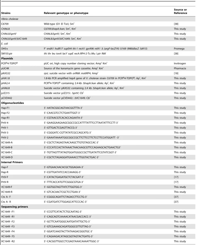
Bacterial strains, plasmids and primers used in this study The bacterial strains used in this study and their relevant properties are summarized in Table 1. All the strains were maintained at280uC in 30% glycerol in tryptic soy broth (TSB, Difco laboratories). For protease purification, a DhapADprtV mutant ofV. choleraeO1 CHA6.8DprtVwas used. Antibiotics were used at the following concentrations unless otherwise indicated: ampicillin (Am), 100mg/ml; streptomycin (Sm), 100mg/ml;
carbenicillin (Cb), 100mg/ml; kanamycin (Km), 50mg/ml forE. coliand 40mg/ml forV. cholerae.
Construction of ahapAknock -out mutant inVibrio choleraeO1 strain C6709
The bacterial mutant was constructed by replacing the hapA gene with its deletion-insertion alleleDhapA::kanusing published methods [18]. One such mutant showing a SmrKmrphenotype
was selected for further study and designated CHA6.8 (Table 1). The in-frame hapA gene deletion in the strain C6709 was confirmed by PCR withhapAinternal primers (Table 1).
Construction of aDhapADprtV V. choleraeO1 strain,
CHA6.8DprtV
The bacterial mutant was constructed by double crossover method using the construct as mentioned previously [17]. The knock out mutant (CHA6.8DprtV) was confirmed withprtVinternal primers (Table 1).
Construction ofDhapADprtVDVC1649V. choleraeO1
strain CHA6.8DprtVDVC1649
A VC1649 in-frame deletion mutant was constructed using published methods [18,19]. Several colonies were purified from the plates, tested for Cb sensitivity and then analyzed for the deletion and confirmed with internal primers for hapA, prtV, VC1649andctx(Table 1).
Azocasein assay
Casein was chosen as the substrate to assay proteolytic activity. The substrate-enzyme mixture was incubated at 37uC and the reaction was terminated with 10% trichloroacetic acid after 1 hour. The precipitated protein was removed by centrifugation (12,0006g for 4 mins), and the supernatant was transferred to a clean tube containing 525 mM NaOH. Absorbance was measured at 440 nm using a Smartspec spectrophotometer (Bio-Rad, Hercules, CA). Substrate with buffer and substrate with inhibitors were used as negative controls.
Skim milk assay
Single colonies of V.cholerae O1 strains C6709, CHA6.8, CHA6.8DprtV and CHA6.8DprtVDVC1649 were streaked onto nutrient agar (NA) plates containing 1.5% skim milk and incubated at 37uC overnight. Protease activity was detected by clearing of the opaque milk proteins incorporated into the NA.
Inhibition of protease activity
The azocasein assay was done with 30mg of ammonium
sulphate precipitated proteins from culture supernatants of C6709, CHA6.8, CHA6.8DprtVand CHA6.8DprtVDVC1649.The prote-ase activity of crude proteins was also tested for inhibition with 25 mM PMSF, 10 mM EDTA and 10 mM 1,10- phenanthroline. Twenty-five mM Tris-HCl and 25 mM Tris-HCl in the presence of 25 mM PMSF, 10 mM EDTA and 10 mM 1,10- phenanthro-line were used as negative controls. The NB (non-binding) fraction (5mg) of the CHA6.8DprtVstrain with protease inhibitors (10 mM EDTA, 10 mM EGTA, 25 mM PMSF, 10 mM EDTA with 25 mM PMSF, 1mg/ml aprotinin, 28 mM E-64, 1mg/ml
leupeptin and 10 mM 1,10- phenanthroline) were incubated for 30 mins at 37uC and assayed by azocasein assay. Twenty-five mM Tris-HCl was used as a negative control. The protease activity with EDTA was measured both in the presence and absence of 10 mM CaCl2. The mean with standard deviation of three
experiments was considered for data analysis.
Partial purification of a novel protease from strain CHA6.8DprtV
Table 1.Bacterial strains, plasmids, primers and oligonucleotides used in this study.
Strains Relevant genotype or phenotype
Source or Reference
Vibrio cholerae
C6709 Wild-type (O1 El Tor); Smr [38]
CHA6.8 C6709DhapA::kan; Smr, Kmr This study
CHA6.8DprtV CHA6.8DprtV; Smr, Kmr This study
CHA6.8DprtVDVC1649 CHA6.8DprtVDVC1649; Smr, Kmr This study
E. coli
DH5a F’endA1 hsdR17 supE44 thi-1 recA1 gyrA96 relA1D(argF-lacZYA) U169 (W80dlacZDM15) Promega SM10lpir thi thr leu tonA lacY supE recA::RP4-2-Tc::Mulpir R6K [38]
Plasmids
PCRH4-TOPOH pUC ori, high copy number cloning vector; AmprKmr Invitrogen
pUC4K Source of the kanamycin gene cassette; AmprKmr Pharmacia
pKAS32 rpsLsuicide vector withoriR6KmobRP4; Ampr [18]
pHA1.8 1.8-kb PCR amplifiedhapAgene ofV. choleraestrain C6709 in PCRH4-TOPOH; Apr, Kmr This study
pHA2.4 PCRH4-TOPOHcontaining 2.4-kbDhapA::kan allele; Apr, Kmr This study
pHA6.8 Suicide vector pKAS32 containing 2.4 kbDhapA::kanallele; Apr, Kmr This study
pJZ215 Suicide vector pJZ215::DprtV; Cbr This study
pCVD442 Suicide vector pCVD442::DVC1649; Cbr This study
Oligonucleotides
Hap-F1 59-AATACGGCAGTAACGGTTTA-39 This study
Hap-F2 59-CAACGTCCTCTGAATTGGT-39 This study
Hap-R1 59-CGTAACGTCACACCAGAATA-39 This study
PrtV-4 59-GAAGGAAGAAGCGGCCGCCATTTTATTTCCTTAATATTTCCTT-39 This study
PrtV-1 59-GTTGACTCGAGTTACCG-39 This study
PrtV-3 59-CGGGATC-CGTTATATCGCCAGCATG-39 This study
PrtV-2 59-GAAATAAAATGGCGGCCGCTTCTTCCTTCTCCTTCCATGGATT -39 This study
VC1649-A 59-CGCTCTAGAGTAACAAGCTTGTGTAGCCAC-39 This study
VC1649-B 59-CCCATCCACTATAAACTAACAAGCGTTCCAGAAGCACTGAACTG39 This study
VC1649-C 59-TGTTAGTTTATAGTGGATGGGCCGCTTGATATTCGTATCGGT-39 This study
VC1649-D 59-CGCTCTAGAGGATGAAACCTTGGTACTGAC-39 This study
Internal Primers
Hap-F 59-GTGAACAACACGCTGGAGAA-39 This study
Hap-R 59-CGTTGATATCCACCAAAGG-39 This study
PrtV-F 59-CATACTGAGATGCTCTACGAT-39 [17]
PrtV-R 59-TTTCACCATGTTCGGGCGTGA-39 [17]
VC1649-F 59-GGTGGTAGTTATCTTGGTGG-39 This study
VC1649-R 59-GTCACAACTCGCTCCTGAA-39 This study
Ctx A- F 59-CGGGCAGATTCTAGACCTTCCTG-39 [37]
Ctx A- R 59-CGATGATCTTGGAGCATTCCCAC-39 [37]
Sequencing primers
VC1649 -F1 59-CCGTTCATACTCTGCAATAG-39 This study
VC1649 -R1 59-CAGCAGTCAAAACATAACGACCACC-39 This study
VC1649 -F2 59-GCTTCAATGGGCAATGATATTGCTG-39 This study
VC1649 -F3 59-GTCGAAAGCAGATGGGCGTTGTTAG-39 This study
VC1649 -F4 59-GGATCAAGTACTTATAAGACGGGTGC-39 This study
VC1649- F5 59-CAGAAGACATAGCGGTAGTACTGATG-39 This study
VC1649 -R2 59-CACGGTTGGCCTCGAGTAAACAAAATTGGC-39 This study
culture supernatant was precipitated with 60% saturated ammo-nium sulphate. After centrifugation at 11,9736g for 20 min at 4uC, the pellet was re-suspended in 25 mM Tris-HCl buffer, pH 7.4. Re-suspended proteins were dialyzed against the same buffer, concentrated by Amicon filtration (Millipore Co, Bellerica MA) and loaded onto an ion exchange chromatography column (DE-52; Whatman, Kent, UK) pre-equilibrated with 25 mM Tris-HCl buffer, pH 7.4. Proteins eluted in the unbound fraction were designated as the non-binding fraction (NB). The proteins bound to the DE-52 column were eluted in the presence of NaCl (0.1 and 0.3 M). Fractions constituting the peaks NB, 0.1 M#1, 0.1 M#2 and 0.3 M were pooled, dialyzed, concentrated and examined for protease activity by azocasein assay. The columns were run on a BioLogic Duo Flow Chromatographic system (Bio-Rad, Hercules, CA).
Native PAGE
The proteins were separated by electrophoresis on a 10% native polyacrylamide gel according to the procedures described by Davis et al [20] in the absence of SDS and 2-mercaptoethanol. Protein samples were mixed with sample buffer containing 10% glycerol, 0.05% bromophenol blue and Tris-HCl pH-6.8, resolved in the gel and bands were visualized by staining with Coomassie brilliant blue.
Protein identification by MS peptide mapping and sequencing analysis
The major band observed in the native PAGE of the non-binding pooled fraction from the DE-52 column was excised from the Coomassie blue stained gel and analysed on a Bruker Autoflex III MALDI TOF/TOF instrument at Alphalyse, Odense, Den-mark. The peptide mixture was analyzed in positive reflector mode for accurate peptide mass determination and 5–10 of the peptides were selected for analysis by MS/MS fragmentation for partial peptide sequencing. The MS and MS/MS spectra were combined and used for a database search in an in-house protein database using the Mascot software. The peptides used for the identification are highlighted in the sequence. Peptides confirmed by MS/MS sequencing are shown in bold.
DNA sequencing
The VC1649 gene (ORF) was amplified by using primers 59CCGTTCATACTCTGCAATAG39and59 CACGGTTGGCC-TCGAGTAAACAAAATTGGC39. The resultant PCR product was analyzed by 0.7% agarose gel electrophoresis. The sequencing was done on an ABI 3130 DNA analyzer (Applied Biosystems, Foster City, CA) and the sequences were aligned, analyzed using Clustal X and NCBI/BLAST programs. The nucleotide sequence data reported in this paper will appear in the DDBJ/EMBL/ GenBank nucleotide sequence database with the accession number AB572560.
Rabbit ileal loop assay
The rabbit ileal loop (RIL) assay was performed in young New Zealand White rabbits (2 kg) essentially by the method described by De and Chatterjee [21]. Culture supernatants (one ml) and washed bacterial cells (109cfu/ml) of C6709, CHA6.8, CHA6.8DprtV, CHA6.8DprtVDVC1649 grown in tryptic soy broth were inoculated in rabbit ileum. Tryptic soy broth was used as negative control in the above assay. The partially purified protease at a concentration of 50mg (non-binding pooled fraction eluted
from DE-52 column) from CHA6.8DprtVstrain, similar concen-tration of protease inhibited with 25 mM PMSF and EDTA and 25 mM Tris-HCl with PMSF and EDTA (negative control) in a
volume of 1 ml were also assayed in RIL assay. The animals were sacrificed after 18 hrs and the enterotoxic response was deter-mined by measuring the fluid accumulation (FA) ratio, which is the ratio of the volume of fluid accumulated in the intestinal loop to the length of the loop. A ratio of greater than 1.0 indicated a strong positive response, while a negative response was defined as FA ratio of less than 0.2.
Histopathological studies
Tissue samples (2 cm in length) from RIL assays were collected and placed in 10% neutral-buffered formalin for histopathological analysis. Tissues were embedded in paraffin and processed following the standard protocol. Sections (3 to 4mm thick) prepared with a Leica rotary microtome were stained with hematoxylin and eosin and examined by light microscopy. Photographs were taken under different magnifications with a Leica DMLB microscope (Solms, Germany), equipped with a digital imaging system.
Results
Construction ofDhapAandDhapADprtV V. choleraeO1
mutant strains
The in-framehapAdeletion in the strain C6709 was confirmed by PCR using internal primers (Table 1). The CHA6.8DprtV mutant was constructed by the double crossover method as described previously (Vaitkevicius et al 2006). The knock out mutant CHA6.8DprtVwas confirmed usingprtV specific internal primers (Table 1).
Protease activity in C6709,CHA6.8 and CHA6.8DprtV
strains
The proteins from culture supernatants of the above strains were precipitated with ammonium sulphate and after dialysis, 30mg of
All strains included in this study were also tested for protease activity in a skim milk assay. As shown in Fig. 1B, the zone of proteolysis created by C6709 on skim milk agar was clear, indicating complete degradation of the milk proteins. With the other two strains, CHA6.8 and CHA6.8DprtV, the zone of proteolysis was hazy, indicating that not all of the milk proteins were degraded. These results suggest that the substrate specificity of proteases in CHA6.8, and CHA6.8DprtVmay be different from that of C6709, which secretes HAP.
Partial purification of protease from the strain CHA6.8DprtV
The ammonium sulphate precipitated proteins from culture supernatants of CHA6.8DprtV were loaded onto an anion-exchange chromatography column (DE-52). The proteins in the non-binding fraction of the column (Fig. 2A) were pooled and
concentrated. The bound proteins were eluted with 0.1 M (Fig. 2B) and 0.3 M (Fig. 2C) NaCl, dialyzed against 25 mM Tris-HCl buffer and concentrated. When protease activity in the NB, 0.1 M and 0.3 M NaCl eluted fractions were tested by azocasein assay, the major protease activity was present in the NB fraction (Fig. 2D). The NB-pooled fraction was concentrated and run on a native PAGE (Fig. 2E). The major protein band was excised and analyzed by MS/MS sequencing (Fig. 2E). The sequences highlighted showed homology with a 59-kDa serine protease encoded by the gene VC1649 (Fig. 2F). The sequence GDSGGP (underlined) flanks the serine residue in trypsin-like serine proteases (Fig. 2F).
The presence of a calcium-dependent serine protease To determine the nature of the partially purified protease from CHA6.8DprtVeluted in the non-binding fraction of a DE52 anion-Figure 1. Protease activity assay.A) Azocasein assay with 30mg of ammonium sulphate precipitated proteins from culture supernatants of C6709, CHA6.8, CHA6.8DprtV and CHA6.8DprtVDVC1649 and inhibition test with 25 mM PMSF, 10 mM EDTA and 10 mM 1,10- phenanthroline. Negative controls were (1) 25 mM Tris-HCl and 25 mM Tris-HCl in the presence of (2) 25 mM PMSF, (3) 10 mM EDTA and (4) 10 mM 1,10-phenanthroline. The values shown are the means with standard deviations from three experiments. B) Skim milk assay for detection of protease in C6709, CHA6.8, CHA6.8DprtVand CHA6.8DprtVDVC1649strains.
exchange column, we performed protease inhibition assays with several inhibitors (Fig. 2G). The protease was partially inhibited in the presence of EDTA (60.3%), EGTA (59.2%) and PMSF (60.3%). The partially purified protease was completely inhibited when PMSF and EDTA are used together (Fig. 2G). There was significantly less inhibition of protease activity in the presence of 1,10- phenanthroline (9%), aprotinin (10.5%), leupeptin (8.7%) and E64 (1.8%). Although EDTA inhibited protease activity by
60.3%, EDTA in the presence of CaCl2inhibited activity by only
3.6% inhibition (Fig. 2G). The serine protease secreted by CHA6.8DprtVis a calcium-dependent serine protease.
Construction of CHA6.8DprtVDVC1649 deletion mutant
The CHA6.8DprtVDVC1649 deletion mutant was constructed as described in the text and confirmed by internal primers for hapA, prtVand VC1649 as shown in Fig. 3A. PCR with internal
Figure 2. Partial purification and identification of protease.Chromatographic profile of ammonium sulphate precipitated crude proteins from culture supernatants of CHA6.8DprtVstrain loaded onto an anion exchange column (DE-52). A) Proteins eluted in the non-binding fraction (NB), B) proteins eluted with 0.1 M NaCl, C) proteins eluted with 0.3 M NaCl,+/2shows presence or absence of protease activity, D) azocasein assay with pooled samples (30mg) NB, 0.1 M#1, 0.1 M#2, 0.3 M and crude proteins. E) Native PAGE profile (lane 1) of crude proteins of CHA6.8DprtVstrain and (lane 2) of partially purified protease (NB) from DE-52 column. The marked protein band was analyzed by MS/MS sequencing and the peptides highlighted showed homology with a 59-kDa trypsin-like serine protease encoded by VC1649. F) The underlined GDSGGP are the amino acid sequences around the serine residue present in trypsin-like serine proteases. G) Protease inhibition test of NB fraction (5mg) with protease inhibitors 10 mM EDTA, 25 mM PMSF, 25 mM PMSF and 10 mM EDTA, 10 mM EDTA and 20 mM CaCl2, 10 mM EGTA, 1mg/ml aprotinin, 28 mM E64, 1mg/ml
leupeptin and 10 mM 1,10-phenanthroline incubated for 30 mins at 37uC. Residual protease activity was assayed by azocasein assay. Twenty-five mM Tris-HCl was used as a negative control. The values shown are the means with standard deviations from three experiments.
primers for the ctx gene confirmed the presence of ctx in both the C6709 and the CHA6.8DprtVDVC1649 strains. PCR with internal primers for hapA, prtV and VC1649 in strain CHA6.8DprtVDVC1649 showed the absence of any band by agarose gel electrophoresis (Fig. 3A) and also confirmed the absence of the deleted gene sequences. On the other hand, strain C6709 showed the presence of PCR products with the internal primers confirming the presence ofhapA, prtVand VC1649 genes (Fig. 3A). A native PAGE profile of crude proteins from CHA6.8DprtV and CHA6.8DprtVDVC1649 showed the absence of a 59-kDa band in the CHA6.8DprtVDVC1649 strain (Fig. 3B).
The VC1649 gene sequence showed complete homology with the published sequence of VC1649 fromV. choleraeO1 biovar El Tor strain N16961 (NCBI).
We initially started the study to characterize the proteases present in the CHA6.8DprtV strain. Partial purification of proteases from CHA6.8DprtV strain showed the presence of a 59-kDa trypsin-like serine protease encoded by the VC1649 gene. The major protease present in the CHA6.8DprtVstrain is a serine protease, but protease activity in CHA6.8DprtVDVC1649, in which the 59-kDa serine protease is not secreted, increased by 11.4% (Fig. 1A). These results indicated that besides HAP, PrtV and a 59-kDa serine protease, there are still other proteases secreted by the CHA6.8DprtVDVC1649 strain. PMSF and EDTA inhibited protease activity in CHA6.8DprtVDVC1649 by 64.5% and 46.2%, respectively, whereas 1,10-phenanthroline only inhibited protease activity by 6.2% suggesting the presence of another serine protease (Fig. 1A). Skim milk assay with CHA6.8DprtVDVC1649 still showed residual protease activity (Fig. 1B).
The serine protease induces a hemorrhagic fluid response in RIL
To study the role of the 59-kDa serine protease in virulence, 50mg of the partially purified protease was injected into the rabbit
ileum, which induced significant hemorrhagic fluid accumulation (FA ratio 1.2+/20.2, n = 3, Fig. 4A). When a similar concentra-tion of the protease was incubated in the presence of PMSF and EDTA and injected into the rabbit ileum, there was a significant decrease in fluid accumulation (FA ratio 0.3+/20.05, n = 3, Fig. 4A). Histopathological analysis of the rabbit ileum revealed that the protease caused extensive damage to all the layers of the mucosa. There was damage to the villus structure, which was completely destroyed. We observed gross damage of the villus surface structure with hemorrhage in all layers of the mucosa (Fig. 5A). On the other hand, analysis of the ileal tissues treated with the protease in presence of both PMSF and EDTA, revealed normal microvillus structure with no gross alteration in villus structure, although the villus lamina propria was slightly dilated and RBCs had accumulated in a few places in the basal area (Fig. 5B). PMSF and EDTA completely inhibited protease activity (Fig. 4A), but still we observed some residual effect in the rabbit ileal loop. This effect could be due to some other domain in the protease, which may not be its proteolytic domain, and could be responsible for causing damage to the ileal tissue. Tissues treated with 25 mM Tris-HCl and PMSF+ EDTA did not cause fluid accumulation in RIL (FA ratio 0.12+/20.002 n = 3, Fig. 4A) and histopathology of the ileal tissue showed normal microvillus structure (Fig. 5C).
One ml of culture supernatant of C6709, CHA6.8 and CHA6.8DprtV strain induced significant fluid accumulation (Fig. 4B) in RIL model (FA ratio 1.1+/20.3, n = 3; 1.08+/20.2, n = 3; and 1.02+/20.2, n = 3 respectively) where as CHA6.8 DprtVDVC1649 strain which is devoid of the serine protease gene and sterile tryptic soy broth, TSB (negative control) did not induce any fluid accumulation (Fig. 4B) (FA ratio 0.11+/20.005, n = 3 and 0.09+/20.002, n = 3 respectively). Almost similar results were observed when one ml of washed bacterial cells (109 cfu/ml) of C6709, CHA6.8 and CHA6.8DprtV induced significant fluid accumulation (FA ratio 1.2+/20.35, n = 3; 1.1+/20.3, n = 3; and 1.0+/20.2, n = 3 respectively). Bacterial cells of CHA6.8DprtV DVC1649 strain did not induce any fluid accumulation (FA ratio 0.15+/20.005, n = 3). Histopathological studies of ileal tissues treated with culture supernatant from C6709 strain showed presence of hemorrhage in all layers of the gut mucosa especially
Figure 3. Confirmation of knock out mutant.A) PCR amplification with internal primers forctx(lane 1),hapA(lane 3),prtV(lane 5) and VC1649 (lane 7) in strain C6709 and forctx(lane 2),hapA(lane 4),prtV (lane 6) and VC1649 (lane 8) in strain CHA6.8DprtVDVC1649. The primer sequence used in the above experiment is given in Table 1. (MW) denotes molecular weight marker (20 kb-75 bp marker, Fermentas). B) Native PAGE (10%) with ammonium sulphate precipitated proteins of C6709 (lane 1), CHA6.8 (lane 2), CHA6.8DprtV (lane 3) and CHA6.8DprtVDVC1649 (lane 4). The * shows the protein band with sequence homology to the 59-kDa serine protease (VC1649). This band is absent in strain CHA6.8DprtVDVC1649 (lane 4).
in the sub-mucosal layer (Fig. 5D). Ileal tissues treated with culture supernatant from CHA6.8 strain showed widely dilated villi with rupture at places with gross hemorrhage and inflammatory cells in mucosa and sub-mucosa (Fig. 5E). Ileal tissues treated with culture supernatant of CHA6.8DprtVstrain also showed dilated villi with gross hemorrhage in all layers of the mucosa (Fig. 5F). The same section at a higher magnification 40X showed ruptured villi with hemorrhage and inflammatory cells in mucosa and sub-mucosa (Fig. 5G). The ileal tissues treated with culture supernatant of
CHA6.8DprtVDVC1649 strain protease showed villous architec-ture almost normal with minimum hemorrhage in mucosa and sub-mucosa (Fig. 5H). TSB treated ileal tissue showed normal gut mucosa (Fig. 5I).
Discussion
Bacterial proteases are an important virulence factor in a variety of organisms, causing massive tissue damage which may aid the
bacteria in host cell entry [22]. The major protease secreted byV. choleraeis HAP, a member of a large family of metalloproteases. HAP is produced by both non-pathogenic and pathogenic species,
including the elastase of P. aeroginosa [23]. It acts on potentially relevant substrates like mucin, fibronectin, lactoferrin and the A subunit of CT [8,10]. In our earlier studies, we demonstrated that
Figure 5. Histopathological study of ileal tissues.Panels show photomicrographs of histology of rabbit ileal loop tissue after treatment with A) Partially purified serine protease fromV. choleraestrain CHA6.8DprtVshowing hemorrhagic fluid accumulation (Fig. 4A, NB). Gross damage of the villus surface structure was observed with hemorrhage in all layers of the mucosa. Magnification, 20X. B) Almost normal villous architecture observed in ileal tissues treated with 50mg of partially purified protease inhibited with 25 mM PMSF and 10 mM EDTA (Fig. 4A, NB+PMSF+EDTA). This photomicrograph shows no gross alteration in villus structure but villus lamina propria are slightly dilated and RBC have accumulated at a few places in the basal area. Magnification, 20X. C) Ileal tissues treated with 25 mM Tris-HCl buffer with PMSF and EDTA (Fig. 4A, control) showed normal villus structure. Magnification 20X. (D) ileal tissues treated with culture supernatant from C6709 strain showed presence of hemorrhage in all layers of the gut mucosa specially in the submucosal layer, Magnification 20X. E) ileal tissues treated with culture supernatant from CHA6.8 strain showed widely dialated villi with rupture at places with gross hemorrhage and inflammatory cells in mucosa and submucosa, Magnification 20X. F) Ileal tissues treated with culture supernatant of CHA6.8DprtVstrain also showing dilated villi with gross hemorrhage in all layers of the mucosa. Magnification 20X. G) The same section in higher magnification 40X showing ruptured villi with hemorrhage and inflammatory cells in mucosa and submucosa. (H) ileal tissues treated with culture supernatant from CHA6.8DprtVDVC1649 strain showing villous architecture almost normal with minimum hemorrhage in mucosa and submucosa. (I) TSB treated ileal tissue showing normal gut mucosa.
HAP may play an important role in the pathogenesis of ctx -negativeV. choleraenon-O1, non-O139 strains [12]. We have also shown that the processed 35-kDa form of HAP induces a dose-dependent hemorrhagic response in the RIL assay, a decrease in the intestinal short circuit current (Isc) in an Ussing chamber, and a cell rounding effect on HeLa cells. Fullner et al., reported that the deletion ofhapinV. choleraedid not affect the production of IL-6, or macrophage inflammatory protein 2 in a murine pulmonary model, and the hap mutant was more virulent than its wild-type parental strain, although the mechanism was not clear [14]. When V. cholerae hapAmutants were tested in theC. eleganskilling assay, hapAdeleted strains were not attenuated compared to wild-typeV. cholerae O1 [17]. The culture supernatant from a hapA mutant contained proteins bands encoded by the ORFs VCA0812, VCA0813, and VCA0223 [24], as determined by mass spectrom-etry. The protein products are a leucine aminopeptidase-related protein, leucine aminopeptidase (Lap) [25], and the PrtV protease [26], respectively. When DprtV, Dlap, and DlapX mutants were tested in the C. elegans assay, the DprtV mutant was completely attenuated compared to the wild-type strain. The PrtV protein is a factor required for theV. choleraelethal infection ofC. elegans[17]. Earlier studies with hapA mutant strains showed that deletion of this gene still produces some extracellular proteolytic activity [5] Measurements of proteolytic activity against azocasein indicated that 10–20% of total activity in culture supernatants was abolished by the DprtVmutation [17]. In the same study, deletion ofhapA reduced the total protease activity to 10% of the wild-type level. HAP, being the major protease inV. choleraeO1, could mask the other secretory proteases. As shown earlier, strain C6709 does not exhibit serine protease activity, but in absence ofhapAthe serine protease is secreted by strain CHA6.8. Our results suggest that the expression of proteases in V. cholerae may follow a cascade of events. HAP controls secretion of the 59-kDa serine protease, which in turn may control secretion of other proteases. Further experiments are being done to confirm these results.
The role of proteases other than HAP can best be studied in hapAmutant strains. The objective of our study was to identify the protease present in the DhapA, DprtV V. cholerae O1 strain CHA6.8DprtVand study the role of this protease in pathogenesis. The protease was partially purified and its activity was observed in the non-binding fraction of an anion exchange column. The major band of this partially purified protease, as visualized by native PAGE, was analyzed by mass peptide sequencing and found to be homologous to a trypsin-like serine protease encoded by the VC1649 gene. The serine protease also had the GDSGGP sequence normally associated with trypsin-like serine protease [27]. Interestingly, when the nature of this partially purified protease was studied using inhibitors, it was observed that EDTA, EGTA and PMSF could partially inhibit its protease activity. Protease activity was completely inhibited in the presence of PMSF and EDTA together. The specific metalloprotease inhibitors, like 1, 10-phenanthroline, could not inhibit the protease activity, nor could the other inhibitors like E-64, aprotinin, leupeptin and bestatin. EDTA with CaCl2 failed to inhibit
protease activity. Our results showed that the protease present in thehapA,prtV-deleted strain is a calcium-dependent serine protease. In an earlier study by Young and Broadbent [16], 100 strains ofV. choleraeEl Tor from different parts of the world were screened for protease production by a rapid assay with gelatin agar plates. Based on protease production, the strains were classified as high, medium and low protease producers. Protease I activity (as shown by PMSF inhibition) was detected only in low protease producers, whereas protease II activity (marked stimulation by EDTA) was associated with the high protease producers. Protease III activity
(EDTA inhibition) was difficult to detect in the presence of large amounts of protease II, but it was seen in some low protease producers. It is possible that the absence of protease I in the high protease producers is simply due to masking of this activity by the much larger amounts of protease II. Filtrates from the low protease producing strain 1621 contained predominantly type I protease activity, which is sensitive to serine protease inhibitors such as PMSF and the lima bean trypsin inhibitor. Activation of CT by limited proteolysis of the A subunit is also sensitive to serine protease inhibitors [28,29], and it seems likely, therefore, that this involves the type I protease. These results were, however, carried out with strain 569B, a low protease producer in which type I protease is readily detectable. This report clearly suggests that in high protease producers, in which EDTA can inhibit activity, the protease could be hemagglutinin protease; in strains in which HAP is not secreted, the serine protease could be the major protease. The 59-kDa serine protease could be the major protease in classical strains like 569 B. Molecular genetic analysis of classical biotypeV. choleraestrains that caused cholera outbreaks in 1942 in Russia showed that these strains contain the gene hapA, demonstrated by PCR, but produce no soluble HAP [30]. It would be interesting to study the presence and role of the 59-kDa serine protease in such classicalV. choleraeO1 strains.
The genus Vibrio consists of many pathogenic species that includeV. cholerae,V. parahemolyticus,V. vulnificus, V. mimicusandV. fluialis [31]. In addition to toxins and hemolysin produced by vibrios, protease is also recognized as one of the pathogenic factors in some Vibrio species [3]. The proteases in vibrios are divided into two main groups, the zinc metalloproteases and the serine proteases. V. cholerae and V. vulnificus secrete proteases belonging to the thermolysin family of metalloproteases which have a zinc ion and are immunologically cross-reactive with each other [3]. There are no studies on the role of serine protease inV. cholerae. Existence of the thermolysin family of zinc metallopro-teases has not been recognized inV. parahemolyticus, although the production of other kinds of proteases including serine proteases have been reported [32]. A 50-kDa serine protease designated as VPPI (Vibrio parahemolyticus protease I) was purified from the culture supernatant of a clinical strain of Vibrio parahemolyticus [32]. VPPI activity was inhibited by EDTA, EGTA and serine protease inhibitors, but not when EDTA was incubated in the presence of CaCl2 suggesting that it is a calcium- dependent
Our results show the presence of several proteases inV. cholerae, such as HAP, PrtV, 59-kDa serine protease and other novel proteases. The serine protease from aDhapADprtV V. choleraeO1 strain induced hemorrhagic response in rabbit ileal loop. The strains used for rabbit ileal loop experiments were grown in tryptic soy broth under conditions which are not optimal for CT production (Results not shown). Although Young and Broadbent [16] had earlier reported that strain 569B secretes a protease that is inhibited by PMSF, our study may be the first to demonstrate the presence of a novel 59-kDa serine protease inV. choleraeand its role in hemorrhagic response in RIL model. Studies have shown wide variation in extracellular protease production among different strains of V. cholerae [16]. Among V. cholerae El Tor strains, there was a 100-fold variation in protease production and
the two classical strains tested differed in protease production by a factor of 80 [16]. Further studies are in progress to characterize the expression levels of this 59-kDa serine protease inV. choleraestrains of both El Tor and classical biotypes.
Acknowledgments
We are grateful to Dr Amit Ghosh for constructive analysis of the work.
Author Contributions
Conceived and designed the experiments: AS SNW AP. Performed the experiments: AS SE HK PKR DRS RKB SNW AP. Analyzed the data: AS SNW AP. Contributed reagents/materials/analysis tools: MKC SNW AP. Wrote the paper: AS SNW AP.
References
1. Hase CC, Finkelstein RA (1993) Bacterial extracellular zinc-containing metalloproteases. Microbiol Rev 57: 823–837.
2. Harrington DJ (1996) Bacterial collagenases and collagen degrading enzymes and their potential role in human disease. Infect Immun 64: 1885–1891. 3. Miyoshi S, Shinoda S (2000) Microbial metalloproteases and pathogenesis.
Microbes Infect 2: 91–98.
4. Finkelstein RA, Hanne LF (1982) Purification and characterization of the soluble hemagglutinin (cholera lectin) produced byVibrio cholerae. Infect Immun 36: 1199–1208.
5. Hase CC, Finkelstein RA (1991) Cloning and nucleotide sequence of theVibrio choleraehemagglutinin/protease (HA/protease) gene and construction of an HA/ protease-negative strain. J Bacteriol 173: 3311–3317.
6. Mel SF, Fullner KJ, Wimer-Mackin S, Lencer WI, Mekalanos JJ (2000) Association of protease activity inVibrio choleraevaccine strains with decreases in transcellular epithelial resistance of polarized T84 intestinal epithelial cells. Infect Immun 68: 6487–6492.
7. Wu Z, Nybom P, Magnusson KE (2000) Distinct effects of Vibrio cholerae haemagglutinin/protease on the structure and localization of the tight junction-associated proteins occludin and ZO-1. Cell Microbiol 2: 11–17.
8. Booth BA, Boesman-Finkelstein M, Finkelstein RA (1984) Vibrio cholerae hemagglutinin/protease nicks cholera enterotoxin. Infect Immun 45: 558–560. 9. Toma C, Honma Y, Iwanaga M (1996) Effect ofVibrio choleraenon-O1 protease on lysozyme, lactoferrin and secretory immunoglobulin A. FEMS Microbiol Lett 135: 143–147.
10. Finkelstein RR, Boesman-Finkelstein M, Holt P (1983)Vibrio choleraehemagglutinin/ lectin/protease hydrolyzes fibronectin and ovomucin: F.M. Burnet revisited. Proc Natl Acad Sci U S A 80: 1092–1095.
11. Rodriguez BL, Rojas A, Campos J, Ledon T, Valle E, et al. (2001) Differential interleukin-8 response of intestinal epithelial cell line to reactogenic and nonreactogenic candidate vaccine strains ofVibrio cholerae. Infect Immun 69: 613–616.
12. Ghosh A, Saha DR, Hoque KM, Asakuna M, Yamsaki S, et al. (2006) Enterotoxigenicity of mature 45-Kilodalton and processed 35-Kilodalton forms of hemagglutinin protease purified from a cholera toxin gene-negativeVibrio choleraenon-O1, non-O139 strain Infect Immun 74: 2937–2946.
13. Finkelstein RA, Boesman-Finkelstein M, Chang Y, Hase C (1992)Vibrio cholerae hemagglutinin/protease, colonial variation, virulence and detachment. Infect Immun 60: 472–478.
14. Fullner KJ, Boucher JC, Hanes MA, Haines III GK, Meehan BM (2002) The contribution of accessory toxins of Vibrio cholerae O1 El Tor to the proinflammatory response in a murine pulmonary cholera model. J Exp Med 195: 1455–1462.
15. Zhou X, Gao DQ, Michalski J, Benitz JA, Kaper JB (2004) Induction of Interleukin-8 in T84 cells by Vibrio cholerae. Infect Immun 72: 389–397. 16. Young DB, Broadbent DA (1982) Biochemical characterization of extracellular
proteases fromVibrio cholerae. Infect Immun 37: 875–883.
17. Vaitkevicius K, Lindmark B, Ou G, Song T, Toma C, et al. (2006) AVibrio choleraeprotease needed for killing ofCaenorhabditis eleganshas a role in protection from natural predator grazing. Proc Natl Acad Sci U S A 103: 9280–9285. 18. Skorupski K, Taylor RK (1996) Positive selection vectors for allelic exchange.
Gene 169: 47–52.
19. Zhu J, Miller MB, Vance RE, Dziejman M, Bassler BL, et al. (2002) Quorum sensing regulators control virulence gene expression inVibrio cholerae. Proc Natl Acad Sci U S A 99: 3129–3134.
20. Davis BJ (1964) Disc electrophoresis II. Method and application to human serum proteins. Ann N Y Acad Sci U S A 121: 427.
21. De SN, Chatterjee DN (1953) An experimental study of the mechanism of action ofVibrio choleraeon the intestinal mucous membranes. J Pathol Bacteriol 66: 559–562.
22. Milton DL, Norqvist A, Wolf-Watz H (1992) Cloning of a Metalloprotease Gene Involved in the Virulence mechanism of Vibrio anguillarum. J Bacteriol 174: 7235–7244.
23. Hase CC, Finkelstein RA (1990) Comparison of theVibrio choleraehemagglutinins and thePseudomonas aeruginosaelastase. Infect Immun 58: 4011–4015. 24. Peterson JD, Umayam LA, Dickinson T, Hickey EK, White O (2001) The
comprehensive microbial resource. Nucleic Acids Res 29: 123–125. 25. Toma C, Honma Y (1996) Cloning and genetic analysis of theVibrio cholerae
Aminopeptidase gene. Infect Immun 64: 4495–450.
26. Ogierman MA, Fallarino A, Riess T, Williams SG, Attridge SR (1997) Characterization ofVibrio choleraeEl Tor lipase operon lipAB. J Bacteriol 179: 7072–7080.
27. Maurizi MR, Clark WP, Kim SH, Gottesman S (1990) Clp P represents a unique family of serine proteases. J Biol Chem 265: 12546–12552.
28. Gill DM, Rappaport RS (1979) Origin of the enzymatically active A1 fragments of cholera toxin. J Infect Dis 139: 674–680.
29. Mekalanos JJ, Collier RJ, Romig WR (1979) Enzymatic activity of cholera toxin II. Relationship to proteolytic processing, disulfide bond reduction and subunit composition. J Biol Chem 254: 5855–5861.
30. Sriminova NI, Cheldyshova NB, Zadnova SP, Kutyrev VV (2004) Molecular-genetic peculiarities of classical biotypeVibrio cholerae, the etiological agent of the last outbreak Asiatic cholera in Russia. Microb Pathog 36: 131–139. 31. Blake PA, Weaver RE, Hollis DG (1980) Diseases of humans (other thanVibrio
cholerae) caused byVibrios. Annu Rev Microbial 34: 341–367.
32. Ishihara M, Kawanishi A, Watanabe H, Tomochika K, Miyoshi S, et al. (2002) Purification of a serine protease ofVibrio parahaemolyticusand its characterization. Microbial Immunol 46: 299–303.
33. Lee CY, Cheng MF, Yu MS, Pan MJ (2002) Purification and characterization of a putative virulence factor, serine protease, from aVibrio parahemolyticus. FEMS Microbiol Lett 209: 31–37.
34. Wang J, Sasaki T, Maehara Y, Nakao H, Tsuchiya T, et al. (2008) Variation of extracellular proteases produced by Vibrio vulnificus clinical isolates: Genetic diversity of the metalloprotease gene (vvp), and serine protease secretion by vvp-negative strains. Microb Pathog 44: 494–500.
35. Biosca EG, Amaro C, Esteve C, Alcaide E, Garay E (1991) First record ofVibrio vulnificusbiotype 2 from diseased European eel, Anguilla Anguilla L. J Fish Dis 14: 103–109.
36. Miyoshi S (2006)Vibtio vulnificusinfection and metalloprotease. J Dermatology 33: 589–595.
37. Fields PI, Popovic T, Wachsmuth K, Olsvik O (1992) Use of polymerase chain reaction for detection of toxigenicVibrio cholerae O1 strains from the Latin American cholera epidemic. J Clin Microbiol 30: 2118–2121.