Dorsal defect and Cross Finger Flap
Covering the Dorsal Finger Defect with Reverse Cross Finger Flap
Ters Çapraz Parmak Flebi ile Dorsal Defekt Kapatılması
DOI: 10.4328/JCAM.4133 Received: 22.11.2015 Accepted: 17.12.2015 Printed: 01.03.2016 J Clin Anal Med 2016;7(2): 262-4
Corresponding Author: Erdal Uzun, Ortopedi ve Travmatoloji Bolumu, Kayseri Eğitim ve Araştirma Hastanesi, Kayseri, Turkey.
T.: +90 3523368884 E-Mail: [email protected]
Özet
Tendon gapi olan ya da olmayan ekstansör yüz parmak yaralanmalarında rekons
-trüksiyon cerrahlar için ciddi zorluk oluşturur. Cerrahide lokal, bölgesel ya da ser
-best lepler kullanılabilse de sonuçlar çok yüz güldürücü olmayabilir. Bu yazıda ten
-don kaybının eşlik ettiği 3.parmak Dd (Digit Dorsal) 1, Dd2 and Dd3 düzeylerin
-deki ekstansör yüz yaralanmasını sunuyoruz. Tendon defekti palmaris longus kası
tendonu kullanılarak rekonstrükte edilen, cilt defekti ise ters çapraz parmak le
-bi ile kapatılan hastada kozmetik ve fonksiyonel açıdan iyi ve tatmin edici sonuç
elde edildi.
Anahtar Kelimeler
Flep; Ters; Çapraz Parmak; Ekstansör Zon
Abstract
Reconstruction of inger extensor zone defects with or without tendon gaps still
remains a challenge for surgeons. Although surgical treatments may difer, and
range from the use of local, regional, to free laps, the outcomes for all cases are
not satisfactory. In this case report, we present a case of a 3rd inger extensor
side crush injury including a defect of Dd (Digit Dorsal) 1, Dd2 and Dd3 defects of
extensor zones with tendon gap. Tendon gap was reconstructed using m. palmaris
longus tendon grat and the defect was covered with reversed cross-inger lap
(random pattern) with good cosmetic and excellent functional results.
Keywords
Flap; Reverse; Cross Finger; Extensor Zone
Kaan Gürbüz, Erdal Uzun, Fırat Ozan Orthopedic and Traumatology Clinic, Kayseri Research and Training Hospital, Kayseri, Turkey
Introduction
Reconstruction of digit extensor zone defects with or without
tendon gaps are challenging cases for surgeons, since these
injuries include both extensor mechanism reconstruction and tissue coverage. Various types of random pattern or free laps have been reported in the literature [1,2]. For adequate cover
-age of dorsal inger wounds, tubed laps raised from the chest, abdominal, inguinal, and forearm areas are realiable, but these laps will be bulky and the techniques ofer poor cosmetic and functional results [3]. For a variety of reasons such as minimal donor site deformity, good pliability, the thinness, and the sim
-plicity, and rapidity of the procedure, adipofascial laps con
-stitute an excellent option [1]. In this case report, we describe complex tissue and tendon defects of 3rd inger reconstruction with m. palmaris longus grat and reversed cross-inger lap
(random pattern).
Case Report
A 22-year-old male mechanic presented ater a crush injury to his dominant let hand 3rd inger, resulting in a composite tis
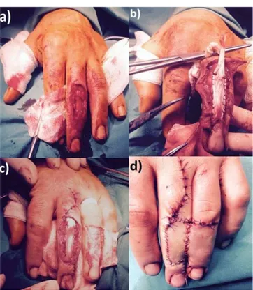
-sue defect at the Dd1, Dd2 and Dd3 defects of extensor tendon zones with tendon gap. In the physical examination, there was a wide, contaminated tissue and tendon defect at the dorsum of the 3rd inger. Neurovascular examination was normal in the afected hand (Figure 1a).
Under axillary block anesthesia, the wound was irrigated and the necrotic tissues were debrided meticulously. Ater the in
-traoperative detailed evaluation it was decided to perform reversed cross-inger lap with tendon reconstruction. Then m. palmaris longus tendon grat was harvested and used for recontructing the extensor mechanism of the 3rd inger (Fig
-ure 1b). The planned index inger lap was de-epithelialized and relected to cover the defect in the adjacent 3rd inger (Figure 1c). With careful dissection via the aid of magniiying loupes, the split thickness skin grat was created at the donor site. That was later used to cover the defect on the index inger. Then the dissected de-epithelialized lap covered the dorsal defect on the adjacent inger (Figure 1d). Ater the de-epithelialized lap was relected to cover the defect in the adjacent inger, the relected lap was covered with split thickness skin grat that had been removed earlier from the inguinal site. Then the donor site was covered with the dissected skin (Figure 1d).
The lap division was performed ater two weeks and the wounds were closed with retention sutures (Figure 2a,b). Ater the division, the patient was allowed early rehabilitation and encouraged to use a full range of motion. Through the follow-up period there was no dehiscence, infection, or complication re
-lated to lap survival. At the most recent follow-up, the range of motion was within normal limits without any extension gap at the afected site and the patients was happy with the esthetic
appearance of the hand (Figure 2c,d,e).
Discussion
Trauma associated with the exposure of digital deep structures can lead to signiicant functional loss, so, most of the time, prompt sot tissue coverage is provided. Skin or sot tissue de
-fects in the dorsal area of the inger oten constitute diiculties
for surgical procedures during treatment and the association
with deep structures becomes more challenging. At present there are many difering reconstruction procedures and it is oten diicult to ind an exact nomenclature to suit all cases. Various laps include: the homodigital subcutaneous lap [4], the proximally based axial digital artery transposition lap [5], the reverse digital artery lap [6], and the de-epithelialized inger lap [2]. Also, by the modiied techniques of reverse cross-inger laps [7], dorsal digital defects can be reconstructed. In this current study we used a de-epithelialized cross-inger lap that was irst described as a reversed dermis lap by Paki
-am [2] and modiied by Al-Qattan [7]. We let the subcutaneous fatty tissue on the dermis in which the dorsal vessels are situ -Figure 1. (a) Irrigated and meticulously debridated wound shows 3rd inger exten
-sor tendon zones Dd1, Dd2 and Dd3 defects with total tendon loss. (b, c) Harvest
-ed M. Palmaris Longus tendon grat was us-ed for reconstructing the 3rd inger extensor mechanism. (d) De-epithelialized and relected index inger reverse cross lap to cover the defect in the adjacent inger.
Figure 2. (a) Intraoperative view, before the division. (b) Ater two weeks from the division (c,d,e) A year ater the surgery, the range of motion was within normal limits without any extension gap at the afected site and the patients was happy with the esthetic appearance of the hand.
Journal of Clinical and Analytical Medicine | 263
ated. Ater 14 days, we separated the lap without any problem. Then, as early as the irst postoperative day ater separation, we allowed the patient to start rehabilitation to prevent exten
-sor tendon adhesion and skin grat contraction.
Although the need for another surgery for the separation of
the digits and inger stifness may be the major concern in terms of joint motion with this technique, it is an easy lap and an excellent alternative for achieving early coverage of cuta
-neous wounds at the dorsal aspect of the long ingers. Also, early rehabilitation can prevent extensor tendon adhesion and decrease stifness incidence [8]. At the most recent follow-up, the range of motion for both ingers were within normal limits without any extension gap at the afected site and the patients was happy with the esthetic appearance of the hand following
reconstruction.
In conclusion, the reversed cross-inger subcutaneous lap is an easy lap to perform and to cover defects on the dorsum of pha
-lanx and is an excellent option for coverage of wounds. Hand
therapy is crucial during the healing phase of a de-epithelialized
cross-inger lap and for the speedy recovery of all hand func -tions.
Competing interests
The authors declare that they have no competing interests.
References
1. Fejjal N, Belmir R, El Mazouz S, Gharib N, Abbassi A, Belmahi A. Reversed cross inger subcutaneous lap. A rapid way to cover inger defects. Indian J Plast Surg
2008;41(1):55-7.
2. Pakiam AI. The reversed dermis lap. Br J Plast Surg 1978;31:131–5. 3. Tark KC, Chung S, Shin KS, Lee YH. Rhomboid design for tubed inguinal lap in ingertip reconstruction. Ann Plast Surg 1996;36:354–9.
4. Voche P, Merle M. The homodigital subcutaneous lap for cover of dorsal inger defects. Br J Plast Surg 1994;47:435–9.
5. Fleegler EJ, Weinzweig N. The versatile axial pattern digital transposition lap. J Hand Surg 1988;13:494–500.
6. Lai CS, Lin SD, Chou CK, et al. A versatile method for reconstruction inger defects: reverse digital artery lap. Br J Plast Surg 1992;45:443–53.
7. Al-Qattan MM. Technical modiications and extended applications of the distally based adipofascial lap for dorsal digital defects. Ann Plast Surg 2004;52:168–73. 8. Shao X, Chen C, Zhang X, Yu Y, Ren D, Lu L. Coverage of ingertip defect using a dorsal island pedicle lap including both dorsal digital nerves. J Hand Surg Am
2009;34:1474-81.
How to cite this article:
Gürbüz K, Uzun E, Ozan F. Covering the Dorsal Finger Defect with Reverse Cross
Finger Flap. J Clin Anal Med 2016;7(2): 262-4.
| Journal of Clinical and Analytical Medicine 264