r
A
a
l
þ
a
t
n
ý
i
r
j
m
ir
a
O
O
h
r
c
i
r
g
a
in
e
a
s
e
R
l
Cengiz Çokluk, Enis Kuruoğlu, Abdullah Hilmi Marangoz, Keramettin Aydın Ondokuzmayıs University, Medical Faculty, Department of Neurosurgery, Samsun, Turkey Ulnar Sinir Sıkışmasında Ultrasonografik Nörografi / Ultrasonographic Neurography for Ulnary Nerve Entrapment
Prechannel Entrapment of the Ulnary Nerve by an
Intermuscular Arcade: The Importance of Ultrasonographic
Neurography in the Demonstrating of Compression Site
Ulnar Sinirin İntermusküler Bağ Tarafından Kanal
Öncesi Sıkıştırılması: Sıkışma Yerinin Gösterilmesinde
Ultrasonografik Nörografinin Önemi
DOI: 10.4328/JCAM.1354 Received: 25.10.2012 Accepted: 19.11.2012 Printed: 01.07.2014 J Clin Anal Med 2014;5(4): 291-3
Corresponding Author: Cengiz Çokluk, Ondokuzmayis University Medical Faculty Department of Neurosurgery, samsun, Turkey. T.: +90 3623121919/3086 F.: +90 3624576041 E-Mail: [email protected]
Özet
Amaç: Ultrasonografik nörografi tekniği periferik sinir yaralanmaları ve sıkış-ma nöropatilerinde daha önceden tanımlanmış bir yöntemdir. Periferik sinir-lerin sık görülen tuzak nöropatileri daha önceden belirtilmiştir. Gereç ve Yön-tem: Bu yazıda ulnar sinirin nadir görülen bir sıkışması ve illustrative bir ol-guda ultrasonografik nörografi bulguları sunulmaktadır. Olgu Beyin Cerrahi-si Kliniğine ulnar Cerrahi-sinir tuzak nöropatiCerrahi-si bulgularıyla başvurmuştur. Bulgular: Elektrofizyolojik muayene ulnar sinirin ulnar kanala girmeden önce sıkışma bulgularını vermiştir. Ameliyattan önce hasta ultrasonografik nörografi yön-temiyle incelenmiştir. Ultrasonografik nörografi incelemesi ulnar sinirin ulnar kanala girmeden 5 cm önce sıkışma bulgularını göstermiştir. Hasta ameliyat edilmiştir. Ameliyat bulguları ultrasonografik nörografiden elde olunan bul-gularla benzerlik göstermiştir. Tartışma: Bu yazıda klinik pratikte bu şekilde-ki patolojilerin gösterilmesinde ultrasonografik nörografinin rolü ile klinik ve ameliyat bulguları sunulmaktadır.
Anahtar Kelimeler
İntermusküler Bağ; Ulnar Sinir; Ultrasonografik Nörografi; Periferik Sinir Cer-rahisi; Tuzak Nöropatisi
Abstract
Aim: Ultrasonographic neurography is a described method in the cases with peripheral nerve injury and entrapment. The usual entrapment of the pe-ripheral nerves had been described previously. Material and Method: In this report it was presented the unusual entrapment of the ulnar nerve and the neurographic findings in an illustrative case. The case was admitted to our neurosurgery department with the symptoms of ulnar nerve entrapment. Re-sults: Electrophisiologic examination revealed that ulnary nerve entrapment before entering ulnary nerve canal. We examined the patient by using ultra-sound assisted neurographic examination before operation. Ultrasonograph-ic neurography demonstrated ulnary nerve compression 5 cm proximal por-tion from its entrance into the ulnary nerve canal. The patient was operated. Operative findings were similar with those of ultrasonographic neurography. Discussion: In this report we present clinical and operative findings of this case including the role of ultrasonographic neurography in the demonstrat-ing these type of pathologies in the clinical practice.
Keywords
Intermuscular Arcade; Ulnar Nerve; Ultrasonographic Neurography; Periph-eral Nerve Surgery; Entrapment Neuropathy
Ulnar Sinir Sıkışmasında Ultrasonografik Nörografi / Ultrasonographic Neurography for Ulnary Nerve Entrapment
2
Introduction
Neurological findings and electrodiagnostic tests are the gold
standards in the diagnosing of peripheral nerve entrapments
and making decision about the selection of treatment
modal-ity [1-3]. Intraoperative ultrasound has been used in the
neu-rosurgical procedures, especially in the localising of cystic and
solid lesions, for several years. Some previous studies had been
conducted to evaluate peripheral nerves by ultrasound [4-8]. In
these studies, the authors described normal and pathologic
ul-trasonographic appearance of peripheral nerves [4-8].
This clinical study was designed to evaluate the feasibility of
presurgical and intraoperative ultrasound assisted
neuroexami-nation in the localisation of entrapped field, determineuroexami-nation of
the type of the entrapment in a patient with entrapment
neu-ropaty of ulnary nerve by an intermuscular arcade.
Material and Method
55 year-old man had beed referred to our neurosurgery
depart-ment with the semptoms of ulnary nerve entrapdepart-ment.
Electro-phisiologic examination showed the ulnary nerve entrapment
before entering its canal. We examined the patient by using
ultrasound assisted neurographic examination before operation
(Figure 1). Ultrasonographic neurography demonstrated that
the ulnary nerve compressed at the point of 5 cm proximal to its
entrance into the ulnar canal (Figure 1). The patient was
operat-ed. Operative findings showed the presence of intermuscular
ar-cade compressed to ulnary nerve (Figure 2). Operative findings
were similar with those of ultrasonographic neurography. The
arcade was cut off by using a scissor. The chronical impression
site on the nerve was inspected by using operative microscope
after removing of intermusculary arcade (Figure 3).
Postopera-tive period was uneventful. The patient was discharged after 3
days from the operation.
Discussion
Ulnar neuropathy at the elbow is the second most common
en-trapment neuropathy of the upper limb, next to carpal tunnel
syndrome. The ulnar nerve entrapment is most frequently seen
in the region of the retroepicondylar (ulnar) groove resulting
from various pathological processes. Anatomically, the ulnary
nerve travels from the axilla after emerging from the brachial
plexus into the medial aspect of the anterior compartment of
the arm. It then traverses the intermuscular
(intercompartmen-tal) septum, passes through the arcade of Struthers, and
con-tinues its path in the posterior aspect of the arm down to the
elbow and submerges into the cubital tunnel posterior to the
medial epicondyle [9]. The ulnary nerve may become entrapped
at the arcade or accross the intermuscular septum. Sir John
Struthrs, a whale anatomist, described the arcade in Scotland
in 1854 [9].
Electrodiagnostic tests can precisely distinguish the severity of
entrapment. Sometimes electrodiagnostic tests may not yield
reliable information about the location of the entrapment site,
because a variety of parameters may affect this examination
[4]. In the same way, electrodiagnostic tests can not give any
information about the position of the nerve, the presence of
a neuroma and excessive scar tissue arount the nerve. In this
perspective, we need an image guided tool preoperative and
intraoperative period in the surgical treatment of entrapment
Figure 1. Ultrasonographic neurography shows ulnary nerve and compression site(UN: Ulnary nerve, Arrows show the compression site).
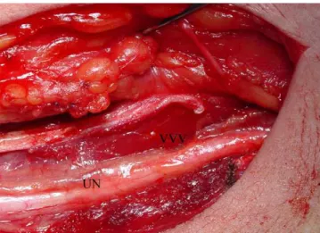
Figure 2. Intraoperative photo shows intermuscular arcade and ulnary nerve (IMA: Intermuscular arcade, UN: Ulnary nerve).
Figure 3. The appearance of compression site in the nerve (Arrows shows the compression site, UN: Ulnary nerve).
| Journal of Clinical and Analytical Medicine
292
Ulnar Sinir Sıkışmasında Ultrasonografik Nörografi / Ultrasonographic Neurography for Ulnary Nerve Entrapment
3
neuropathies for precice localisation of entrapped site and
de-termination of the type lesion. We theorized that sonographic
neuroexamination may help us in the localization and type of the
lesion. The electrodiagnostic test revealed that the entrapped
site was located proximal of the ulnar canal. Sonographic
neu-roexamination showed the precise location of the entrapment.
Our case demonstrated that ultrasonographic
neuroexamina-tion is a useful diagnostic test in the making-decision of the
treatment of ulnary nerve neuropaty. Sonographic
neuroexami-nation may also be used as an image guided tool during the
surgical treatment of peripheral nerve entrapment neuropathy.
Competing interests
The authors declare that they have no competing interests.
References
1. Gentili F, Hudson AR, Midha R. Peripheral nerve injuries: Types, causes, and grad-ing. In: Wilkins RH, Rengachary SS (eds). Neurosurgery. Vol 3. New York: McGraw-Hill, 1996: 3105-14.
2. Kline DG. Surgical repair of peripheral nerve injury. Muscle Nerve 1990; 13(9):843-52.
3. Kline DG, Hudson AR. Acute injuries of peripheral nerves. In: Youmans JR (ed) Neurological surgery Vol 4. Philadelphia: W. B. Saunders Company, 1990:p.2423-510.
4. Peer S, Bodner G, Meirer R, Willeit J, Piza-Katzer H. Examination of postoperative peripheral nerve lesions with high-resolution sonography. AJR 2001;177(2):415-9. 5. Martinoli C, Bianchi S, Dahmane M, Pugliese F, Bianchi-Zamorani MP, Valle M. Ultrasound of tendons and nerves. Eur Radiol 2002;12(1):44-55.
6. Martinoli C, Bianchi S, Giovagnorio F, Pugliese F. Ultrasound of the elbow. Skel-etal Radiol 2001;30(11):605-14.
7. Martinoli C, Serafini G, Bianchi S, Bertolotto M, Gandolfo N, Derchi LE. Ultraso-nography of peripheral nerves. J Peripher Nerv Syst 1996; 1(3):169-78. 8. Peer S, Kovacs P, Harpf C, Bodner G. High-resolution sonography of lower ex-tremity peripheral nerves. Anatomic correlation and spectrum of disease. J Ultra-sound Med 2002;21(3):315-22.
9. von Schroeder HP, Scheker LR. Redefining the arcade of Struthers. J Hand Surg 2003;28(6):1018-21.
Journal of Clinical and Analytical Medicine |