DOI: 10.1590/0004-282X20160010
ARTICLE
Clinical and neurophysiological features of
the hereditary neuropathy with liability to
pressure palsy due to the 17p11.2 deletion
Aspectos clínicos e neurofisiológicos de pacientes com a neuropatia hereditária com a
susceptibilidade à pressão associada à deleção 17p11.2
Aline Pinheiro Martins de Oliveira, Raquel Campos Pereira, Patrícia Toscano Onofre, Vanessa Daccach Marques, Gilberto Brown de Andrade, Amilton Antunes Barreira, Wilson Marques Junior
he hereditary neuropathy with liability to pressure palsies (HNPP) is an autossomal dominant disorder characterized by recurrent sensory and motor mononeuropathies that tend to occur at entrapment sites, although occasional patients present a generalized neuropathy1,2,3. Most cases of HNPP
are associated to PMP22 gene deletion4
. Point mutations in the same gene occasionally cause HNPPand at least 26 small mutations have been found5
. he same region is duplicated in
Charcot–Marie–Tooth disease type 1A (CMT1A)6, the most
frequent inherited neuropathy.
In contrast to CMT1A, in which nerve conduction slowing is uniform along the entire nerve length5,7,8,9, HNPP is characterized
by multifocal or segmental conduction abnormalities1,10,11,12,13,
being necessary a correct distinction from other acquired and treatable neuropathies. Diagnosis of HNPP is also important for
correct prognostic evaluation and genetic counseling.
Universidade de São Paulo, Faculdade de Medicina de Ribeirão Preto, Departamento de Neurociências e Ciências do Comportamento, Ribeirao Preto SP, Brazil.
Correspondence: Wilson Marques Jr; Departamento de Neurociências e Ciências do Comportamento, Faculdade de Medicina de Ribeirão Preto / USP; Avenida Bandeirantes, 3900; Campus da USP; 14049-900 Ribeirão Preto SP, Brasil; E-mail: [email protected]
Conflict of interest: There is no conlict of interest to declare.
Support: We wish to thank all staff of the Clinical Neurophysiology Unit and of the Neurogenetic Laboratory for their valuable support. Received 22 April 2015; Received in inal form 12 September 2015; Accepted 01 October 2015.
ABSTRACT
The hereditary neuropathy with liability to pressure palsies (HNPP) is an autossomal dominant disorder manifesting recurrent mononeuropathies. Objective: Evaluate its clinical and nerve conduction studies (NCS) characteristics, searching for diagnostic particularities. Method: We reviewed the neurological manifestations of 39 and the NCS of 33 patients. Results: Family history was absent in 16/39 (41%). The onset complaints were weakness in 24, pain in 6, sensory deicit in 5 and paresthesias in 4. Pain was seen in 3 other patients. The following neuropathy patterns were found: multiple mononeuropathy (26), mononeuropathy (7), chronic sensorimotor polyneuropathy (4), chronic sensory polyneuropathy (1) and unilateral brachial plexopathy (1). NCS showed a sensorimotor neuropathy with focal conduction slowing in 31, two had mononeuropathy and another brachial plexopathy. Conclusion: HNPP presentation is variable and may include pain. The most frequent pattern is of an asymmetrical sensory and motor neuropathy with focal slowing at speciic topographies on NCS.
Keywords: hereditary neuropathy with liability to pressure palsies, peripheral neuropathy, nerve conduction study, pain.
RESUMO
A neuropatia hereditária com susceptibilidade à pressão (HNPP) é uma doença autossômica dominante que manifesta mononeuropatias recorrentes. Objetivo: Avaliar as características clínicas e os estudos da condução nervosa (ECN) procurando particularidades diagnósticas. Método: Revisamos as características clínicas de 39 e os ECN de 33 pacientes. Resultados: História familiar ausente em 16/39 (41%). As manifestações iniciais foram: fraqueza em 24, dor em 6, déicit sensitivo em 5 e parestesias em 4. Dor foi referida por outros 3 pacientes. Os seguintes padrões de neuropatia foram observados: mononeuropatia múltipla (26), mononeuropatia (6), polineuropatia sensitivo-motora (4), polineuropatia sensitiva (1) e plexopatia braquial unilateral (1). Os ECN mostraram uma neuropatia sensitivo-motora com redução focal da velocidade de condução em 31, dois tinham mononeuropatia e outro plexopatia braquial. Conclusão: A apresentação da HNPP é variável e pode incluir dor. O padrão mais frequente é o de uma neuropatia sensitivo-motora assimétrica com alentecimentos focais da condução em topograias especíicas nos ECN.
Sporadic cases are sometimes found3,14. hey may result
from de novo mutations3, incomplete family history, adop
-tion, false paternity and no recognition of minor manifesta
-tions as signs of neuropathy.
he classic phenotype is the occurrence of acute or sub
-acute painless mononeuropathy after minor trauma2,14.
Patients frequently complaint of paresthesias after a short period of time in positions that result in nerve compression. he prognosis is relatively benign and most mononeuropa
-thies resolve spontaneously. Severe injuries are avoided if the patient learns to protect his nerves15.
Atypical presentations as chronic sensory-motor polyneu
-ropathies have been described in young3, adults1,2 and seniors11
. Around 30% of the patients have chronic symptoms such as cramps, paresthesias and myalgia induced by exercise11
. Pain was considered rare2,16, but recent studies described pain in pa
-tients with upper limbs mononeuropathies17,18 and occasion
-ally neuropathic pain may be found17. Some patients present
musculoskeletal pain that meet the ibromyalgia criteria, po
-tentially delaying diagnosis19.
here is no consensus for the neurophysiology of HNPP. Verhagen et al.20 proposed that the most discriminating ind
-ings (99% accuracy) were slowing of conduction velocity (CV) of the ibular nerve from the ibula head to the ankle, slowing of the ulnar nerve conduction at the elbow and prolongation of the distal motor latency (DML) of at least one of the ibular nerves. Mouton et al.2 and Gouider et al.15
found that after 15 years of age all patients present prolonged DML and reduction of the wrist sensory CV of the median nerve and prolongation of the DML or reduction of the CV of at least one of the ibular nerves. Cruz-Martinez et al.21 found that the DML of the median nerve,
the sensory CV of the median nerve at the wirst and the sensory CV of the sural nerves were abnormal in all afected individuals21.
Guidelines to the diagnosis of HNPP were proposed by Dubourg et al.16: a) bilateral increase in DML of the median
nerves associated with reduced CV in median sensory nerve at the palm-wrist segment; b) at least one of the ibular nerves should have increased DML or reduced CV; c) CV of the ul
-nar nerve at the elbow segment is frequently reduced; d) CV in the lower limb nerves may be moderately reduced and e) SNAP amplitudes may be reduced, mostly in the upper limbs. Infante et al.3 proposed that the neurophysiological evalua
-tion of patients suspected of having HNPP should include the motor conduction in, at least, two anatomical sites of nerve compression, particularly the ulnar nerve at the elbow and the study of the sural nerve3.
Li et al.12 showed that there is a unique electrophysiological
pattern in HNPP, characterized by severe distal slowing in some nerves, multifocal conduction slowing at the sites of entrapment and mildly reduced CV in other segments. Distal sensory con
-duction velocities are usually difusely abnormal. hese indings were consistent with those of some previous reports1,2,3,10,11,22
.
he distal slowing was more frequent in median and ibular nerves, more liable to pressure palsies and repetitive trauma.
Interestingly, the distal latencies to more proximal muscles did not present slowing, a strong argument against HNPP being a distal myelinopathy as suggested by others1,22.
Luigetti et al.23 proposed that HNPP should be suspect
-ed in patients with a demyelinating neuropathy associat-ed to carpal tunnel syndrome, plus another motor conduction abnormality (CV of the ulnar nerve at the elbow, DML of the ulnar nerve, CV of the ibular nerve) plus a sensory nerve conduction abnormality in a nerve not prone to compression (sural or radial nerves). Following this criteria over a 20-year period, they genetically conirmed the diagnosis of HNPP in more than 70% of the suspected cases.
In order to shed more light about the clinical and the elec
-trophysiologic patterns of HNPP we analyzed a group of our patients with the 17p11.2 deletion.
METHOD
We have included patients with HNPP carrying the 17p11.2-12 deletion regularly followed at our institution. hose with other causes of neuropathy, including alcoholism, mal
-nutrition, vitamin B12 deiciency and systemic diseases, as diabetes and other endocrinopathies, were not considered. heir records were evaluated retrospectively. Ethics approval was obtained from the Ethics Committee at the HCFMRP-USP.
he patients were evaluated with special emphasis on the neuromuscular system. Electromyography (EMG) evalua
-tion was performed in 33 patients, using surface stimula-tion and recording for the motor conduction studies and surface stimulation and needle recording electrodes for sensory con
-duction studies. Compound muscle action potentials (CMAP) were recorded from median, ulnar, ibular and posterior tibial nerves. he following characteristics were studied: amplitude, motor conduction velocity (MCV), distal motor latency (DML) and minimal F-wave latency. he following segments were studied: a) median nerve: wrist to antecubital fossa and an
-tecubital fossa to axilla; b) ulnar nerve: wrist to below elbow, below to above elbow, above elbow to axilla; c) ibular nerve: ankle to below ibular head and below to above ibular head; and d) posterior tibial nerve: ankle to popliteal fossa.
Conduction block (CB) was deined as a 50% or greater decrease in negative peak amplitude and area of the proximal CMAP. Focal slowing at a compression site was considered present when a decrement ≥ 10 m/s was identiied. Compound sensory nerve action potentials (SNAP) were recorded ortho
-dromically from median (index inger-wrist and palm-wrist) and ulnar (ifth inger-wrist and palm-wrist) nerves and an
-tidromically from radial (forearm-base of the thumb), sural (midcalf-lateral malleolus) and supericial ibular (leg-ankle) nerves. he following parameters were analyzed: SNAP ampli
-tude, sensory CV (SNCV), duration and morphology.
institution. Undetected potentials were generally classiied as abnormal. Statistical analysis was carried on with the SPSS Statistic version 17.0 software (SPSS Inc.). Means and per
-cents were compared using the Student’s t-test or Q-square test, and diferences were considered signiicant at p ≤ 0.05.
RESULTS
hirty-nine patients (21 men and 18 women), including a mother and daughter, fulilled our inclusion criteria. Family history was absent in 16/39 (41%). he mean age of disease onset was 24 years (5 to 76 years), while the mean age of the irst evaluation was 32 years (6 to 77 years). he time of onset to evaluation was on average 8.2 years.
he irst spontaneous complaints were localized muscu
-lar weakness in 24 patients (61%), pain in 6 patients (15%), sensory loss in 5 (13%) and paresthesias in 4 (10%) (Table 1). At least one episode of acute paralysis of a speciic nerve was reported by 18 patients (46%). 28 episodes of paralysis were reported, 19 in common ibular nerve, 6 in radial and ulnar nerves, respectively, and 2 in the axillary nerve. Position re
-lated sensory symptoms were reported by 12 patients (31%) and a history of compression or precipitant factors preceding the paralysis in 19 patients (48%), including playing volleyball or soccer, climbing stairs, driving vehicles, prolonged time crouched, physical efort, waking up, horse fall.
Painless symptoms were reported by 30 patients (77%). In addition to the 6 patients that complained of pain as the initial symptom, other 3 patients reported pain at some point
of their disease, triggered by physical efort (2 patients) and without a precipitating factor (1 patient). Of the 6 patients that reported pain as initial symptom, 3 presented a slowly progressive painful neuropathy predominantly afecting the lower limbs in the set of a chronic sensorimotor polyneurop
-athy on the neurological examination and 3 manifested pain in the set of a multiple mononeuropathy (1 patient with slow
-ly progressive pain in the territory of ulnar nerve; 1 patient with slowly progressive pain in the left lower limb; 1 patient with pain in the lower limbs triggered by the maintenance of a position for a long period of time). Pain was referred by both, adults (7 patients) and teens (14 and 16 years old).
Pes cavus was present in 5 patients (13%), ankle jerks were absent in 8 (20%) and distal atrophy was seen in 8 (20%). Two of the patients with atrophy had a sensorimotor polyneuropathy. In the remaining patients with muscle atro
-phy there was a nerve-selective distribution, most of times involving ulnar and/or median and/or ibular nerves. Nerve thickening was rare (4 patients-10%).
None of our patients had peripheral facial nerve pal
-sy. One patient presented a transient involvement of the trigeminal nerve and other one an episodic involvement of the eyelid branch of the oculomotor nerve.
On neurological evaluation, 26 patients (66.7%) had a mul
-tiple mononeuropathy pattern, 7 (17.9%) presented an isolat
-ed mononeuropathy, 4 (10.2%) had a chronic sensorimotor polyneuropathy, 1 (2.6%) developed a predominant sensory polyneuropathy and 1 (2.6%) patient developed a unilateral brachial plexopathy.
he most common clinical course was intermittent, comprising a succession of acute attacks with complete or partial recovery (31 patients - 79.5%). hese patients had recurrent mononeuropathies, often from diferent nerves. Progressive evolution, with no obvious luctuations, was observed in 8 patients (20.5%), whose neuropathy pattern included multiple mononeuropathy, chronic sensorimotor polyneuropathy or purely sensory polyneuropathy on neu
-rologic examination (Table 2).
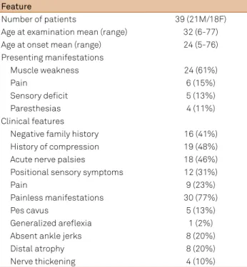
Table 1. Clinical indings in 39 hereditary neuropathy with liability to pressure palsies patients with the PMP22 deletion.
Feature
Number of patients 39 (21M/18F)
Age at examination mean (range) 32 (6-77)
Age at onset mean (range) 24 (5-76)
Presenting manifestations
Muscle weakness 24 (61%)
Pain 6 (15%)
Sensory deicit 5 (13%)
Paresthesias 4 (11%)
Clinical features
Negative family history 16 (41%)
History of compression 19 (48%)
Acute nerve palsies 18 (46%)
Positional sensory symptoms 12 (31%)
Pain 9 (23%)
Painless manifestations 30 (77%)
Pes cavus 5 (13%)
Generalized arelexia 1 (2%)
Absent ankle jerks 8 (20%)
Distal atrophy 8 (20%)
Nerve thickening 4 (10%)
F: female; M: male.
Table 2. Clinical and electrophysiological patterns.
Clinical pattern N (%)
Multiple mononeuropathy 26 (66.7)
Isolated mononeuropathy 7 (17.9)
Sensorimotor polyneuropathy 4 (10.2)
Sensory polyneuropathy 1 (2.6)
Unilateral brachial plexopathy 1 (2.6)
Total 39 (100)
Electrophysiological pattern N (%)
ASMNFS 30 (91.0)
Single mononeuropathy 2 (6.0)
Brachial plexus + ASMNFS 1 (3.0)
Total 33 (100)
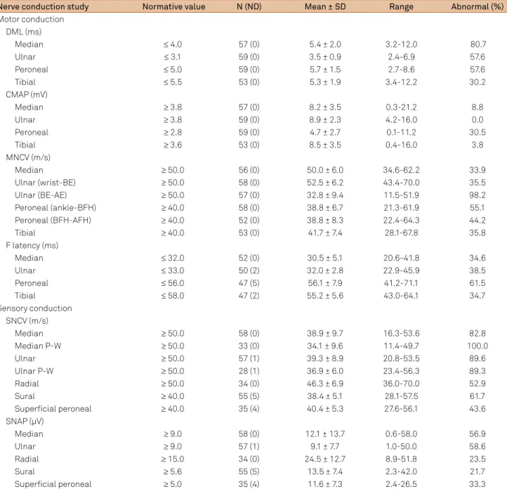
Nerve conduction studies were performed on 253 motor and 237 sensory nerves. Six patients underwent EMG exami
-nation in another hospital. hey have not been included in this study. Despite the clinical presentation, nerve conduction stud
-ies showed a pattern of sensory and motor neuropathy with focal conduction slowing in 31 patients, including a patient with unilat
-eral brachial plexopathy and focal conduction slowing (Table 2). Two patients had isolated mononeuropathy (a 13-year-old boy with radial neuropathy with persistent CB at the arm segment and one 6 years-old boy with deep ibular neuropathy).
he most afected nerves in our patients were: motor ul
-nar nerve (CV slowing at the elbow segment in 98.2% of the nerves), sensory ulnar nerve (CV slowing at the inger-wrist segment in 89.6%), deep ibular nerve (at least one parameter of NCS abnormal in 83.0% of the nerves, mainly DML or F wave latency or CV), sensory median nerve (CV slowing at the inger-wrist segment in 82.8% and at the palm-wrist seg
-ment in 100%); motor median nerve (DML prolonged out of proportion to the reduction in forearm CV in 80% of the nerves), sural nerve (CV slowing in 61,7%) and supericial radial nerve (CV reduction in 52.9%). (Table 3).
he DML of the median nerve was more frequently pro
-longed than the DML of the ulnar nerve (p = 0.03), ibular nerve (p = 0.03) and tibial nerve (p < 0.001). DML of the ul
-nar and ibular nerves were signiicantly more frequently prolonged than those of the tibial nerve (p = 0.002).
Temporal dispersion was observed in SNAP of median (4 times), ulnar (once) and sural nerves (once), and in CMAP of the median and ulnar nerves (once), tibial posterior and ibular nerves (4 times) inside and outside entrapment sites. CB of the ulnar nerve at the elbow segment was observed 7 times, once in the ibular nerve at the ibula neck and once at the leg segment and once in the radial motor nerve at spiral groove. his last patient had a persistent CB at this site.
DISCUSSION
he characteristics of the population with HNPP we stud
-ied seem to be similar to other stud-ied populations2,11,14,15,23.
In most patients, disease onset occurred in the irst three de
-cades of life, although the extremes were quite large, ranging from 5 to 76 years old in our patients. Additionally, the pro
-portion between males and females approach to 1, as was seen in a population of Brazilian patients with CMT1A that we have studied previously9. Some authors however found a
male predominance (male/female = 4:3) and a signiicantly earlier onset in men2.
Family history was positive in only 23 cases (59%). In previ
-ous studies the percentage of sporadic cases were variable3,14.
his inding probably relects the wide range of the clinical manifestations, that may be very mild or even absent3,10,15 and
has important clinical implications as family history very fre
-quently is not a clue to the inal diagnosis.
In 24 of the 39 patients (61.5%), the irst clinical manifes
-tation was painless muscle weakness and at least one epi
-sode of acute nerve paralysis was reported by 18 patients. his is the classic phenotype: a painless acute or subacute mononeuropathy2,14. Among the 18 patients with acute pa
-ralysis, 12 reported some precipitating factor, what highlights the importance of environmental factors for development of the clinical manifestations. Six patients reported pain as the initial manifestation. In only one patient pain was associated to an episode of acute nerve paralysis. In the remaining 5 pa
-tients pain heralded a chronic neuropathy. Another 5 pa-tients reported numbness and another 4 mentioned paresthesias as the initial symptoms.
Apart from the 6 patients who reported pain as the ini
-tial symptom, other 3 patients began the clinical picture with subacute or chronic muscle weakness but reported pain at some point of their evolution. he pain manifested in these cases was neuropathic, either focal associated to ulnar neu
-ropathy, or afecting the lower limbs without a recognized precipitating factor.
Pain is rarely reported in HNPP2,16 and is considered a
very uncommon in the acute episodes of nerve paralysis2,
but may be the initial manifestation of the disease17 or a
chronic component of this neuropathy18,19. Interestingly, it
has been recently described a HNPP family due to a point mutation, whose main manifestations were pain and pares
-thesias, without recurrent palsies18. Ours observations cor
-roborates the importance of considering HNPP in patients presenting pain, even in younger ages, as has happened in our population.
Cranial nerve involvement was rare in our population. We have seen a patient with paresthesias in the territory of the trigeminal nerve and another with unilateral transient paral
-ysis of the eyelid branch of the oculomotor nerve. It seems that this is the pattern in most studies2,20,21,24. Interestingly,
we have previously described a HNPP patient that developed dysphagia25. Other rare manifestations in our patients were
pes cavus and nerve thickening, as seems to be the case in
most studies3,15. Clinically, most patients of our patients pre
-sented a pattern of multiple mononeuropathy or mononeu
-ropathy as seems to occur in most series1,2,3,13,14. However, 4
patients presented a chronic sensorimotor polyneuropathy and one patient had a pure sensory polyneuropathy, both patterns are very rare in HNPP3,11,23.
In our study, one patient had unilateral clinical and neu
-rophysiological impairment of the upper trunk of the brachial plexus. his is a very uncommonly referred presentation that is more frequent in women2 and that should be diferentiated
from acute brachial neuritis26 and other plexopathies that are
usually painful27.
On nerve conduction studies,most of our patients (94.0%) presented an EMG pattern of an asymmetric sensorimotor neuropathy with focal slowing of nerve conduction, suggest
clinically had a plexopathy and 5 of our patients with mono
-neuropathy presented this pattern on neurophysiology, that seems to be the rule in the literature1,2,3,12,13. Very occasional
-ly, NCS shows only a mononeuropathy2,21,23. he patient that
showed clinically a sensory polyneuropathy also presented in NCS an asymmetric demyelinating sensory and motor neu
-ropathy with focal slowing of the ulnar nerves at the elbow segment and temporal dispersion of the right ibular and tib
-ial nerves at the leg segment.
In our patients, sensory CV slowing was a common fea
-ture, being more frequent in the ulnar, median, sural and radial SNAP. In addition, abnormalities in sensory CV were more frequent than those of motor CV outside the sites of
compression as previously described3,12. It should be stated,
however, that the SNAP usually are evaluated at the proximal regions due to technical diiculties, including physiological temporal dispersion and phase cancelation.
On motor conduction studies, DML was proportionate
-ly more afected than CMAP amplitudes and CV outside of sites of compression and also than F-wave latency, as previ
-ously described1,12,13. he most afected DML were those of
the median, ulnar and deep ibular nerves, specially that of the median nerve, specially prone to pressure palsies and/or repetitive trauma12,13. On entrapment sites, almost half of the
patients (19 patients, 48%) referred some history of com
-pression or precipitant factor prior the beginning of the
Table 3. Nerve conduction indings of 33 patients with hereditary neuropathy with liability to pressure palsies.
Nerve conduction study Normative value N (ND) Mean ± SD Range Abnormal (%)
Motor conduction DML (ms)
Median ≤ 4.0 57 (0) 5.4 ± 2.0 3.2-12.0 80.7
Ulnar ≤ 3.1 59 (0) 3.5 ± 0.9 2.4-6.9 57.6
Peroneal ≤ 5.0 59 (0) 5.7 ± 1.5 2.7-8.6 57.6
Tibial ≤ 5.5 53 (0) 5.3 ± 1.9 3.4-12.2 30.2
CMAP (mV)
Median ≥ 3.8 57 (0) 8.2 ± 3.5 0.3-21.2 8.8
Ulnar ≥ 3.8 59 (0) 8.9 ± 2.3 4.2-16.0 0.0
Peroneal ≥ 2.8 59 (0) 4.7 ± 2.7 0.1-11.2 30.5
Tibial ≥ 3.6 53 (0) 8.5 ± 3.5 0.4-16.0 3.8
MNCV (m/s)
Median ≥ 50.0 56 (0) 50.0 ± 6.0 34.6-62.2 33.9
Ulnar (wrist-BE) ≥ 50.0 58 (0) 52.5 ± 6.2 43.4-70.0 35.5
Ulnar (BE-AE) ≥ 50.0 57 (0) 32.8 ± 9.4 11.5-51.9 98.2
Peroneal (ankle-BFH) ≥ 40.0 58 (0) 38.8 ± 6.7 21.3-61.9 55.1
Peroneal (BFH-AFH) ≥ 40.0 52 (0) 38.8 ± 8.3 22.4-64.3 44.2
Tibial ≥ 40.0 53 (0) 41.7 ± 7.4 28.1-67.8 35.8
F latency (ms)
Median ≤ 32.0 52 (0) 30.5 ± 5.1 20.6-41.8 34.6
Ulnar ≤ 33.0 50 (2) 32.0 ± 2.8 22.9-45.9 38.5
Peroneal ≤ 56.0 47 (5) 56.1 ± 7.9 41.2-71.1 61.5
Tibial ≤ 58.0 47 (2) 55.2 ± 5.6 43.0-64.1 34.7
Sensory conduction SNCV (m/s)
Median ≥ 50.0 58 (0) 38.9 ± 9.7 16.3-53.6 82.8
Median P-W ≥ 50.0 33 (0) 34.1 ± 9.6 11.4-49.7 100.0
Ulnar ≥ 50.0 57 (1) 39.3 ± 8.9 20.8-53.5 89.6
Ulnar P-W ≥ 50.0 28 (1) 36.9 ± 6.0 23.4-56.3 89.3
Radial ≥ 50.0 34 (0) 46.3 ± 6.9 36.0-70.0 52.9
Sural ≥ 40.0 55 (5) 38.4 ± 5.1 28.1-57.5 61.7
Supericial peroneal ≥ 40.0 35 (4) 40.4 ± 5.3 27.6-56.1 43.6
SNAP (μV)
Median ≥ 9.0 58 (0) 12.1 ± 13.7 0.6-58.0 56.9
Ulnar ≥ 9.0 57 (1) 9.1 ± 7.7 1.0-50.0 58.6
Radial ≥ 15.0 34 (0) 24.5 ± 12.7 8.9-51.8 23.5
Sural ≥ 5.6 55 (5) 13.5 ± 7.4 2.3-42.0 21.7
Supericial peroneal ≥ 5.0 35 (4) 11.6 ± 7.3 2.4-26.5 33.3
symptoms, mainly in the ibular, ulnar and radial nerves, and less in the posterior tibial.
SNAP and CMAP amplitude reduction was not as fre
-quent as CV abnormalities. Median and ulnar SNAP and deep ibular nerves CMAP were the most afected, as happened in other studies3,20,22.
Temporal dispersion was seen in sensory (median, ulnar and sural) and motor nerves (median, ulnar, posterior tibial and ibular) in segments susceptible to compression and also not susceptible segments. CB were observed in ulnar, ibular and radial nerves, being more frequent in the ulnar nerve at the elbow segment (7/57-12%). he frequencies of CB de
-scribed in previous studies vary greatly, from 6 to 22%28.
In summary our study showed that the classical clinical pre
-sentation of HNPP is the most frequent, but alternative presen
-tations occur, including mononeuropathies, polyneuropathies and painful neuropathies. Pain should not exclude HNPP diag
-nosis. he electrophysiological features are much more homo
-geneous, characterized by a sensory and motor demyelinating multiple mononeuropathy with focal slowing of CV. he most frequent abnormalities on NCS were: prolonged DML of the me
-dian and ulnar nerves; CV slowing of ulnar motor nerve CMAP at the elbow segment; prolonged distal latency, reduced CV and prolonged minimal F-wave latency of the deep ibular nerve; re
-duced amplitude and CV of the median and ulnar SNAP; and re
-duced CV of the sural and supericial radial nerves.
References
1. Amato AA, Gronseth GS, Callerame KJ, Kagan-Hallet KS, Bryan WW, Barohn RJ. Tomaculous neuropathy: a clinical and electrophysiological study in patients with and without 1.5Mb deletions in chromosome 17p11.2. Muscle Nerve. 1996;19(1):16-22. doi:10.1002/(SICI)1097-4598(199601)19:1<16::AID-MUS3>3.0.CO;2-B
2. Mouton P, Tardieu S, Gouider R, Birouk N, Maisonobe T, Dubourg O et al. Spectrum of clinical and eletrophysiologic features in HNPP patients with the 17 p11.2 deletion. Neurology. 1999;52(7):1440-6. doi:10.1212/WNL.52.7.1440
3. Infante J, Garcia A, Combarroso O, Mateio JI, Berciano J, Sedano MJ et al. Diagnostic strategy for familial and sporadic cases of neuropathy associated with 17p11.2 deletion. Muscle Nerve. 2001;24(9):1149-55. doi:10.1002/mus.1126
4. Chance PF, Alderson MK, Leppig KA, Lensch MW, Matsunami N, Smith B et al. DNA deletion associated with hereditary neuropathy with liability to pressure palsies. Cell. 1993;72(1):143-51. doi:10.1016/0092-8674(93)90058-X
5. Li J, Parker B, Martyn C, Natarajan C, Guo J. The PMP-22 gene and its related diseases. Mol Neurobiol. 2013;47(2):673-98. doi:10.1007/s12035-012-8370-x
6. Lupski JR, Oca-Luna RM, Slaugenhaupt S, Pentao L, Guzzetta V, Trask BJ et al. DNA duplication associated with Charcot-Marie-Tooth disease type 1A. Cell. 1991;66(2):219-32. doi:10.1016/0092-8674(91)90613-4
7. Li J, Krajewski K, Lewis RA, Shy ME. Loss-of-function phenotype of hereditary neuropathy with liability to pressure palsies. Muscle Nerve. 2004;29(2):205-10. doi:10.1002/mus.10521
8. Lewis RA, Sumner AJ, Shy M. Electrophysiological features of inherited demyelinating neuropathies: reappraisal in the era of molecular diagnosis. Muscle Nerve. 2000;23(10):1472-87. doi:10.1002/1097-4598(200010)23:10<1472::AID-MUS3>3.0.CO;2-#
9. Marques W Jr, Freitas MR, Nascimento OJM, Oliveira AB, Calia L, Melo A et al. 17p duplicated Charcot-Marie-Tooth 1A. Characteristics of a new population. J Neurol. 2005;252(8):972-9. doi:10.1007/s00415-005-0797-9
10. Uncini A, Guglielmo G, Muzio A, Gambi D, Sabatelli M, Mignogna T et al. Differential electrophysiological features of neuropathies associated with 17p11.2 deletion and duplication. Muscle Nerve. 1995;18(6):628-35. doi:10.1002/mus.880180610
11. Pareyson D, Scaioli S, Taroni F, Botti S, Lorenzetti D, Solari A et al. Phenotypic heterogeneity in hereditary neuropathy with liability to pressure palsies associated with chromosome 17p11.2-12 deletion. Neurology. 1996;46(4):1133-7.
doi:10.1212/WNL.46.4.1133
12. Li J, Krajewski K, Shy ME, Lewis RA. Hereditary neuropathy with liability to pressure palsy. The electrophysiology its the name. Neurology. 2002;58(12):1769-73. doi:10.1212/WNL.58.12.1769
13. Hong YH, Kim M, Kim HJ, Sung JJ, Kim SH, Lee KW. Clinical and electrophysiologic features of patients with 17p11.2 deletion. Acta Neurol Scand. 2003;108(5):352-8. doi:10.1034/j.1600-0404.2003.00132.x
14. Lenssen P. Hereditary neuropathy with liability to pressure palsy: phenotypic differences between patients with the common deletion and a PMP22 frame shift mutation. Brain. 1998;121(8):1451-8. doi:10.1093/brain/121.8.1451
15. Gouider R, LeGuern E, Gugenheim M, Tardieu S, Maisonobe T, Leger JM et al. Clinical, eletrophysiologic and molecular correlations in 13 families with hereditary neuropathy with liability to pressure palsies and a cromossome 17p11.2 deletion. Neurology. 1995;45(11):2018-23. doi:10.1212/WNL.45.11.2018
16. Dubourg O, Mouton P, Brice A, LeGuern E, Bouche P. Guidelines for diagnosis of hereditary neuropathy with liability to pressure palsies. Neuromuscular Disord. 2000;10(3):206-8. doi:10.1016/S0960-8966(99)00103-0
17. Nogués M, Barroso F, Rivero A, et al. Pain: unusual presentation of hereditary neuropathy with liability to pressure palsies (HNPP). Posters/Clin Neurophysiol. 2006;117:S121-336.
18. Yurrebaso I, Casado OL, Barcena J, Perez de Nanclares G, Aguirre U. Clinical, electrophysiological and magnetic resonance indings in a family with hereditary neuropathy with liability to pressure palsies caused by a novel PMP22 mutation. Neuromusc Disord. 2014;24(1):56-62. doi:10.1016/j.nmd.2013.09.005
19. Yilmaz U, Bird T, Carter G, Wang LH, Weiss MD. Pain in hereditary neuropathy with liability to pressure palsy: an association with ibromyalgia syndrome? Muscle Nerve. 2015;51(3):385-90. doi:10.1002/mus.24331
20. Verhagen WI, Gabreels-Festen AA, Wensen PJ, Joosten EM, Vingerhoets HM, Gabreëls FJ et al. Hereditary neuropathy with liability to pressure palsy: clinical, electroneurophysiological and morphological study. J Neurol Sciences. 1993;116(2):176-84. doi:10.1016/0022-510X(93)90323-Q
21. Cruz-Martinez A, Bort S, Arpa J, Duarte J, Palau F. Clinical, genetic and electrophysiologic correlation in hereditary neuropathy with liability to pressure palsies with involvement of PMP22 gene at chromosome 17p11.2. Eur J Neurol. 1997;4(3):274-86. doi:10.1111/j.1468-1331.1997.tb00347.x
23. Luigetti M, Grande AD, Conte A, Lo Monaco M, Bisogni G, Romano A et al. Clinical, neurophysiological and pathological indings of HNPP patients with 17p12 deletion: a single-centre experience. J Neurol Sci. 2014;341(1-2):46-50. doi:10.1016/j.jns.2014.03.046
24. Merejota P, Silander K, Kalinmo H, Aula P, Meretoja A, Savontaus ML. Epidemiology of hereditary neuropathy with liability to pressure palsies (HNPP) in south western Finland. Neuromuscul Disord. 1997;(8):529-32. doi:10.1016/S0960-8966(97)00100-4
25. Lorenzoni PJ, Scola RH, Cardoso J, Kay CS, Fugmann EA, Marques W Jr et al. Swallowing dysfunction in hereditary neuropathy with
liability to pressure palsies. Arq Neuropsiquiatr. 2008;66(4):898-900. doi:10.1590/S0004-282X2008000600027
26. Malamut RI, Marques W, Sumner AJ. Postsurgical Idiopathic Brachial Neuritis. Muscle Nerve. 1994;17(3):320-4. doi:10.1002/mus.880170310
27. Simmons Z. Electrodiagnosis of brachial plexopathies and proximal upper extremity neuropathies. Phys Med Rehabil Clin N Am. 2013;24(1):13-32. doi:10.1016/j.pmr.2012.08.021