ABCD Arq Bras Cir Dig
original article
2013;26(2):101-106ANATOMIC VARIATIONS IN THE SURGICAL ANATOMY OF THE
THORACIC ESOPHAGUS AND ITS SURROUNDING STRUCTURES
Variações anatômicas na anatomia cirúrgica do esôfago torácico e suas estruturas circundantes
Guilherme F. TAKASSI1, Fernando A. M. HERBELLA1, Marco G. PATTI2
From the 1Department of Surgery, Escola Paulista de Medicina, Federal University of São Paulo, São Paulo, SP, Brazil and 2Department of Surgery, Pritzker School of Medicine, University of Chicago, Chicago,IL, USA
HEADINGS - Esophagus. Vagus nerve. Thoracic duct. Recurrent laryngeal nerve.
ABSTRACT - Background: Esophagectomy is a challenging procedure due to: a) it is a complex operation; b) it is linked to very high morbidity and mortality rates; c) surgical anatomy of the esophagus is very peculiar. The anatomic variations that can be
unexpectedly found during an operation may cause complications and inluence the
outcome. Aim: To review the anatomic basis for esophagectomy focusing on anatomic variations found in the mediastinal structures based on literature review and cadaver dissection. Methods: Literature related to the surgical anatomy of the esophagus and mediastinal structures was reviewed. Also, a total of 20 fresh (embalmed, non-preserved, time of death under 12 h) human cadavers were dissected. There were 16 male and mean age was 53 ± 23 years. Results: Anatomic variations for aorta, azygos system, pleura, vagus nerve, lymph nodes and thoracic duct were documented. Conclusions: The organs and structures of the mediastinum may frequently present anatomic variations.
Some of these may be clinically signiicant during an esophagectomy. Because only a part of them may be identiied before the operation with the current imaging tools,
surgeons must be aware of these anatomic variations.
RESUMO – Racional: A esofagectomia é procedimento difícil, devido a: a) ser operação complexa; b) apresentar alta morbidade e mortalidade; c) a anatomia cirúrgica do esôfago é muito peculiar. As variações anatômicas que podem ser encontradas inesperadamente
durante uma operação podem causar complicações e inluenciar os resultados. Objetivo:
Revisar a base anatômica para esofagectomia destacando as variações encontradas nas estruturas mediastinais através de revisão de literatura e dissecção de cadáveres. Métodos:
A literatura relacionada com a anatomia cirúrgica das estruturas esôfago e mediastino foi revista. Além disso, um total de 20 cadáveres humanos frescos (não embalsamados, não preservados e com tempo de morte com menos de 12 h) foram dissecados. Dezesseis eram do sexo masculino com idade média de 53±23 anos. Resultados: Variações anatômicas de aorta, sistema ázigos, pleura, nervo vago, linfonodos e ducto torácico foram documentadas. Conclusões: Os órgãos e estruturas do mediastino podem, frequentemente, apresentar variações anatômicas. Algumas delas podem ser clinicamente
signiicativas durante esofagectomia. Devido a que apenas uma parte dessas variações são identiicadas antes da operação com os meios de imagens atuais, os cirurgiões devem
estar cientes da possibilidade dessas variações anatômicas.
Correspondence:
Fernando A. M. Herbella, e-mail: herbella.dcir@epm.br
Financial source: Guilherme akassi obtained funding from the National Research Council - CNPq, federal support agency research in Brazil
Conlicts of interest: none
Received for publication: 24/09/2012 Accepted for publication: 15/01/2013
DESCRITORES - Esôfago. Nervo vago. Ducto torácico. Nervo laríngeo recorrente.
INTRODUCTION
S
urgical resection is a key component in the treatment of esophageal cancer and is the only modality associated with a real chance to cure the disease5. Furthermore, surgical resection of the esophagus is also an option in the treatment of benign diseases, such as esophageal perforations and achalasia.Previous studies reviewed the basic surgical anatomy of the esophagus 20. However, it is believed that the specialist must have knowledge of the anatomic variations that can be unexpectedly found during an esophagectomy.
This study aims to review the anatomic basis for esophagectomy focusing on anatomic variations found in the mediastinal structures based on a review of the literature and cadaver dissection.
METHODS
Literature review
Literature related to the surgical anatomy of the esophagus and mediastinal structures was reviewed. Papers concerning clinical series and anatomic dissections were considered. A PubMed search was conducted limiting the search to English language. Emphasis was given on anatomic variations. A formal systematic review was not performed.
Cadaver dissection
A total of 20 fresh (non-embalmed, non-preserved and with time of death under 12 h) human cadavers were studied. There were 16 male cadavers and mean age was 53 ± 23 years. Victims of trauma to the neck or torso, or those likely to have esophageal diseases were excluded from the study. The esophagus and anatomic structures were dissected in loco and assessed through an ample incision with sternectomy and removal of the heart and pericardium.
RESULTS
Esophagus
The esophagus enters the thorax at the level of the sternal notch and leaves to the abdomen at the level of T10 or T11. It is located in the posterior mediastinum whose boundaries are the pericardium and trachea anteriorly, the pleuras laterally, and the vertebrae posteriorly. The axis of the esophagus is slightly curved to the right in the thorax.
There is no variation in this normal anatomy. However, the axis of the esophagus may be altered in pathological condition such as advanced achalasia, leading to a higher chance of pleural damage during an esophagectomy, especially in the right side11. Recently, some studies have also shown a change in the esophageal posterior-anterior axis with esophageal shortening in advanced kyphosis 22.
Pleura
The parietal pleura lines both thoracic cavities
and relects medially isolating the mediastinum and
comprising its lateral wall.
An elongated recess of pleura intervenes between the esophagus and the azygos vein on the right side below the pulmonary veins, contacting the right side of
the esophagus, as noticed in high resolution magnetic resonance images. This recess has clinical implications as it might be associated with a higher chance of pleural damage during the dissection of the mid-thoracic esophagus.
Distally the left pleura is closer to the esophagus. As such, distal esophageal perforations commonly drain to the left pleural and the left pleura is more frequently injured during operations involving the distal esophagus 15.
No important anatomic variations are noteworthy.
Aorta
The esophagus has a close relationship with the descending aorta and its arch. The esophagus, which spirals around the aorta, is located in a right anterolateral position superiorly, becomes anterior in the lower chest, and goes to a left anterolateral position at the level of the diaphragm.
The esophageal arteries (four or ive in number),
arise from anterior wall of the aorta 8. These arteries divide into small branches before entering the esophageal wall so that minimal blood loss during a transhiatal esophagectomy can be achieved as long as they are interrupted close to the esophageal wall 18.
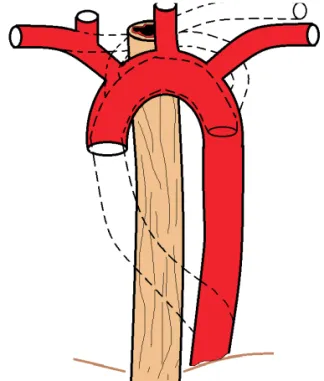
Variations of the branches of the aortic arch are not too rare and may be present in the adult population and found during clinical series of esophageal resections (Figure 1). A right aortic arch may render the esophagectomy more
dificult, especially the dissection of the right paratracheal
lymph nodes 29. Patients with esophageal cancer undergo a routine thoracic computerized tomography and the aortic branches variations can be diagnosed before the operation. In that case, a left thoracotomy, not a right one, is the route of choice 29, sometimes associated to a median sternotomy 16. Aberrant subclavian arteries are also at risk of injury during an esophagectomy 19.
Dysphagia lusoria is caused by vascular compression of the esophagus. It has been described after the following anatomic variations: right or left aberrant subclavian arteries that crosses between the esophagus and the vertebral column in 80% of cases, in 15% of cases it runs between the esophagus and the trachea, and in 5% of cases it passes anterior to both the trachea and esophagus; or vascular rings as in a left ligamentum arteriosum 17.
Were found no anatomic variations in the cadavers dissected.
Azygos vein
The azygos vein originates in the abdomen and it enters the thorax through the aortic hiatus in the diaphragm, and passes along the right side of the vertebral column to the fourth thoracic vertebra, where it arches forward over the esophagus and the root of the right lung, and ends in the superior vena cava. In the aortic hiatus, it lies with the thoracic duct on the right side of the aorta; in the thorax it lies upon the intercostal arteries, on the right side of the aorta and thoracic duct, and is partly covered by pleura 8.
The hemiazygos vein also begins in the abdomen. It enters the thorax, through the left crus of the diaphragm, and, ascending on the left side of the vertebral column, as high as the 9th thoracic vertebra, passes across the column, behind the aorta, esophagus, and thoracic duct, to end in the azygos vein 8.
The accessory hemiazygos vein descends on the left side of the vertebral column, and varies inversely in size with the highest left intercostal vein. It either crosses the body of the eighth thoracic vertebra to join the azygos vein or ends in the hemiazygos 8.
The azygos system anatomy is of interest during an esophagectomy since it is resected in an en-bloc esophagectomy 26. Some authors, however, believe the resection of the azygos system is not considered essential during a radical esophagectomy. The arch of the azygos is divided even for benign cases to allow a better exposure of the proximal mediastinal esophagus.
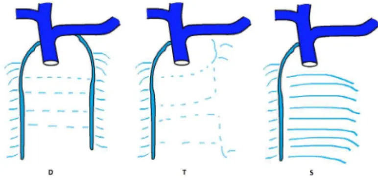
Variations of the azygos system are uncountable and related to the origin of the veins or the communication between the left and right side system 24(Figure 2). However, the clinical importance of these variations is negligible since they can be promptly recognized during an esophagectomy and comprise small caliber vessels that can be easily ligated without any consequences. Most of the clinical important variations occur in the setting of the vena cava (either superior or inferior) anomalies such as duplication7 and total or partial agenesia 2 when a grossly dilatation of the azygos system can be present.
Vagus nerve
Traditionally, the surgical anatomy of the vagus was of interest for peptic ulcer surgery. Currently, esophageal surgeons are worried about preserving the vagus during an esophagectomy, since part of the postoperative
comorbidities may be linked to the vagotomy 1.
Classic anatomy textbooks describe the vagus in the mediastinum as a plexus surrounding the esophagus 8. Our group showed in a previous paper one or more vagal trunks bilaterally in 30 dissections13 (Figure 3). Interestingly, the vagal trunks were preserved after a vagal-sparing esophagectomy was performed in the cadavers 13.
Recurrent laryngeal nerve
The recurrent laryngeal nerve is the most signiicant
branch of the vagus during an esophagectomy. The right recurrent nerve originates at the origin of the right subclavian artery behind the sternoclavicular joint, loops around the artery and ascends to the neck 28. The left recurrent nerve originates at the inferior border of the aortic arch, them it loops around the aorta and ascends to the neck 28. The nerve on either side ascends in the groove between the trachea and esophagus, passes under the lower border of the inferior
FIGURE 2 - Anatomic variations of the azygos system according to Sieb 1934. D - double column type; T - transitional type; S - single column type
constrictor of the pharynx, and enters the larynx behind the articulation of the inferior cornu of the thyroid cartilage with the cricoid8.
The recurrent nerve can easily be damaged at his course in the neck due to the proximity of the esophagus. In the mediastinum, the left nerve is at risk at the level of the aortic arch during the thoracic lymphadenectomy in the aortopulmonary window or along the left paratracheal
groove. The right nerve is usually not in the operative ield.
Several studies have described the surgical anatomy of the laryngeal recurrent nerve in the neck, since it is a major concern during thyroid surgery. There is a paucity of studies dealing with the anatomy of the laryngeal nerve in the thorax. Anatomic variations, however, are uncommon. Non-recurrence has been reported in less than 1% of the cases 3, and in these cases the nerve does not have a thoracic course and is automatically protect from injury. It maybe associated to aberrant subclavian artery or aorta abnormalities. The recurrent laryngeal nerve may exist as two bundles on one side 2.
In all 20 cases dissected, the left laryngeal recurrent nerve arises from the vagus trunk and loops around the aortic arch near the ligamentum arteriosum. On the right side, the authors found four patterns of anatomic variations (Figure 4): type 1: no recurrence - two cases (10%), laryngeal nerve arises from vagus trunk and have no recurrence; type 2: recurrence at the subclavian artery – three cases (15%), laryngeal nerve arises from vagus trunk and loops around the subclavian artery; type 3: recurrence at the brachiocephalic trunk - 12 cases (60%), laryngeal nerve arises from vagus trunk and loops around brachiocephalic trunk; type 4: doubling or tripling - two cases (10%) laryngeal nerve arises from vagus trunk and have two branches, one loops around brachiocephalic trunk another loops around subclavian artery and one case (5%) laryngeal nerve arises from vagus trunk and have three branches, one loops around the brachiocephalic trunk and two loops around subclavian artery.
Lymph nodes
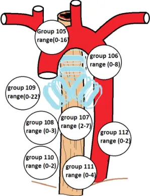
Anatomy textbooks show a regular disposition of nodes not seen by surgeons, but very few studies describe a surgically-oriented mediastinal lymph node distribution. Moreover, there is no standardization of
the classiication and nomenclature of mediastinal lymph nodes. Although various classiications have
been developed by societies and individual authors 12, none has gained unanimous acceptance.
Different anatomical studies, including a previous study by our group 12, showed an inconstant and variable distribution in the number and location of lymph nodes. Clinical studies also show an outstanding variance in the number of nodes resected in the mediastinum during an esophagectomy and lymphadenectomy for cancer, an average per patient ranging from 13 to 38 23. According to Skinner in 1983 26, the number of lymph nodes in each patient varied from 24 to 127.
Some authors and medical societies proposed a minimum number of lymph nodes to be resected in an adequate lymphadenectomy. While the Union for International Cancer Control (UICC) and some authors propose a minimum of six lymph nodes22, others raised this number to 12 6 or even 23 21. Our previous study showed an average of 16 lymph nodes per case in
cadavers without cancer supporting a igure higher
than six (Figure 5).
RVN: right vagus nerve. LVN: left vagus nerve. Arrow: recurrent laryngeal
nerves
FIGURE 4 - Anatomic variation of the mediastinal laryngeal recurrent nerves. type 1: no recurrence;type; 2: recurrence at the subclavian artery; type; 3: recurrence at the brachiocephalic trunk; type 4: doubling or tripling
Thoracic duct
The thoracic duct originates from the cranial part of the cisterna chili in the abdomen. With the aorta on its left and the azygos vein on its right, the thoracic duct passes through the aortic hiatus of the diaphragm. It maintains this relationship as it passes through the posterior mediastinum. The thoracic vertebrae, right intercostal arteries, and terminal portions of the hemiazygos and accessory hemiazygos veins are posterior to the thoracic duct; the esophagus, diaphragm and pericardium are anterior to it. At the level of T7, the thoracic duct travels
obliquely behind the esophagus to the level of the ifth
thoracic vertebra. At T5, it reappears from behind the esophagus to continue its upward journey on the left of the esophagus and medial to the pleura up to the neck 8. Variations are very common in its end in the neck25.
Jacobsson in 1972 14 investigated 122 autopsy cases to study the anatomy and pathology of the thoracic duct. The thoracic duct always started below the diaphragm, passing through the posterior mediastinum. In 1%, the duct had a completely plexiform structure.
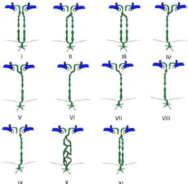
Major variations of the thoracic duct (Figure 6) include doubling, tripling, left-sidedness, and right or bilateral termination, as well as the rare azygos vein termination. In a few instances, the abdominal components of the trunk may pass upward to both sides or only to the left of the aorta. In fact, more than half of the cases do not have a typical presentation of the duct 10.
The major interest in the surgical anatomy of the thoracic duct during an esophagectomy is the possibility of
inadvertent injury to the duct and subsequent chylothorax.
The intraoperative identiication of the injury and the duct itself may be dificult. Mass ligation of the duct including
all tissue between the aorta, spine, esophagus and pericardium is recommended by some authors in cases of suspect lesion of the duct or even routinely. The ligation of
the duct is safe and a randomized trial showed a signiicant
decrease in postoperative chylothorax with routine ligation.
Mass ligation is preferred over identiication and individual
ligation since duplication or a plexiform duct is found in a
very signiicant number of individuals 4.
DISCUSSION
Some peculiar characteristics of the surgical anatomy of the esophagus must be summarized: (1) important organs surround the esophagus, (2) the esophagus crosses the neck, the chest, and the abdomen, (3) the lymphatic distribution is abundant, (4) the organs and structures of the mediastinum may frequently present anatomic variations, and (5) the classic anatomic description of some organs and structures is different from clinical presentation. Halsted can be quoted regarding the view provided by the dissection of a patient during an operation: There is a gap between the
surgeon and pathologist which can be illed only by the
surgeon. The pathologist seldom has the opportunity to see diseased conditions as the surgeon sees them 9.
Moreover, there is no standardization of the classiication
and nomenclature of some structures.
Most of the variations reviewed may be clinically
signiicant during an esophagectomy especially that only a part of them may be identiied before the
operation with imaging tools. This is very true in regards to the lymph nodes, thoracic duct and recurrent nerve. As previously stressed, a complete lymphadenectomy is advocated by some authors. The search for lymphnodes must be careful since lymph nodes distribution is erratic and they may be located within areas close to other important organs susceptible to anatomic variations themselves. Thoracic duct injury may be fatal. Again, a
careful and planned identiication and dissection of the
duct is necessary and the same principles should be adopted to the recurrent nerve in order to avoid surgical technique-related morbidity during an esophagectomy.
We conclude that surgeons must be aware of the anatomic variations of the organs and structures of the mediastinum when performing an esophagectomy.
CONCLUSION
The organs and structures of the mediastinum may frequently present anatomic variations. Some of
these variations may be clinically signiicant during
an esophagectomy; however, only a part of them may
be identiied before the operation with imaging tools.
Surgeons must be aware of these anatomic variations.
FIGURE 6 - Anatomic variations of the mediastinal thoracic duct adapted from Davis 1915 - types I-III: persistence of the right duct concomitant with the left duct with left, right or bilateral termination; types IV, VI and VIII: presence of the right duct alone with left, right or bilateral termination; types V, VII and IX: thoracic duct with left, right or bilateral termination; type X:
REFERENCES
1. Dubecz A, Sepesi B, Salvador R, Polomsky M, Watson TJ, Raymond DP, Jones CE, Litle VR, Wisnivesky JP, Peters JH. J Surgical resection for locoregional esophageal cancer is underutilized in the United States. Am Coll Surg. 2010 Dec;211(6):754-61. Epub 2010 Oct 25. 2. Patti MG, Gantert W, Way LW. Surgery of the esophagus. Anatomy
and physiology. Surg Clin North Am. 1997 Oct;77(5):959-70. 3. Herbella FA, Aquino JL, Stefani-Nakano S, Artifon EL, Sakai P,
Crema E, Andreollo NA, Lopes LR, de Castro Pochini C, Corsi PR, Gagliardi D, Del Grande JC. Treatment of achalasia: lessons learned with Chagas’ disease. Dis Esophagus. 2008;21(5):461-7.
4. Polomsky M, Siddall KA, Salvador R, Dubecz A, Donahue LA, Raymond D, Jones C, Watson TJ, Peters JH. Association of kyphosis and spinal skeletal abnormalities with intrathoracic stomach: a link toward understanding its pathogenesis. J Am Coll Surg. 2009 Apr;208(4):562-9.
5. Joris JL, Chiche JD, Lamy ML. Pneumothorax during laparoscopic fundoplication: diagnosis and treatment with positive end-expiratory pressure. Anesth Analg. 1995 Nov;81(5):993-1000. 6. Gray, H. Anatomy of the Human Body. Philadelphia: Lea & Febiger,
1918; Bartleby.com, 2000.
7. Liebermann-Meffert DM, Luescher U, Neff U, Rüedi TP, Allgöwer M. Esophagectomy without thoracotomy: is there a risk of intramediastinal bleeding? A study on blood supply of the esophagus. Ann Surg. 1987 Aug;206(2):184-92.
8. Yamamoto Y, Watanabe Y, Horiuchi A, Yoshida M, Sugishita H, Nakagawa H, Yukumi S, Sato K, Kawachi K. Esophageal cancer resection associated with a right aortic arch after descending aortic graft replacement. Hepatogastroenterology. 2009 Mar-Apr;56(90):395-7.
9. Kinoshita Y, Udagawa H, Kajiyama Y et al. Esophageal cancer and right aortic arch associated with a vascular ring. Dis Esophagus 1999; 12: 216–18.
10. Pantvaidya GH, Mistry RC, Ghanekar VR, Upasani VV, Pramesh CS. Injury of an aberrant subclavian artery: a rare complication of video assisted thoracoscopic esophagectomy. Ann Thorac Cardiovasc Surg. 2005 Feb;11(1):35-7.
11. Levitt B, Richter JE. Dysphagia lusoria: a comprehensive review. Dis Esophagus. 2007;20(6):455-60.
12. Skinner DB. En bloc resection for neoplasms of the esophagus and cardia. J Thorac Cardiovasc Surg. 1983 Jan;85(1):59-71.
13. Sieb, GA. The azygos system of veins in American White and American Negroes, including observations on the inferior caval venous system. Am. J. Physiol. Anthrop. 1934; 19:39-163.
14. Gesase AP, Fabian FM, Ngassapa DN. Double superior vena cava presenting with anomalous jugular and azygos venous systems. Indian Heart J. 2008 Jul-Aug;60(4):352-8.
15. Bergman RA, Aii AK, Miyauchi R. Illustrated Encyclopedia
of Human Anatomic Variation. Available online http://www. anatomyatlases.org/AnatomicVariants/AnatomyHP.shtml. Accessed Jan 17th, 2012.
16. Banki F, Mason R J, DeMeester S R et al. Vagal-sparing esophagectomy: a more physiologic alternative. Ann Surg 2002; 236: 324–35.
17. Herbella FA, Regatieri CV, Moreno DG, Matone J, Del Grande JC. Vagal integrity in vagal-sparing esophagectomy: a cadaveric study. Dis Esophagus. 2006;19(5):406-9.
18. Wang J, Li J, Liu G, Deslauriers J. Nerves of the mediastinum. Thorac Surg Clin. 2011 May;21(2):239-49, ix.
19. Cannon RC. The anomaly of nonrecurrent laryngeal nerve:
identiication and management. Otolaryngol Head Neck Surg
1999;120:769-71.
20. Herbella FA, Del Grande JC, Colleoni R; Japanese Society for Disease of the Esophagus. Anatomical analysis of the mediastinal lymph
nodes of normal Brazilian subjects according to the classiication
of the Japanese Society for Diseases of the Esophagus. Surg Today. 2003;33(4):249-53.
21. Shen Y, Zhang Y, Tan L, Feng M, Wang H, Khan MA, Liang M, Wang Q. Extensive Mediastinal Lymphadenectomy During Minimally Invasive Esophagectomy: Optimal Results from a Single Center. J Gastrointest Surg. 2012 Jan 19.
22. Tachibana M, Kinugasa S, Hirahara N, Yoshimura H. Lymph
node classiication of esophageal squamous cell carcinoma and
adenocarcinoma. Eur J Cardiothorac Surg. 2008 Aug;34(2):427-31. 23. Dutkowski P, Hommel G, Böttger T, Schlick T, Junginger T. How
many lymph nodes are needed for an accurate pN classiication
in esophageal cancer? Evidence for a new threshold value. Hepatogastroenterology. 2002 Jan-Feb;49(43):176-80.
24. Peyre CG, Hagen JA, DeMeester SR, Altorki NK, Ancona E, Grifin
SM, Hölscher A, Lerut T, Law S, Rice TW, Ruol A, van Lanschot JJ, Wong J, DeMeester TR. The number of lymph nodes removed predicts survival in esophageal cancer: an international study on the impact of extent of surgical resection. Ann Surg. 2008 Oct;248(4):549-56.
25. Skandalakis JE, Skandalakis LJ, Skandalakis PN. Anatomy of the lymphatics. Surg Oncol Clin N Am 2007; 16:1-16.
26. Jacobsson SI. Clinical anatomy and pathology of the thoracic duct: an investigation of 122 cases. Stockholm (Sweden): Almqvist & Wiksell; 1972.
27. Hematti H, Mehran RJ. Anatomy of the thoracic duct. Thorac Surg Clin. 2011 May;21(2):229-38, ix.