Recebido em 12.03.2001. / Received in March, 12thof 2001.
Aprovado pelo Conselho Consultivo e aceito para publicação em 23.03.2002. / Approved by the Consultive Council and accepted for publication in March, 23thof 2002. * Trabalho realizado no Hospital das Clínicas da Universidade Federal de Pernambuco / Work done at "Hospital das Clínicas da Universidade Federal de Pernambuco"
1 Médica Dermatologista / Mestranda do Departamento de Medicina Tropical -UFPE / M.D., Dermatologist and Master of Tropical Medicine, Tropical Medicine Department, UFPE. 2 Médica Dermatologista/ Professora da UFPE / M.D., Dermatologist and professor at UFPE.
3 Odontóloga / Mestranda em Patologia Bucal/ Odontologist and Master of Buccal Pathology. 4 Cirurgião Gastroenterológico/ Professor da UFPE / Surgeon in Gastroenterology, professor at UFPE. 5 Gastroenterologista / Professor da UFPE / Gastroenterologist, professor at UFPE
6 Médico Dermatologista/Professor da UFPE / M.D., Dermatologist, professor at UFPE
7 Virologista do Instituto Ludwig de Pesquisa sobre o Câncer/SP / Virologist at the Ludwig Cancer Research Institute/São Paulo 8 Médica Dermatologista / Médica Dermatologista / M.D., Dermatologist.
©2002by Anais Brasileiros de Dermatologia
Síndrome de Cowden - relato de um caso
*Cowden's Syndrome- A new case report
*Patrícia de Barros Guimarães
1Adeíza de Alencar Branco
2Elaine Carvalho
3Francisco Eduardo Lima
4José Roberto Almeida
5Josemir Belo dos Santos
6Luisa Villa
7Sílvia Helena Rodrigues
8Roberta Siqueira
8Tatiana De Perreli
8Resumo: A síndrome de Cowden (SC) ou síndrome de múltiplos hamartomas (SMH) é
genodermato-se rara de herança autossômica dominante e expressividade variável. É caracterizada por múltiplas lesões hamartomatosas de origem ectodérmica , mesodérmica e endodérmica. O órgão mais acometido é a pele, e as lesões mucocutâneas estão presentes em proporção que varia de 99 a 100% dos casos. Esses sinais precedem o desenvolvimento do câncer em vários anos, servindo como importantes mar-cadores clínicos na identificação de pacientes com alto rico para desenvolver câncer da mama e tireói-de. Devido a associações com malignidades internas o diagnóstico precoce é essencial. O locus gênico para SC foi identificado no cromossomo 10q22-23. As mutações no gene supressor tumoral, PTEN/MMAC1, localizado no cromossomo 10q23, têm sido implicadas no desenvolvimento do câncer mamário. Os autores relatam um caso dessa rara entidade. Trata-se de paciente do sexo masculino com quadro clínico característico dessa síndrome.
Palavras-chave: neoplasias; Síndrome do Hamartoma Múltiplo.
Summary: Cowden's Syndrome (CS) or Multiple Hamartoma Syndrome (MHS) is a rare genoderma-tosis of autossomal-dominant inheritance with variable expressivity. It is characterized by multiple hamartomatous lesions of ectodermal, mesodermal and endodermal origins. The organ system that most consistently manifests this syndrome is the skin. Mucocutaneous lesions are present in 99 to 100% of cases. These signs precede the development of cancer by several years, and they serve as impor-tant clinical markers for identification of patients at high risk for malignancies of the breast or thyroid. Because of its potentially serious associations with internal malignancy, early and accurate diagnosis is essential. The gene locus for CS has been identified as chromosome 10 q22-23. Mutations in the human tumor suppressor gene, PTEN/MMAC1, located on the 10q23 chromosome, have been implicated in the development of breast cancer. The authors report a case of this rare entity, dealing with a male patient with the clinical characteristics of this syndrome.
Key words: neoplasms; Hamartoma Syndrome, Multiple.
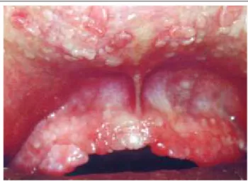
Figure 1: Multiple papules in the gingivae, and upper and lower lips. Aspect similar to small "round pebbles." Figura 1: Múltiplas pápulas nas
gengivas, nos lábios superior e inferior. Aspecto semelhante
ao de pequenas "pedras arredondadas"
INTRODUÇÃO
Em sua pesquisa sobre doenças internas, o médico é, freqüentemente, auxiliado por sinais externos de um proces-so subjacente.3Considerando que a difusão de seu
conheci-mento pode ser de utilidade no diagnóstico de anormalidades internas, apresenta-se um caso de síndrome de Cowden (SC). A SCé genodermatose rara,4,5de herança
autossômi-ca dominante e expressividade variável. Caracteriza-se por múltiplos hamartomas, de origem ectodérmica, mesodérmi-ca e endodérmimesodérmi-ca.1,2,4,5-8Embora a maioria dos tumores que
se desenvolvem seja benigna, a prevalência de malignidade é maior do que na população em geral, sobretudo em carci-nomas mamários e tireoideanos. Tal síndrome foi descrita pela primeira vez por Lloyd e Dennis,2,5,6em 1963, e
deno-minada SCem referência a sua paciente3, 4Rachel Cowden,
que faleceu de carcinoma mamário.2 Weary e
t al.,3,7 em
1972, reportaram cinco casos adicionais e sugeriram a
denominação síndrome de múltiplos hamartomas (SMH).
Desde sua primeira descrição, foram documentados menos de 200 casos.4,13Os relatos são crescentes à medida
que aumenta o conhecimento da doença. Mesmo assim,
Gentry et al.3 acreditam que sua freqüência seja mais
comum do que os relatos de literatura, devido à expressivi-dade variável existente nessa genodermatose. Dessa forma,
muitos casos não são corretamente diagnosticados.3,10 Os
sexos são igualmente afetados,1 embora alguns autores
acreditem que a doença seja mais comum no feminino.7Os
caucasianos formam a maioria dos pacientes relatados.2
RELATO DO CASO
Foi atendido no ambulatório de dermatologia do
Hospital das Clínicas-PE, paciente com 38 anos, do sexo
masculino, pele tipo III (segundo classificação de Fitzpatrick), que nunca trabalhou nem freqüentou escola, natural e procedente de Olinda, PE.
Apresentava múltiplas pápulas assintomáticas con-fluentes, nas gengivas, nos lábios superior e inferior e no dorso da língua, com aparência e textura semelhantes à de pequenas "pedras arredondadas" (Figura 1).
Observou-se, ainda, lipoma solitário na região frontal, fácies adenóide, aumento do
diâmetro craniano e deformi-dades ósseas nos tornozelos, além de retardo mental mode-rado. O exame físico dos apa-relhos cardiovascular, digesti-vo, genitourinário e sistema nervoso mostrou-se normal.
INTRODUCTION
In her research on internal diseases, the medical doctor is often assisted by the external signs of an
underl-ying process.3
Given that diffusion of her knowledge may be useful for the diagnosis of internal abnormalities, a case report is presented here of Cowden's syndrome.
CSis a rare genodermatosis,4,5
of autossomal-domi-nant inheritance with variable expressivity. It is characteri-zed by multiple hamartomatous lesions of ectodermal,
mesodermal and endodermal origins.1,2,4-8
While most CS
tumors that develop are benign, the prevalence of malig-nancy is higher than in the tumor population in general, especially in breast and thyroid carcinomas. This syndrome
was first described by Lloyd and Dennis,2,5,6
in 1963, and
called SC in reference to their patient,3,4
Rachel Cowden,
who died from breast cancer.2
In 1972, Weary et al. 3, 7
repor-ted five additional cases and suggesrepor-ted the name of
Multiple Hamartoma Syndrome (MHS).
Ever since its first description, less than 200 cases
have been documented.4,13
Reports grow in number as
kno-wledge of the disease increases. Even so, Gentry et al.3
believe that frequency is more common than what is repor-ted in the literature, due to the existing variable expressivity of this genodermatosis. The result is that many cases are not
correctly diagnosed.3,10
Both sexes are equally affected,1
though some authors believe the disease to be more
com-mon in females.7
The majority of the patients reported are
caucasians. 2
CASE REPORT
A 38-year-old male patient was attended to at the Dermatology Outpatients Clinic of the Hospital das
Clíni-cas in Pernambuco State (PE), due to type III skin
(accor-ding to Fitzpatrick's classification). The patient had never been employed nor had he ever attended school. He was
born in Olinda, PE, where he currently resides.
The patient showed multiple asymptomatic confluent papules in the gingivae, upper and lower lips, and on the back of the tongue, with an appearance and texture similar to small "round pebbles" (Figure 1).
diges-Figura 2: lesão com aspecto polipóide com hiperplasia epitelial. Papilomatose com estroma conjutivo rico em vasos, com leve infiltração
linfo-histiocitária
tive and genitourinary organ and systems, and the nervous system were normal.
During childhood, the patient had undergone orthope-dic surgery in the ankles. He had suffered complete dental extraction a few years earlier due to cavities, according to information gathered at the moment of the physical examina-tion. Medical history taking was incomplete, because neither the patient nor his mother responded adequately to the inquiry. Information on family antecedence was also compro-mised since the patient does not have any contact with his relatives, and nor does his genitor, who moreover did not show clinical characteristics of the syndrome being studied.
Complementary Tests
Routine hemogram and biochemical laboratory tests were normal.
RXthorax and the computerized axial tomography
(CT scan) of the thoracic spin did not show significant
alterations, nor did the laryngoscopy and USG breast
biopsy.
Biopsy of the oral papules showed evidence of a polypoid lesion with epithelial hyperplasia at the expense of hyperkeratosis and acanthosis. Papilloma with conjunctive stroma rich in vessels, with a light lymphohistiocytic infil-trate, compatible with fibroepithelial papilloma (Figure 2). The electron microscope and immunohistology reveal an
absence of HPV.
In the thyroidian investigation, T3, T4 and TSH
hormone doses were administered, which were normal;
the glandular USG showed evidence of 0.3 cm
hypoe-coic nodules in the right lobe; the punction - nodule biopsy with a cytological test - revealed Hashimoto's thyroiditis.
In the colonoscopy polyps were found in the rec-tum, transversal colon, ascen-dant colon, and cecum (Figure 4); the histopathologic study of intestinal lesions demonstrated hyperplastic and adenomatous polyps.
In brain magnetic resonance with contrast a suggestive image was found of angioma cavernoma, located at the level of the left frontal region and having the classic 'popcorn-like' aspect. There was no lesion found suggestive of hamarto-ma (Figure 5).
Em seus antecedentes pessoais registra-se na infância cirurgia ortopédica nos tornozelos. Sofreu extração dentária completa, há alguns anos, devido a cáries, segundo informações colhidas no momento do exame. A anamnese foi incompleta, pois nem o pacien-te, nem sua genitora respondiam adequadamente às indagações.
Os antecedentes familiares também foram prejudica-dos, porque o paciente não tem contato algum com seus parentes, a não ser com sua genitora, que não apresentou características clínicas da síndrome em estudo.
Exames Complementares
Os exames laboratoriais de rotina, hemograma e bio-química, mostraram-se normais.
O RXde tórax e a tomografia axial computadorizada torácica não demonstraram alterações significativas, bem
como a laringoscopia e USGdas mamas.
A biópsia das pápulas orais evidenciou lesão poli-póide com hiperplasia epitelial à custa de hiperqueratose e acantose. Papilomatose com estroma conjuntivo rico em vasos, com leve infiltração linfo-histiocitária, compatível com papiloma fibroepitelial (Figura 2). A microscopia ele-trônica e a imuno-histologia revelaram ausência do HPV.
Na investigação tireoidiana realizou-se dosagem dos
hormônios T3,T4 e TSH, que se apresentaram normais; a
USGglandular evidenciou nódulo hipoecóico de 0,3cm no
lobo direito; a punção - biópsia do nódulo com o exame citológico - revelou tireoidite de Hashimoto.
Na avaliação do trato gastrointestinal (TGI) realizou-se a esofagogastroduodenoscopia, que demonstrou polipose esofagia-na, gástrica e duodenal, além de hérnia de hiato e esofagite erosi-va grave (Figura 3). O exame histopatológico revelou gastrite crônica ativa e pólipo com características de hamartoma vascular.
Na colonoscopia foram encontrados pólipos no reto, no cólon transverso, no cólon ascen-dente e ceco (Figura 4); o estudo histopatológico das lesões intesti-nais demonstrou pólipo hiperplásico e pólipo adenomatoso.
Na ressonância magnética encefálica com contraste encontrou-se imagem sugestiva de angioma cavernoso (cavernoma), localizado na região frontal esquerda e com o clássico aspecto popcorn-like. Não
foi encontrada lesão sugestiva de
Figure 5: Image suggestive of angioma cavernosa. Classical 'popcorn-like' aspect. Figura 5: Imagem sugestiva de
angioma cavernoso. Aspecto clássico popcorn-like
hamartoma (Figura 5).
Com base nesses achados clínicos e patológicos, o paciente foi diagnosticado como portador da síndrome de Cowden e está sendo acompanhado nas clínicas dermato-lógica, endocrinodermato-lógica, gastroenterológica e neurológica.
DISCUSSÃO
A SCé genodermatose rara com penetrância autossô-mica dominante e expressividade variável.4 É representada
por múltiplas lesões hamartomatosas e neoplasias da pele, mucosas, mamas, tireóide, trato gastrointestinal, entre outras anormalidades congênitas.2,4,6Os hamartomas são definidos
como malformações de aspecto tumoral, compostos por ele-mentos que normalmente fazem parte do organismo daqueles que se desenvolvem, mas que têm uma disposição errônea.10
O órgão que mais freqüentemente manifesta sintomas dessa síndrome é a pele,3 sendo os achados mucocutâneos os
mais característicos1,2e constantes,2,4,5,6 reportados em
propor-ção que varia de 99 a 100% dos casos. Como precedem em muitos anos o desenvolvimento do câncer, servem como importantes marcadores clínicos na identificação de pacien-tes com alto risco de
maligni-dades nas mamas e
tireói-de.1,4,5,8,13
As lesões mucocutâ-neas mais características são múltiplos triquilemomas faciais, papilomas orais e queratose acral.
Os triquilemomas, que representam hamartomas de
Based on these clinical and pathological fin-dings, the patient was diagnosed as a carrier of Cowden's syndrome and is being followed up at the der-matology, endocrinology, gastroenterologic and neuro-logical clinics.
DISCUSSION
CSis a rare genodermatosis with
autossomal-domi-nant penetration and variable expressivity.4
It is represented by multiple hamartomatose lesions and neoplasias of the skin, mucose, breast, thyroid, and gastrointestinal tract,
among other congenital abnormalities.2,4,6
The hamartomas are defined as tumor-like malformations, consisting of ele-ments normally part of the organism in which it develops,
but having an erroneous disposition.10
The organ most frequently manifesting the symptoms of this syndrome is the skin, the mucocutaneous findings of which
are the most characteristic1,2
and constant,2,4,5,6
reported in a pro-portion varying from 99 to 100% of cases. As they anticipate the development of cancer by several years, they serve as important clinical markers for the identification of patients at high risk of contracting breast and thyroid
malignancies.1,4,5,8,13
The most characte-ristic mucocutaneous lesions are multiple facial trichile-momma, oral papilloma and acral keratosis.
The trichilemommas, representing hamartomas of
hair follicle tissue,2,4,12,13
has
Figura 3: Polipose esofagiana, gástrica e duodenal. Figure 3: Esophageal, Gastric and Duodenal Polyposis.
Figura 4: Pólipos no reto, no cólon transverso, no cólon ascen-dente e ceco. / Figure 4: Polyps in the rectum, transversal colon
a preferential affectation for the face.7
It may be so nume-rous that orifice border and facial contours coalesce, thus
becoming cosmetically unacceptable for the patient.2
They are characterized by 1 to 4 mm papules and nodules and, in most cases, they are manifested before the second or third
decade of life,1,2
though they may occur between the fourth
decade and 75-years of age.2
When multiple, these tumors are patognomonic and are frequently present prior to
inter-nal manifestations.2
Distribution of papules also occurs in
distal extremities,2,7
including the palms of the hands, soles
of the feet, neck and forearms.4
In the case reported, the patient did not present the characteristic trichilemomma.
Typical papilloma is usually between 1 and 3 mm, and affects the gingivae, underside of the tongue, buccal mucous, the palate, alveolar endothelium and tonsillar fossa, often taking on an aspect similar to "round pebbles". The verrucous lesions in the angles of the mouth and oral mucous may mimic perleche hypertrophy or benign
gingi-val hyperplasia.3
The histological form of oral lesions con-siderably lacks specificity, representing essentially
fibroepi-thelial hyperplasia.2, 9
Notwithstanding the predisposition to neoplasia, oral squamous carcinoma is a very rare
compli-cation.5,13
The oral papillomatous lesions of the case studied were multiple and characteristic of the syndrome.
Acral keratosis is present in more than half of all
patients.2,4,5
It occurs on the dorsal surface of the hands and feet, resembling spotted keratosis with a central depression.
2 It belongs clinically to verruca vulgaris,2,1
yet
papilloma-virus has not been detected until now.13
In addition to these characteristic mucocutaneous findings, a variety of lesions has been regularly described
in CS, including achrocordon, lipoma, angiolipoma,
heman-gioma, xanthomatosis, vitiligo and café-au-lait spots.1
In the case report the presence was observed of a solitary lipoma in the frontal region.
The importance of this disease arises from the cuta-neous findings and malignant or benign disorders present
in other systems and organs,3
in particular the thyroid gland, breast, gastrointestinal tract and nervous system. These extracutaneous findings are present in 90% of patients, and various congenital abnormalities are also
involved.6
The most frequent extracutaneous manifestation is
thyroid disease,2,7
which may occur in roughly 65% of
patients,4,7,14
affecting both sexes equally.13
Although the lesions are usually of benign nature, like goiter, adenomas, thyroiditis, hypo - and hyperthyroidism and thyroglossal duct cyst, thyroid cancer occurs in proportions varying
from 3 to 10% of all individuals affected.15,16,19
The most fre-quent histological type in this neoplasia is follicular neo-plasia, but papillar neoplasia is more rarely encountered. In the thyroidian investigation of the present case, hypoe-coic nodule was made evident in the right lobe of the gland.
Breast affection translates as a fibrocystic disease,2,4,5
and has been encountered in all of the female patients.3
folículos pilosos,2,4,12,13 atingem preferencialmente a face7 e
podem ser tão numerosos, que coalescem ao redor dos orifícios e contornos faciais, tornando-se cosmeticamente inaceitáveis por parte do paciente.2Caracterizam-se por pápulas e nódulos
de 1 a 4mm e, na maioria dos casos, manifestam-se antes da segunda ou terceira década de vida,1,2embora possam ocorrer
entre os quatro e os 75 anos.2Esses tumores, quando múltiplos,
são patognomônicos e freqüentemente presentes antes das manifestações internas.2Também ocorre distribuição das
pápu-las nas extremidades distais,2,7 incluindo palmas das mãos,
plantas dos pés, pescoço e antebraços.4 No caso relatado, o
paciente não apresentou os triquilemomas característicos. Os papilomas orais típicos usualmente têm entre um e 3mm e afetam as gengivas, o dorso da língua, a mucosa bucal, o palato, o endotélio alveolar e a fossa tonsilar,4
assu-mindo com freqüência aspecto semelhante ao de "pedras arredondadas".1, 2, 9 As lesões verrucosas dos ângulos da
boca e mucosa oral podem mimetizar perleche hipertrófica ou hiperplasia gengival benigna.3O quadro histológico das
lesões orais é bastante inespecífico, representando essen-cialmente hiperplasia fibroepitelial.2, 9A despeito da
predis-posição para neoplasia, carcinoma escamoso oral é compli-cação muito rara.5,13As lesões papilomatosas orais do caso
eram múltiplas e características da síndrome.
A queratose acral está presente em mais da metade dos pacientes,2,4,5ocorrendo na superfície dorsal das mãos e
pés, assemelhando-se a queratoses puntadas com depressão central.2Aparenta, clinicamente, verruga vulgar,2,13porém, o
papiloma vírus humano não foi detectado até o momento.13
Em adição a esses achados mucocutâneos caracterís-ticos, uma variedade de lesões tem sido descrita com regu-laridade na SC,2, 5incluindo acrocórdons,5lipomas,
angioli-pomas, hemangiomas, xantomas, vitiligo e manchas
café-au-lait.1 No caso relatado observou-se a presença de um
lipoma solitário na região frontal.
A importância dessa doença decorre dos achados cutâneos e alterações malignas ou benignas presentes em outros sistemas e órgãos,3em particular glândula tireóide,
mama, trato gastrointestinal, sistema nervoso. Esses acha-dos extracutâneos estão presentes em 90% acha-dos pacientes, e várias anormalidades congênitas também estão envolvidas.6
A mais freqüente manifestação extracutânea é a doença da tireóide,2,7que pode ocorrer em
aproximadamen-te 65% dos pacienaproximadamen-tes,4,7,14 afetando igualmente ambos os
sexos.13Apesar de as lesões serem usualmente de natureza
benigna,6,7como bócio, adenomas, tireoidite, hipo e
hiperti-reoidismo e cisto de ducto tireoglosso, o câncer de tireóide ocorre em proporção que varia de três a 10% de todos os indivíduos afetados.15,16,19O tipo histológico mais freqüente
nessa neoplasia é o folicular; mais raramente, encontra-se o papilar.20Na investigação tireoidiana do presente caso
evi-denciou-se nódulo hipoecóico no lobo direito da glândula. A afecção mamária traduz-se por doença
fibrocísti-ca2,4,5e tem sido encontrada em todos os pacientes do sexo
ocorrendo em torno de 80% dos casos.2, 3, 4,7,13O carcinoma da
mama é a mais séria conseqüência na SC,2acometendo
pro-porção que varia de 30 a 50% das mulheres afetadas;7, 13, 15, 19
25% das pacientes têm risco de acometimento bilateral da
neoplasia.13 A idade média de aparecimento do câncer
mamário é 40 anos, o que é mais precoce do que a maligni-dade não associada à síndrome.2, 13 A doença fibrocística é
sempre precedente nas pacientes que desenvolvem carcino-ma da carcino-macarcino-ma. No caso original, descrito por Lloyd e Dennis, a paciente foi submetida a mastectomia bilateral devido a uma doença fibrocística inflamatória ulcerativa. Exames microscópicos do tecido excisado revelaram focos de proli-feração indistinguíveis de carcinoma intraductal.3
Papi-lomatose florida intraductal e hiperplasia ductal atípica, mar-cadores de alto risco para câncer da mama, freqüentemente são reportados nos substratos histopatológicos mamários em
mulheres com SC. Pacientes com a síndrome devem ser
acompanhadas por mastologista e, freqüentemente, submeti-das a autoexames e exames especializados, incluindo mamo-grafia. Alguns autores indicam a mastectomia bilateral pro-filática.2, 4, 13 Nos pacientes do sexo masculino pode ser
encontrada ginecomastia,2porém nenhum caso de câncer da
mama em homens foi descrito associado à SC.2,13
As anormalidades no TGIcompreendem lesões
poli-póides que podem desenvolver-se em qualquer parte de seu segmento, como cólon, retossigmóide, estômago, duodeno, intestino delgado e esôfago, em ordem decrescente de aco-metimento;14o ânus também pode ser afetado.2, 13Tais
póli-pos são tipicamente pequenos, múltiplos e sésseis. O quadro histológico das lesões gastrointestinais pode ser hamarto-matoso, lipohamarto-matoso, linfohamarto-matoso, inflamatório, hiperplásico e, ocasionalmente, adenomatoso.7O potencial degenerativo
desses pólipos é pequeno.2, 8 Poucos casos de carcinoma
colônico têm sido reportados, e ainda não foi estabelecido um status pré-maligno nessa síndrome, com relação ao TGI.14A esofagogastroduodenospia e a colonoscopia
reali-zadas na avaliação do caso relatado demonstraram polipose esofagiana, gástrica, duodenal, colônica, cecal e retal.
O diagnóstico diferencial da polipose na SC inclui
polipose familiar, síndrome de Gardner, síndrome de Peutz Jeghers, síndrome de Turcot, síndrome de Cronkit Canada e múltiplos pólipos adenomatosos. Outros achados incluem divertículos colônicos, hamartomas e cistos hepáticos.3,13
O sistema nervoso pode ser acometido de diversas maneiras:3, 4ganglioneuromas e neurofibromas sintomáticos
ou assintomáticos, que podem ser encontrados
incidental-mente ou em associações com lesões cutâneas.3
Recen-temente a doença de Lhermitte-Duclos (DLD) foi
conside-rada manifestação neurológica da SC.7,12,13,15,19Caracteriza-se
pela proliferação peculiar de células ganglionares no cere-belo, levando à substituição de células granulares e células de Purkinje, o que representa um hamartoma do tecido neu-ral.2Os pacientes costumam apresentar sinais de
hiperten-são intracraniana, hidrocefalia, disfunção cerebelar e de nervos cranianos.12, 13Koch
et al.12recomendam o exame de
Fibroadenomas are also very common, occurring in
rou-ghly 80% of cases.2,3,4,7,13
Breast cancer is the most serious
consequence in CS.7,13,15,19
It involves a proportion ranging from 30 to 50% of women affected. 25% of patients are at
risk of contracting bilateral affliction of the neoplasia.13
The average age at which breast cancer appears is 40 years, which is earlier than for the malignancy not associated with
the syndrome.2,13
Fibrocystic disease is always a precedent in patients who go on to develop breast cancer. In the origi-nal case, described by Lloyd and Dennis, the patient was submitted to a bilateral mastectomy due to an inflammatory ulcerated fibrocystic disease. Microscopic tests of the exci-sed tissue revealed focuses of proliferation
indistinguisha-ble from intraductal carcinoma. 3
Intraductal florida papil-loma and atypical ductal hyperplasia, high risk markers for breast cancer, are often reported in the histopathological
mammary substrate in women with CS. Patients with the
syndrome have to be followed up by a mastologist, and often submitted to self-examinations and specialized exami-nations, including mammography. Some authors indicate
bilateral prophylactic mastectomy.2,4,13
In male patients
gynecomastia may be found,2
however no case of breast
cancer in men has been described in association with CS.2,13
The abnormalities in TGI include polypoid lesions
that may develop at some point in its course, like
rectosig-moid colon, duodenum, Delgado intestine and esophagus,14
in decreasing order of affliction; the anus may be affected
as well.2,13
Such polyposes are typically small, multiple and sessile. The histological form of the gastrointestinal lesions may be hamartomous, lipomatous, lymphomatous,
inflam-matory, hyperplastic and occasionally adenomatous.7
The
degenerative potential of these polyps is low.2,8
Few cases of colon carcinoma have been reported, and a premalignant status of this syndrome has still not been established in
rela-tion with TGI.14
The esophagogastroduodenoscopy and colonoscopy performed in the case report demonstrate eso-phageal, gastric, duodenal, colonic, cecal and rectal poly-poses.
The differential diagnosis of polyposis in CSincludes
familiar polyposis, Gardner's syndrome, Peutz Jeghers's syndrome, Turcot's syndrome, Cronkhite-Canada syndrome and multiple adenomatosous polyposis. Other findings
included colonic cysts, hamartomas and hepatic cysts.3,13
The nervous system may be affected in diverse ways; 3,4
ganglioneuroma and symptomatic or asymptomatic neuro-fibroma, which may be found incidentally or in association
with cutaneous lesions. 3
Recently Lhermitte-Duclos
disea-se (DLD) was considered a neurological manifestation of
CS.7,12,13,15,19
It is characterized by the peculiar proliferation of ganglionar cells in the brain, leading to the substitution of granular cells and Purkinje cells, which represent a
hamartoma of the neural tissue. 2
Patients customarily show signs of intracranial hypertension, hydrocephalus, cerebral
and cranial nerve dysfunction.12,13
Koch et al.12
mainly those who show megalencephaly.12
Other neurologi-cal findings include pseudotumor cerebri, subarachnoid hemorrhage, arterio-venous malformation, meningeoma, and electroencephalographic alterations. The neurological sequelae include low intelligence, motor disturbance and
convulsions.12
In children, only macrocephalus and mental retarda-tion can be found. In the magnetic resonance performed, an image was found to be suggestive of cavernous hemangio-ma in the left frontal region, though without clinical reper-cussions.
The abnormalities in the female genitourinary tract
commonly comprise menstrual irregularities,2,3
ovarian
cysts and leiomyomas.2
Other less frequent tumors are tera-toma, uterine fibromas, adenocarcinomas, urethral and cer-vical carcinomas, vaginal cysts, adenocarcinomas of the
kidney and benign polyposes of the urethra.2,13
In the male genitourinary tract hydrocele, varicocele and hypoplastic
testicules may be found.13
A third of SC patients have skeletal defects among
which one finds increased skull circumference,2,4
characte-rizing one of the most common extracutaneous findings.
Other skeletal alterations found are adenoid facies,5,7
cyphosis, cyphoscoleosis, pectus excavatum, big hands and
feet and syndactyly.2,13
Structural abnormalities of the oral cavity include mandibular and maxilar hypoplasia, high
arch of the hard palate,7
microstomia, hyperplasia of the soft palate and uvula, scrotal tongue, periodontitis and
extensive dental cavities.9,13
Regarding eye alterations, lenticular opacity, hyper-telorism, congenital vascular abnormality of the bottom of the eye, glaucoma, angioid streaks, cerebral pseudotumor,
retinal glioma, and myopy. Yang et al.5
report a case of CS
associated with congenital nystagmus.
In relation to the cardiovascular system, hyperten-sion was observed, as were defects in the atrial septum,
mitral valve prolapse and aortic and mitral insufficiency.7
Nevertheless, it is difficult to know whether these findings
are part of the syndrome or only coincidences.2
Respiratory tract abnormalities associated with CS
are rarely described. Findings consist of hamartomas, larynx polyps, pulmonary cysts, and arterio-venous
malfor-mations.6
Solli et al.6
described a case of multiple bilateral pulmonary lipomatosa. Performing computerized thoracic axial tomography did not demonstrate any significant alte-rations.
Seldom described associations with CS include
merckel cell carcinoma, renal cell carcinoma, cutaneous squamous cell carcinoma, transitional cell bladder
carcino-mas, endometrial adenocarcinoma, lymphocarcino-mas,4,5
leuke-mias5
and malignant melanoma.13,17
In spite of the wealth of clinical manifestations, most
characteristic with CS are mucocutaneous lesions that
translate into a combination of orofacial hamartomas and
acral keratosis.4,5,6
Yet many patients do not show the
com-ressonância magnética do crânio em todos os pacientes,
principalmente os que apresentam megaloencefalia.12
Outros achados neurológicos incluem pseudotumor cere-bral, hemorragia subaracnoídea, malformação arterioveno-sa, meningeoma e alterações eletroencefalográficas. As seqüelas neurológicas compreendem baixa inteligência, dis-túrbio de coordenação e convulsão.12
Em crianças podem ser encontrados apenas macroce-falia e retardo mental. Na ressonância magnética realizada encontrou-se imagem sugestiva de angioma cavernoso na região frontal esquerda, porém sem repercussões clínicas.
As anormalidades no trato genitourinário feminino abrangem, comumente, irregularidades menstruais,2, 3cistos
ovarianos e leiomiomas.2Outros tumores menos freqüentes
são teratomas, fibromas uterinos, adenocarcinomas, carci-nomas de uretra e cérvix, cistos vaginais, adenocarcicarci-nomas de rim e pólipos benignos da uretra.2,13No trato
genitouriná-rio masculino podem ser encontrados ainda hidrocele, vari-cocele e testículos hipoplásicos.13
Um terço dos pacientes com SCtem defeitos esquelé-ticos, entre eles, aumento da circunferência do crânio,2,4que
caracteriza um dos achados extracutâneos mais comuns. Outras alterações esqueléticas encontradas são fácies adenói-de,5,7 cifose, cifoescoliose,
pectus excavatum, mãos e pés
grandes e sindactilia.2,13Anormalidades estruturais da
cavida-de oral incluem hipoplasia mandibular e maxilar, palato cavida-de arco alto,7microstomia, hipoplasia do palato mole e úvula,
língua escrotal, periodenite e cáries dentárias extensas.9, 13
Como alterações oculares podem ocorrer opacifici-dade lenticular, hipertelorismo, anormaliopacifici-dade vascular con-gênita do fundo de olho, glaucoma, estrias angióides, pseu-dotumor cerebral, glioma de retina5e miopia.2Yang
et al.5
relatam um caso de SC associado a nistagmo congênito. Em relação ao sistema cardiovascular,2, 7
verificam-se hipertensão, defeitos no verificam-septo atrial, prolapso de válvula mitral e insuficiência aórtica e mitral.7Entretanto, é difícil
saber se esses achados são parte da síndrome ou apenas coincidentes.2
Anormalidades do trato respiratório associadas com
a SC são raramente descritas. Os achados consistem em
hamartomas, pólipos de laringe, cistos pulmonares e mal-formações arteriovenosas.6 Solli
et al.6 descreveram um
caso de lipomatose pulmonar múltipla bilateral. A tomogra-fia axial computadorizada torácica realizada não demons-trou alterações significativas.
Associações com a SCmenos freqüentemente descritas incluem carcinoma de células de Merckel, carcinomas de célu-las renais, carcinomas cutâneos de célucélu-las escamosas, carcino-mas de células de transição da bexiga, adenocarcinoma do endométrio, linfomas.4, 5leucemias5e melanoma maligno.13, 17
Apesar da riqueza de manifestações clínicas, o que mais caracteriza a SCsão as lesões mucocutâneas, traduzi-das pela combinação de hamartomas orofaciais e queratose acral.4,5,6Muitos pacientes, porém, não apresentam o quadro
Com base nesses aspectos, Salem e Steck,2,5,18 em
1983, propuseram os critérios diagnósticos, dividindo-os em critérios maiores, menores e história familiar (Tabela 1).
O diagnóstico foi considerado definitivo na presen-ça de dois critérios maiores; um critério maior e um menor; um critério maior e história familiar positiva ou dois crité-rios menores e história familiar positiva.
Os aspectos genéticos e moleculares têm sido
investi-gados em profundidade na SC.6,7 Recentemente o gene
PTEN/MMAC1, um gene supressor tumoral, foi localizado no cromossomo 10q 22-2312, 13, 16, 19e responsabilizado pelas
malig-nidades da mama e da tireóide encontradas na SC. As muta-ções no gene PTEN identificadas em familiares e pacientes com SCpodem ser a causa da tendência à proliferação desor-denada dos tecidos, levando à formação de hamartomas.12, 15, 19
A síndrome é herdada com traço autossômico domi-nante, mostrando alto grau de penetrância e grande variação de expressividade,1o que permite encontrar manifestações
discretas nos familiares desses pacientes ou mesmo casos de história familiar negativa. A identificação do gene
envol-vido na SC possibilita o diagnóstico pré-sintomático da
doença.15
O tratamento da SC é controverso e visa principal-mente à melhora do aspecto estético das lesões e à busca ativa de neoplasias associadas.
Os triquilemomas faciais e papilomas orais podem ser excisados com bisturi,2, 21 eletrocirurgia, dermoabrasão
ou nitrogênio líquido,21que traz mais beneficios ao
pacien-te, porém todas as lesões removidas recorrem rapidamente. Aplicações tópicas de tretinoína têm pouco sucesso tera-pêutico.21
Da mesma forma que na pele, a experiência de
plete form, due to variable degrees of penetration in the syndrome.
Based on these aspects, Salem and Steck,2,5,18
in 1983, proposed the following diagnostic criteria, divided into major and minor criteria, and family history. (Table 1).
The diagnosis was considered definitive in the pre-sence of two larger criteria; a major criterion and minor one; a major criterion and positive family history, or two minor criteria and positive family history.
The genetic and molecular aspects in CShave been
investigated in depth.6, 7
The PTEN/MMAC1 gene, a tumor
suppressor gene, was recently localized in chromosome 10q
22-2312,13,16,19
and made responsible by the breast and thyroid
malignancies found in CS. Mutations in the PTENgene
iden-tified in acquaintances and patients with CS may be the
cause behind the tendency for disordenate tissue
prolifera-tion, leading to the formation of hamartomas.12,15,19
The syndrome is inherited with an autossomal-domi-nant trace, exhibiting a high degree of penetration and
large variation of expressivity,1
which enables the identifi-cation of discreet manifestations among the patient's relati-ves or even cases of negative family history. Identification
of the gene involved in CS enables the pre-symptomatic
diagnosis of the disorder.15
CS treatment is controversial and aims principally
toward improving the aesthetic aspect of the lesion and to the active search for associated neoplasias.
Facial trichilemmomas and oral papillomas may be
excised by means of laser,2, 21
electrosurgery, dermoabrasion
or liquid nitrogen,21
which is more beneficial for the patient. However all removed lesions recur quickly. Topical
appli-cations of tritinoin have little therapeutic success.21
Tabela 1: Critérios diagnósticos para a doença de Cowden*
Table 1: Diagnostic criteria for Cowden's disease*
Critério clínico maior
Major clinical criteria
Critério clínico menor
Minor clinical criteria
História familiar de doença de Cowden
Family history of Cowden's disease
a) pápulas cutâneas faciais
a) cutaneous facial papules
b) papilomatose da mucosa oral
b) papillomatosis of oral mucous
a) ceratose acral
a) acral keratosis
b) ceratose palmoplantar
b) palmoplantar keratosis
Diagnóstico definitivo:
Definitive diagnosis:
1 a + 1 b
(1 a ou / or 1 b) + (2 a ou / or 2 b)
(1 a ou / or1 b) + 3
2 a + 2 b +3
Diagnóstico provável:
Probable diagnosis:
1 a ou / or1 b
(2 a ou / or2b) +3
Diagnóstico possível:
Possible diagnosis:
2 a e/ou / and /or2 b
As with the skin, the experiment conducted by
Gentry et al.3
demonstrated that excision of polyps, with ful-guration of the base, does not eradicate the lesions. The sig-moidoscopic exam segment showed evidence in two cases of the recurrence of benign lesions at the same site as previous
neoplasms. Cnudde et al.10
report a case of CSassociated
with psoriasis, in which acitretine was introduced in a 0.75 mg per kg daily dose for treatment of the latter. Regression
of the psoriatic and SC-related cutaneous lesions was
detec-ted over a three-month period, though follow-up to the digestive lesion was not performed. The mucocutaneous manifestations show particular sensitivity to oral retinoids,
10
but the regression of lesions was transitory, with the ini-tial aspect reappearing when the retinoid dose was tapered or the treatment interrupted.
Thivolent et al.11
in turn report a case of a CSpatient
treated with 30 mg daily of etritinate for nine months, which confirmed the spectacular disappearance of cuta-neous manifestations and the marked reduction of rectal polyposis.
To be sure, medical doctors and odontologists must
be able to recognize the presence of CSsigns and symptoms
as clinical markers for the identification of patients at high risk of developing internal neoplasias. The possibility of breast and thyroid carcinoma must always be borne in mind. As such, all of the patients have to be rigorously sub-mitted to a search for hidden malignancies, with an empha-sis on the breasts, thyroid gland, gastrointestinal tract and nervous system. Early recognition and management of these associations may reduce the morbidity and mortality
pre-sent in Cowden's syndrome.5
q
Gentry et al.3 demonstrou que a excisão de pólipos, com
fulguração da base, não erradica essas lesões. O segmento com exame sigmoidoscópico em dois casos evidenciou a recorrência de lesões benignas nos mesmos sítios dos neo-plasmas anteriores.3
Cnudde et al.10 relataram um caso de SC associado a
psoríase, no qual foi introduzida acitretina na dose de 0,75mg/kg/dia para tratamento desta última. Detectou-se regressão das lesões cutâneas, tanto psoriáticas como as rela-cionadas à SC, num período de três meses. Não foi realizado, entretanto, o acompanhamento das lesões digestivas. As mani-festações mucocutâneas mostraram sensibilidade particular aos retinóides orais,10porém a regressão das lesões foi
transitó-ria, com o reaparecimento do aspecto inicial quando se dimi-nuiu a dose dos retinóides ou o tratamento foi interrompido.
Thivolet et al.,11por sua vez, relataram um caso de
paciente com SCtratado com 30mg/dia de etretinato duran-te nove meses, comprovando o desaparecimento espetacu-lar das manifestações cutâneas e a diminuição marcante das poliposes retais.
Certamente, os médicos e odontólogos devem estar aptos a reconhecer a presença dos sinais e sintomas da SC como marcadores clínicos para a identificação de pacientes com alto risco de desenvolver neoplasias internas. A possi-bilidade de carcinoma da mama e tireóide deve estar sem-pre em mente. Sendo assim, todos os pacientes devem ser rigorosamente submetidos à busca de malignidades ocultas, com ênfase para mama, glândula tireóide, trato gastrointes-tinal e sistema nervoso. O reconhecimento precoce e o manuseio dessas associações podem reduzir a morbidade e
mortalidade presentes na síndrome de Cowden.5
q
REFERÊNCIAS/ REFERENCES
1. Champion RH, Burton JL, Ebling FJG. Textbook of Derma-tology. 6th ed. Oxford: Blackwell, 1998;390-91.
2. Mallory SB. Cowden Syndrome(Multiple Hamartoma Syn-drome). Genodermatoses with malignant potential. In: Maddin S, McLean D, eds. Dermatologic Clinics. Philadelphia WB Saunders, 1995;13(1):27-31.
3. Gentry C W, Jr, R Nyles ,et al. Multiple Hamartoma Syndro-me(Cowden Disease). Arch Dermatol 1974;109:521-525. 4. Porter S, Cawson R, et al. Multiple hamartoma syndrome pre-senting with oral lesions. Oral Surg Oral Med Pathol Oral Radiol Endod. 1996;82(3):295-301.
5. Yang J-H, Cheng H-M, et al. Cowden's Disease: Report of the First Case in a Chinese. The Journal of Dermatology 1994;21:415:420.
6. Solli P, Rossi G, et al. Pulmonary abnormalities in Cowden's disease. J Cardiovasc Surg 1999;40:753-755.
7. Lee H-R, Moon Y-S, et al. Cowden's Disease. A Report on the First Case in Korea and Literature Review. J Korean Med Sci. 1997;12(6):570-575.
8. Nudenberg B, Pöex A, Escovich L, G Reeves, Alonso H, Sarano H. Enfermedad de Cowden. Med Cut I. L.A. 1977; 1: 47-52.
9. Neville BW, Damm DD, Allen CM, Bouquot JE. Patologia Oral e Maxilofacial.1a ed. Rio de Janeiro: Guanabara Koogan, 1998:541-43.
10. Cnnude FR, Boulard F, et al. Maladie de Cowden: Traitement par acitrétine. Cas clinique. Ann Dermatol. Venereol. 1996;123: 739-741.
11. Thivolet J, Mauduit G, et al. Maladie de Cowden: Efficacité de L'Étrétinate sur les manifestations cutanées et digestives. Ann Dermatol. Venereol. 1984;111:1017-1018. 12. Koch R, Scholz M, et al. Lhermitte-Duclos disease as a component of Cowden's syn-drome. Case report and review of the literature. J Neurosurg 1999;90:776-779.
13. Schweitzer S, Hogge P J, Grimes M, Bear H D, de Paredes E S. Cowden Disease: A Cutaneous Marker of Increase Risk of Breast Cancer. Case Report. AJR 1999;172:349-351.
14. Cho C K, Sundaram K, Sebastiano S L. Filiform Polyposis of the Small Bowel in a Patient with Multiple Hamartoma Syndrome(Cowden Disease). AJR1999; 173:501-502.
16. De Vivo I, Gertig D M, Nagase S, et al. Novel germiline muta-tions in the PTEN tumour supressor gene found in woman with multiple cancers. J Med Genet 2000;37:336-341.
17. Siegel JM. Cowden's disease: report of a case with malignant melanoma. Cutis 1975;16:255-8.
18. Salem OS, Steck WD. Cowden's disease (multiple hamartoma syndrome). J Am Acad Dermatol 1983;8:686-96.
19. Çelebi JT, Tsou HC, Chen FF, et al. Phenotypic findings of
Cowden syndrome and Bannayan-Zonana syndrome in a family associated with a single germline mutation in PTEN. J Med Gent 1999;36:360-364.
20. Gimm O, Perren A, Weng L-P, et al. Differential Nuclear and Cytoplasmic Expression of PTEN in Normal Thyroid Tissue, and Benign and Malignant Epithelial Thyroid Tumors. American Journal of Pathology. 2000;156(5):1693-1700.
21. Siegel J M. Tuberous Sclerosis ( Forme Fruste) vs Cowden Syndrome. Arch Dermatol 1974;110:476-77.
ENDEREÇO PARA CORRESPONDÊNCIA: / MAILINGADDRESS:
Patricia de Barros Guimarães Rua da Harmonia 430/804