From the Children’s Institute, Hospital das Clínicas, Faculty of Medicine, University of São Paulo.
CASE REPORT
BLUE RUBBER BLEB NEVUS SYNDROME
Daleth Rodrigues, Maria Lucia de Moraes Bourroul, Ana Paula Scoleze Ferrer, Henrique Monteiro Neto, Manoel Ernesto P. Gonçalves and Silvia Regina Cardoso
RHCFAP/3000
RODRIGUES D et al. - Blue Rubber Bleb Nevus Syndrome. Rev. Hosp. Clín. Fac. Med. S. Paulo 55 (1):29-34, 2000.
SUMMARY: The blue rubber nevus syndrome consists of multiple venous malformations in the skin and gastrointestinal tract associated with intestinal hemorrhage and iron deficiency anemia. Other organs may be involved. The causes of this syndrome are unknown. Its most common presentation is in the form of sporadic cases, but dominant autosomal inheritance has been described. It is a condition that affects both sexes equally, and its occurrence is rare in the black race.
We present a case of this syndrome diagnosed in a 11-year-old patient. He had severe anemia and a venous swelling on the trunk. Similar lesions were found in the stomach, bowel, and on his foot.
We emphasize the main clinical aspects: intestine, eyes, nasopharynx, parotids, lungs, liver, spleen, heart, brain, pleura, peritoneum, pericardium, skeletal muscles, bladder, and penis lesions, systemic complications that may occur to these patients which are thrombosis and calcification, as well as consumptive coagulopathy and thrombocytopenia that may occur within the nevi.
DESCRIPTORS: Blue rubber bleb nevus syndrome. Hemorrhage. Anemia. Vascular malformations.
The blue rubber bleb nevus syn-drome was probably first observed by Gascoyen in 1860. He described a con-genital parotid lesion in an adult who died by suffocation after considerable tumor growth. The patient presented several nevi in the skin, and the necropsy revealed similar lesions on the intestinal surface1–5.
In 1958, almost a century later, William Bennet Bean described a con-dition with similar findings: vascular nevi of the skin, gastrointestinal tract hemangiomas, and hemorrhage, lead-ing to iron deficiency anemia. He gave this particular association the name of
“blue rubber bleb syndrome” due to
the bluish color of the nevi, and to their rubbery consistency at palpation2–7.
The typical finding in this disease
is venous malformations, that are present at birth or in the first years of life. They are bluish lesions that range from a few millimeters to several cen-timeters in diameter and that occur preferentially in the trunk and upper extremities. Vascular malformations of the gastrointestinal tract are part of the syndrome, and in most cases lead to extensive hemorrhages with hematemesis, melena, or rectal bleed-ing. At other times, they cause occult and chronic bleeding, manifested by iron deficiency anemia4,5,7.
The causes of this syndrome are
unknown. Its most common presenta-tion is in the form of sporadic cases, but dominant autosomal inheritance has been described. It is a condition that affects both sexes equally, and its occurrence is rare in the black race1,8,9.
Since Bean’s description of the syn-drome, less than 150 cases have been reported in the literature. We describe here a case of the blue rubber bleb ne-vus syndrome in an adolescent who sought medical assistance due to ex-treme pallor for several months.
CASE REPORT
S.A. A., male, brown-skinned, born in the State of Alagoas, Brazil, living in the City of São Paulo for the past five months, fifth child of a non-con-sanguineous couple, sought the Emer-gency Service of the Children’s Insti-tute of the Clinics’ Hospital of the
Fac-ulty of Medicine of São Paulo Univer-sity, due to cutaneous pallor “for the past 3 months” and a growing cystic mass in the left supraclavicular region, present since birth, but that had re-cently been increasing in size. The pa-tient had a history of removal of “cysts” in that part of the body, at 2 months and again at 2 years of age,
with the mass always reappearing in the same place. No surgical pathology results were available from the previ-ous surgeries. One year earlier, the child had been treated for schistoso-miasis, without controls after treat-ment. The patient did not report any type of bleeding, loss of weight, jaun-dice, fatigue, or fever. There was no
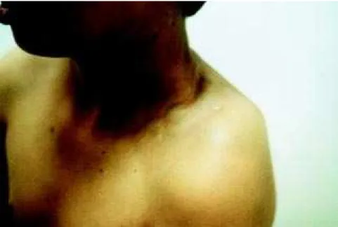
Figure 1 - Cistic lesions on the left supraclavicular area.
family history of the disease. It was not possible to get precise information about previous food intake, but the cur-rent nourishment provided to the child was adequate.
At the physical examination, the boy was found to be in good general condition, extremely pale, hydrated, eupneic, tachycardiac, and without vis-ceromegalies. The examination re-vealed 5 cystic, varicose, bluish, pressoreceptive formations in the left supraclavicular region, with diameters that varied between 2 and 6 cm, and that immediately returned to their original form after digital pressure (Fig. 1). The child also presented a nodule of 2 cm of diameter at the sole of the left foot, in which small caliber vases could be visualized, and another of similar characteristics in the first left toe (Fig. 2).
The admission hemogram revealed: Hb = 4.2 mg/dL, Ht = 15%, VCM = 50, HCM = 14, CHCM = 28, anisocy-tosis, microcytosis and macrocyanisocy-tosis, hypochromia, poikilocytosis, and tear-shaped ovalocytes and erythrocytes. The leukocyte and platelet counts were normal. Before receiving a transfusion of erythrocyte concentrate, blood samples were collected for etiologic analysis of the anemia that showed 12 mcg/dL of iron, 6.5 mcg/L of ferritin, total iron-binding capability of 525 mcg/dL, and normal hemoglobin elec-trophoresis and globular resistance curve.
The Doppler ultrasound examina-tion of the left supraclavicular region showed a mass in the soft tissues of heterogeneous texture, with small anechoic areas, suggesting the pres-ence of liquid. The Doppler image re-vealed that there was discreet flow and vascularization within the mass.
The diagnostic search for a clinical condition presenting the association of vascular malformations with serious iron deficiency anemia led the authors
to consider the blue rubber bleb nevus syndrome and thus to initiate the search for vascular malformations in the gastrointestinal tract by collecting of two samples for the investigation of occult blood in the feces. Both speci-mens were positive. In view of the con-firmation of intestinal bleeding, endo-scopic exams were then performed.
The colonoscopy showed wine-col-ored vascular lesions of about 0.6 cm in the rectum, sigmoid, descendent, and transverse colon, with raised, ir-regular surfaces, which were brittle to the contact with the equipment. In the cecum, near the ileo-cecal valve there was another larger lesion, measuring approximately two cm at its widest di-ameter.
The upper gastrointestinal endos-copy revealed three small wine-colored lesions in the stomach and a larger one in the anterosuperior duodenal wall, measuring two cm at its widest diam-eter.
The investigation of systemic in-volvement was completed with an ab-dominal ultrasound exam, contrasted cranium tomography, eyeground ex-amination and test for consumptive coagulopathy (prothrombin time, total activated thromboplastin time, fibrino-gen dosage and D-dimer), all of which yielded normal findings. The karyo-type collected after consultation with the geneticist also showed a normal re-sult (46 XY).
Endoscopic sclerotherapy of the gastric lesions was initiated and was later also performed in the colon le-sions, yielding good results.
The child was kept on iron replace-ment therapy by mouth and proceeded to show hematimetric rates within nor-mal levels for his age and sex.
To avoid the eventual risks involved in transfusions of blood derivatives, the patient was vaccinated against type B hepatitis.
DISCUSSION
The blue rubber bleb nevus syn-drome is a rare condition in which cu-taneous vascular malformations are as-sociated with gastrointestinal hemor-rhage due to the presence of similar le-sions in the gastrointestinal tract.1
Al-though the literature often denominates these lesions as “hemangiomas”, they are really vascular malformations with regard to their histological characteris-tics. Hemangiomas have diverse char-acteristics including hyperplastic en-dothelium, while the vascular malfor-mations were coated by normal endot-helium. In children, hemangiomas tend to grow considerably during the first 6 months of life, moderately in the sec-ond semester, and decrease in size, from 12 months of age on, and 95% of them will have disappeared by the time the child reaches 10 years of age. Vas-cular malformations do not usually present this behavior10.
In the blue rubber bleb nevus syn-drome, intestinal lesions are more fre-quent in the small intestine, although any anatomical site, from the mouth to the anus, may be affected. When the colon is affected, they are more
fre-quent in the rectal or distal areas9.
Other sites may also be affected, such as the eyes, nasopharynx, parotids, lungs, liver, spleen, heart, brain, pleura, peritoneum, pericardium, skeletal
muscles, bladder, and penis1,2,5,7.
Waybright correlated venous malfor-mations in the sublingual region with similar lesions in the central nervous system. Orthopedic abnormalities, such as bone deformities and hypertrophy, fractures, and articular involvement may also be present1,2,5,7. The patient
In this pathology, the appearance, number, size, and distribution of the lesions may vary. The largest lesion found in our patient was in the trunk, which has been described as a prefer-ential site of occurrence1,10.
The nevi are often present in early childhood, and they may grow, in-crease in number, and lead to compli-cations3,8. Some patients report painful
lesions, which may be due to the con-traction of surrounding smooth muscles1,3,5,7. There are also
descrip-tions of lesions that cause increased sweating, possibly due to the proxim-ity of the nevi to sweat-secreting
glands1,3,7. Our patient presented
le-sions first seen at birth that had been increasing in size, but that never caused local pain or excessive sweating. His major complaints regarded the incon-venience that was related to aesthetics. The reappearance of the lesions after surgical removal, as occurred in this case, has been described in the litera-ture, but their evolution to malignancy has never yet been reported1,5,9.
Thrombosis and calcification, as well as consumptive coagulopathy and thrombocytopenia, may occur within
the nevi1,6,11. but none of these were
found in the patient described here. This syndrome has been detected in successive generations, but that is not its most common presentation. Some au-thors believe that its transmission is pre-dominantly autosomal1–3,7. We could not
identify any similar cases among our patient’s relatives. The child’s mother had presented hematemesis 1 year be-fore the initial consultation, but high di-gestive endoscopy revealed a gastric ul-cer. The patient’s 6 brothers are healthy, and his father died in a car crash.
The main consequence of skin he-mangiomas is aesthetic, since they hardly ever bleed. Surgical excision of the cutaneous lesions is limited to those that occur in areas of higher risk of trauma, as in our patient, for whom surgery was indicated for the lesions in
the sole of the foot. The treatment may be by surgery, by local or systemic corticotherapy, or by laser photocoagu-lation5,7,10,12.
The intestinal nevi, in contrast to the cutaneous nevi, are easily injured and can bleed profusely. The bleeding may be massive, requiring transfusion or surgery. The bleeding may also be occult, and when it is chronic, it may
lead to iron deficiency anemia1,7. Our
patient never presented any visible bleeding, but he certainly suffered chronic blood losses, as indicated by the several nevi found in his gas-trointestinal tract. Symptoms of intes-tinal obstruction may occur due to in-tussusception, secondary to an angi-omatous intestinal involvement1,9,11. We
chose to perform the endoscopic exams of the gastrointestinal tract directly be-cause they are the most reliable diag-nostic means to detect these lesions. Additionally, in certain situations, en-doscopic exams can act therapeutically owing to the sclerosis of the localized lesions7.
Treatment of gastrointestinal le-sions varies according to their extent, localization, and consequences. For patients with small numbers of lesions or with lesions that are too diffuse and having anemia without open bleeding, elective transfusions and iron replace-ment have been recommended. For more significant hemorrhages, surgical resection, endoscopic sclerosis, laser photocoagulation, and therapy with in-terferon have been proposed1,5–7,9. The
management of these patients requires continued follow-up by outpatient con-sultations, in which new intercurrent diseases are evaluated and periodical monitoring of hematimetric rates is performed.
The blue rubber bleb nevus syn-drome must be differentiated from in-herited hemorrhagic telangiectasia (Rendu-Osler-Weber Syndrome). These patients may present palindro-mic epistaxis at the beginning of life
and present telangiectasias in the ado-lescence. In this condition, the gas-trointestinal lesions are similar, but the skin lesions are morphologically and histologically different. They are punc-tiform, red-bluish, and never reach the dimensions of those of the blue rubber bleb nevus syndrome. They affect the mucosa, lips, face, trunk, lungs, intes-tines, fingers, and lower limbs, but the sublingual lesions are very sugges-tive3,7,9.
The Klippel-Trenaunay Syndrome presents varicosities, hypertrophia, and soft tissue and bone deformities, as well as “port wine nevus”, generally located in just one of the extremities. It is also possible to find arteriovenous fistulas3,4,7,9,10.
The Maffucci Syndrome is charac-terized by diffuse vascular malforma-tions in the skin and soft tissues, asso-ciated with bone malformations and chondrodysplasias. Gastrointestinal tract lesions do not occur3,7,9,10.
There are other conditions in which vascular skin lesions are associated with lesions in other organs, such as meninges, in the Sturge-Weber Syn-drome; brain and retina, in the Von-Hippel-Lindau Syndrome; and spinal cord, in the Cobb Syndrome and others3,7.
CONCLUSION
The presentation of this case em-phasizes the importance of an inte-grated approach in which personal and family history, habits, lifestyle and physical evaluation are associated and made compatible. In this case, since the severe iron deficiency anemia did not seem to be a result of the previous alimentary history, the search for a pa-thology that could associate the find-ings of the physical exam was essen-tial to arrive at the diagnosis.
which vascular skin lesions are associ-ated with similar ones in other organs, as well as with other systemic alter-ations or specific complicalter-ations, such
as anemia, consumptive coagulopathy, intestinal obstruction, bone deformi-ties, ocular and central nervous system alterations. Thus, the early detection of
specific situations becomes essential as much for treatment as for genetic coun-seling.
RESUMO RHCFAP/3000
RODRIGUES D e col. - Síndrome de “nevo em bolha de borracha azul”.
Rev. Hosp. Clín. Fac. Med. S. Paulo 55 (1):29-34, 2000.
A síndrome do nevo em bolha de borracha azul “blue rubber bled nevus syndrome” é caracterizada por malformações venosas da pele e trato gastrointestinal, associadas a hemorragia intestinal e anemia ferropriva. Outros órgãos podem estar envolvidos. As causas desta síndrome são desconhecidas e sua apresentação
mais comum é na forma de casos esporádicos, mas herança autossômica dominante já foi descrita. É uma condição que afeta igualmente ambos os sexos e sua ocorrência é rara na raça negra.
Apresentamos um caso desta síndrome diagnosticada em criança de onze anos de idade. Ela apresenta ane-mia grave e tumoração venosa do tronco. Lesões semelhantes foram encontradas no estômago, intestino e um dos pés. São enfatizados os aspectos clínicos principais – lesões no
intestino, olhos, nasofaringe, parótida, pulmões, fígado, baço, coração, cérebro, pleura, peritônio, pericárdio, músculo esquelético, bexiga e pênis – e complicações sistêmicas que podem ocorrer nestes pacientes – trombose e calcificação dos nevus, assim como coagulopatia de consumo e trombocitopenia.
DESCRITORES: Síndrome do
nevo em bolha de borracha azul. Hemorragia. Anemia. Malformações venosas.
REFERENCES
1. MOODLEY M – Blue rubber bled nevus syndrome: case report and review of the literature. Pediatrics 1993; 92:160-162.
2. MC CANNEL C A, HOENIG J, UMLAS J et al. – Orbital lesions in the blue rubber bled syndrome. Ophtalmology 1996; 103:933. 3. JENNINGS M – Case report Blue rubber bled nevus syndrome: an
uncommon cause of gastrointestinal tract bleeding. Gut 1988; 29:1408.
4. CROSHER R F, BLACKBURN C W & DINSDALE R C W – Blue rubber bled nevus syndrome. Br J Oral Maxillofac Surg 1988; 26:160-164.
6. KUNISHIGE M, AZUMA H, MASUDA K et al. – Interferon alpha-2 a therapy for disseminated intravascular coagulation in a patient with blue rubber bled nevus syndrome. Angiology 1997; 48:273-277.
7. GALLO S H & McCLAVE S A - Blue rubber bled syndrome: gastrointestinal involvement and its endoscopic presentation. Gastrointest Endosc 1992; 38:72.
8. McKINLAY J R, KAISER J , BARRETT T L et al. – Blue rubber bled nevus syndrome. Cutis 1998; 62:97-98.
9. OKSUZOGLU B C, OKSUZOGLU G, ULKEM C et al. – Blue rubber bled nevus syndrome. Am J Gastroenterol 1996; 91:780-782.
10. RABINOWITZ L & ESTERLY N B – Vascular birthmarks and other abnormalities of blood vessels and lymphatics. In: SCHACHNER L A et al. – Pediatric Dermatology. New York, Churchill 1995. p. 953-971.
11. WONG Y C, LI Y W & CHANG M H - Gastrointestinal bleeding and paraparesis in blue rubber bled nevus syndrome. Pediatr, Radiol 1994; 24: 600-601.
12. WIRF F A & LOWITT M H - Diagnosis and treatment of cutaneous vascular lesions. Am Fam Physician 1998; 57 (4): 765-773.