Equine Assisted Therapy and Changes in Gait
for a Young Adult Female with Down Syndrome
Katherine J. Coffey Texas Woman’s University
Adam C. Knight Benjamin Wax Mississippi State University
The purpose of this study was to examine the effects of equine assisted therapy on selected gait parameters in a person with Down syndrome. One female
participant with Down syndrome completed two therapeutic horseback riding programs, each consisting of six riding sessions. Specific gait characteristics were analyzed with a trend analysis of the data by examining the means of the different variables. The trend analysis revealed a difference in stride length as well as hip and knee angle. These results indicate that over the course of the two therapeutic horseback riding programs, changes in gait occurred. Therefore, therapeutic horseback riding may have the potential to benefit gait characteristics and stability in young adult females with Down syndrome; however, further research is warranted.
Keywords: hippotherapy, therapeutic horseback riding, equine assisted therapy, individuals with disabilities, locomotor skills
Introduction
Equine assisted therapy is an umbrella term for all forms of therapy that include the assistance of a horse. The two most common forms of equine assisted therapy are therapeutic horseback riding and hippotherapy. Therapeutic horseback riding has been defined by the North American Riding for the Handicapped Association (NARHA, 2008) as the use of equine-oriented activities to achieve therapeutic goals. NARHA recently changed its organizational name and is now known as the Professional Association of Therapeutic Horsemanship International (PATH Intl.). The benefits of therapeutic horseback riding are claimed to be in the areas of therapy, education, sport and recreation, and leisure, with a focus on teaching horsemanship skills. This is in
contrast to hippotherapy, which refers to the equine-assisted movement under the direction of a physical or occupational therapist (American Hippotherapy Association, 2000). Prior research attributes positive benefits to the horse’s mimicking of normal human gait for the participant
(Bertoti, 1988). Bertoti (1991) identified the equine replication of normal human gait to include hip and pelvic rotation, weight shift, and proprioceptive stimulation.
The literature revealed other benefits of equine-assisted therapies; however, many of these studies were not subjected to a rigorous peer-review process (Gilliland & Knight, 2012). Studies in the literature subjected to a peer-review process utilized populations with cerebral palsy (Benda, McGibbon, & Grant, 2003; Bertoti, 1988; MacKinnon et al., 1995; McGibbon, Andrade, Widener, & Cintas, 1998; Sterba, Rogers, France, & Vokes, 2002). Included in the NARHA (2008) accreditation manual are benefits from therapeutic horseback riding programs for individuals with Down syndrome, but it is very cautious when prescribing activities for this population and contains more information about precautions and contraindications than benefits.
Down syndrome occurs in 1 out of every 724 births and is caused by an extra copy of the 21st chromosome (Laney, 2008). Atlantoaxial instability is the main concern of NARHA in the decision to include individuals with Down syndrome in program planning within the spectrum of equine assisted activities. Atlantoaxial instability is a misalignment of the top two vertebrae of the spine, the atlas and the axis (C-1 and C-2). According to the National Down Syndrome Society (2012), atlantoaxial instability occurs in 10 to 20 percent of individuals with Down syndrome; however, actual spine compression resulting from the condition is rare. Other
orthopedic characteristics that are more frequently associated with Down syndrome are ligament laxity and muscle hypotonia, indicating there is a lack of muscle tone and an extreme mobility of the joints. (Rigoldi, Galli, & Albertini, 2011).
The typical gait demonstrated by individuals with Down syndrome has been described as poorly coordinated and less efficient than in individuals without Down syndrome (Rigoldi et al., 2011). The delay in cognitive aspects combined with muscle hypotonia and ligament laxity that are associated with a diagnosis of Down syndrome are thought to be responsible for poorer control in programming actions and timing, as well as a reduction in force production. Cioni, Cocilovo, Rossi, Paci, and Valle (2001) concur with the association between the orthopedic characteristics of muscle hypotonia and joint/ligament laxity, combined with lower levels of cognition, and claim this combination manifests in gait pathologies demonstrated by limitations in range of motion to compensate for lack of balance and coordinated movement.
movement that appears to be used by individuals with Down syndrome to increase stability and improve safety. Carmeli, Ariav, Bar-Yossef, Levy, and Imam (2012) studied the progression of gait in younger compared to older individuals with Down syndrome and identified a variety of strategies that are used to compensate for the lack of coordination and balance and the inherent fear of falling. They identified a trunk stiffening strategy to be performed by older individuals with Down syndrome under dynamic or high postural threat conditions. The older adults in their study performed poorly in functional mobility tasks when compared to the younger individuals suggesting a need for adults with Down syndrome to continue therapeutic activity as an
intervention for deteriorating coordination problems.
There has been a lot of speculation regarding the role of cognitive delay in poor motor planning for individuals with Down syndrome (Cioni et al., 2001; Rigoldi et al., 2011). It is important to note, however, that the cognitive aspect described by a measure of intelligence can only account for a small portion of the gait anomalies, and that the common orthopedic characteristics provide the greatest disadvantage in achieving a normal gait pattern for individuals with Down
syndrome. Individuals with Down syndrome have a stereotypical gait, being described as ‘Chaplinesque’ (Caselli, Cohen-Sobel, Thompson, Adler, & Gonzalez, 1991). However, all individuals with a cognitive delay do not necessarily have Down syndrome, and those without the diagnosis of Down syndrome but with a cognitive delay do not necessarily have the same gait anomalies. Consequently, a large portion of the gait dysfunction for an individual with Down syndrome would be attributed to the common orthopedic characteristics of muscle hypotonia and ligament laxity. Carmeli et al. (2012) suggest several additional assumptions that add to the causes of gait dysfunction for individuals with Down syndrome: differences in lifestyle, trunk stiffness, and fear of falling.
One of the benefits often associated with equine assisted therapy is the improvement in muscle tone (Bertoti, 1988). Other benefits identified for children with cerebral palsy include
improvements in posture, balance, and coordination (Sterba et al., 2002). Copetti et al. (2007) found hippotherapy produced positive changes in the angular behavior of the ankle joint for children with Down syndrome having an average age of 7. Winchester, Kendall, Peters, Sears, and Winkley (2002) found improvements in gross motor function following a therapeutic
horseback riding program for children ages 4-7 with developmental delays. These improvements were maintained for at least seven weeks following therapeutic horseback riding. Rigoldi et al. (2011) stresses the importance of early intervention to modulate positive changes in gait for individuals with Down syndrome, but there is no evidence on the effects of a therapeutic program on an individual with Down syndrome past the age of 15. The purpose of this case study was to determine the effect on gait of a 24-year-old individual with Down syndrome from participation in equine assisted therapy. This information will add to the current body of
literature and add to a greater understanding of gait pathologies in the Down syndrome
for individuals with Down syndrome to help determine if they outweigh the potential risks of participation for this specific population.
Case Study
Method
Participant. This paper presents a single case study of a 24-year-old female with the clinical diagnosis of Down syndrome. She was screened for atlantoaxial instability with negative results prior to the onset of hippotherapy. She originally started participating in hippotherapy as part of her occupational therapy approximately 3 years prior to this study. After participation in
hippotherapy for three years, she graduated to therapeutic horseback riding, so she could focus on riding skills and has a goal of competing in local equine events. The participant agreed to participation in the study, and her mother, acting as legal guardian, gave her written consent to participate in this study and to have the results and conclusions published. The protocols were developed to conform to the Human Subjects Review Board of Mississippi State University.
Study. The primary purpose of this study was to analyze the effect of equine assisted therapy on the participant’s walking gait. Gait was recorded both before and after each horseback riding session to analyze any changes that may have occurred over the course of two separate six-week riding sessions, approximately five months apart. The procedures of this study followed the same protocol as Gilliland and Knight (2012).
Biomechanical Analysis of Gait
Variables. The dependent variables that were also described in Gilliland and Knight (2012) included stride length (meters), step width (meters), ankle angle (degrees), knee angle (degrees), and hip angle (degrees) at both heel strike and toe off, measured at two levels for the independent variable of testing time (Pre = before riding; Post = after riding) and measured at two separate events (Fall 2009 session and Spring 2010 session) for the independent variable of testing session. Stride length was defined as the distance, in meters, from left heel strike to left heel strike. Step width was defined as the distance, in meters, between the centers of the posterior aspect of the participant’s shoe. Joint angles were measured at each joint axis.
Measurement instruments. To document the female’s gait, two Sony Handycam (Sony Corporation, Tokyo, Japan) digital camcorders were used in this investigation. One camera was placed laterally to the participant to film her motion in the sagittal plane, and the second camera was placed anteriorly to the participant to film her motion in the frontal plane.
Experimental Design
Development of the experiment. The experiment was designed to examine the participant’s gait characteristics both before and after each therapeutic horseback riding session and over the course of two programs. This study followed the same design as Gilliland and Knight (2012).
After the participant arrived at the horseback riding center, the participant’s walking gait was recorded prior to the therapeutic riding session. The participant walked 20 meters, turned
around, and walked back to the starting position. The participant was instructed to walk at a self-selected comfortable walking pace. After completing the first gait session, the participant completed a 45-minute therapeutic horseback riding session. Following the riding session, the participant’s gait was recorded again.
Statistical methods. Since this study involved a single case study with one participant, a trend analysis was conducted by graphing the means of the different variables and comparing
differences across time and the two separate testing sessions. Differences were ascertained by a 2 x 2 repeated measures ANOVA (time by session). An alpha significance level of 0.05 was set for this investigation.
Results
Stride Length, Step Length, and Step Width
The trend analysis revealed a difference in stride length. There was a large decrease in stride length between the fall 2009 pretest (stride length < 1.11 m) and the spring 2010 pretest (stride length = .98 m), a decrease in stride length between the fall 2010 posttest (stride length = 1.11 m) and the spring 2010 pretest (.98 m), and a decrease in stride length (p < .05) between the fall 2010 posttest and the spring 2010 posttest.
Step width was only measured for the spring 2010 riding session. There was no difference (p > .05) in step width between the pretest and posttest session. The means and standard deviations for stride length, step length, and step width can be found in Table 1.
Table 1. Mean (+ SD) of the Observed Distances During the Different Testing Sessions
Variable
Fall 2009 PreTest
Fall 2009 PostTest
Spring 2010 PreTest
Spring 2010 PostTest
Stride Length *1.11 (.02) m 1.11 (.06) m 0.98 (.10) m 0.96 (.14) m
Step Width N/A N/A 0.20 (.03) m 0.18 (.26) m
*Indicates a difference in group means identified by trend analysis.
Joint Angles
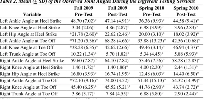
(68.28°) and the 2010 pretest and 2010 posttest. There were differences in left knee angle at toe off (p < .05), with an increase in knee angle between the 2009 pretest (38.20°) and the 2010 pretest (49.46°) and an increase in knee angle between the 2009 posttest (42.62°) and the 2010 pretest. There were also differences (p < .05) in right ankle angle at toe off, with a decrease in ankle angle between the 2009 pretest (72.10°) and the 2010 pretest (51.44°), a decrease between the 2009 pretest compared to the 2010 posttest (54.32°), and a decrease between the 2009 posttest compared to the 2010 posttest. There were no notable differences found in any of the other joint angles. The means and standard deviations for all joint angles can be found in Table 2.
Table 2. Mean (+ SD) of the Observed Joint Angles During the Different Testing Sessions
Variable
Fall 2009 Pre-Test
Fall 2009 Post-Test
Spring 2010 Pre-Test
Spring 2010 Post-Test
Left Ankle Angle at Heel Strike 48.70 (7.02)° 47.14 (4.91)° 36.36 (9.93)° 44.58 (9.41)°
Left Knee Angle at Heel Strike 3.04 (2.06)° 4.86 (2.87)° 6.98 (3.99)° 3.96 (2.83)°
Left Hip Angle at Heel Strike *21.78 (2.60)° 22.62 (2.46)° 20.00 (3.10)° 18.02 (3.92)° Left Ankle Angle at Toe Off *71.20 (5.36)° 68.28 (4.66)° 33.88 (13.23)° 42.56 (10.68)°
Left Knee Angle at Toe Off *38.28 (6.35)° 42.62 (2.66)° 49.46 (3.14)° 46.94 (4.37)°
Left Trunk Angle at Toe Off 10.22 (1.34)° 5.70 (1.82)° 5.34 (4.45)° 5.88 (5.93)°
Right Ankle Angle at Heel Strike 59.60 (7.87)° 64.10 (7.84)° 53.46 (7.56)° 58.28 (12.83)°
Right Knee Angle at Heel Strike 1.46 (1.72)° 1.40 (.86)° 4.00 (2.30)° 2.44 (1.31)°
Right Hip Angle at Heel Strike 16.80 (3.93)° 16.74 (1.95)° 12.48 (6.03)° 14.40 (6.50)° Right Ankle Angle at Toe Off *72.10 (9.16)° 74.00 (3.52)° 51.44 (15.13)° 54.32 (14.99)°
Right Knee Angle at Toe Off 45.40 (6.25)° 45.52 (5.21)° 41.76 (2.90)° 43.74 (2.72)°
Right Trunk Angle at Toe Off 3.86 (3.17)° 7.84 (4.55)° 6.88 (5.80)° 2.90 (2.44)°
*Indicates a difference in group means identified by trend analysis.
Discussion
The purpose of this study was to examine the effects of equine assisted therapy on selected gait parameters in a person with Down syndrome. Rigoldi et al. (2011) indicated that individuals with Down syndrome demonstrate a motion pattern consistent with compensatory strategies to increase stability, while concurrently diminishing the number of body segments involved in gait and thereby increasing joint stiffness. This strategy results in a less functional mechanical structure and is less efficient than normal gait demonstrated by individuals without Down syndrome. Another aspect of the compensatory strategy theory involves increasing gait width. Older individuals with Down syndrome were shown to have a wider gait than younger
Rigoldi et al. (2011) also noted that as individuals with Down syndrome age, stride length has been shown to increase, and joint range of motion, particularly at the knee, to decrease in favor of increasing ankle-generated work. Following horseback riding, the participant in this study demonstrated a decrease in stride length and an increase in knee extension during the 2010 riding program as compared to the 2009 riding program. This could indicate a more stable gait pattern by the reduction in the amount of time the participant spent during single leg support. In the current study, there was an overall decrease in left hip angle at heal strike, creating a more upright posture, and according to Cote, Brunet, Gansneder, and Shultz (2005), adding to stability by maintaining the center of gravity over the base of support. Additionally, the decrease in hip angle at heal strike is consistent with greater hip extensor power to accelerate forward
momentum. Rigoldi et al. (2011) claims as individuals with Down syndrome age, they reveal a reduction in power generation and absorption through a reduction in hip and knee range of motion. Power production was not reviewed in this study; however, future research should be conducted to evaluate this issue in greater detail.
Following horseback riding, the rider in this study demonstrated an increase in left knee angle (increased knee extension) at toe off and a decrease in left ankle angle (increased dorsi-flexion) at toe off which implies a shorter, more stable gait cycle. In contrast, Cioni et al. (2001) found individuals with Down syndrome demonstrate a greater amount of plantar flexor action and knee flexion, possibly caused from an unstable base of support. They continue to explain ankle kinetic dysfunction in individuals with Down syndrome as an expression in part by weak or instable ankle flexor muscles due to muscle hypotonia and ligament laxity. One of the basic postures in horseback riding requires the rider to keep his/her heel down and maintain the foot in a dorsi-flexed position. Consequently, the rider in this study was required to work on ankle strengthening activities along with the core muscles involved in postural control and found benefits in angular kinetics of the ankle joint that concur with Copetti et al. (2007).
Carmeli et al. (2012) explain that older individuals with Down syndrome demonstrate greater difficulty performing motor tasks when compared to younger individuals with Down syndrome. They found balance and coordination to be the most challenging and claim limited participation in daily as well as recreational activities to be a potential cause. Rigoldi et al. (2011) also suggest balance and coordination to be a particular problem for older individuals with Down syndrome and indicate a fear of falling to be an issue concerning gait. Participation in horseback riding has been demonstrated to improve postural control, coordination, and balance (Winchester et al., 2002). The participant in the current study demonstrated changes that led to increased stability and a more efficient gait pattern.
The limitations of this study are aligned with the limitations reported by Gilliland and Knight (2012), primarily being attributed to the nature of a case study. First, there was one female participant with Down syndrome in this investigation; therefore, generalizations to other
present study found encouraging results regarding positive changes in gait for her, although this may have been due to participation in hippotherapy prior to the two years examined during the therapeutic horseback riding in the program. Future research studies should examine if
therapeutic horseback riding increases gait efficiency among a variety of ages and gender of participants with Down syndrome and how long these changes in gait efficiency persist in the posttherapeutic period.
References
Agiovlasitis, S., McCubbin, J.A., Yun, J., Pavol, M.J., & Widrick, J.J. (2009). Economy and preferred speed of walking in adults with and without Down syndrome. Adapted Physical Activity Quarterly, 26, 118-130.
American Hippotherapy Association. (2000). Present use of hippotherapy in the United States.
Retrieved from http://www.americanhippotherapyassociation.org/hippotherapy/present-use-of-hippotherapy/
Benda, W., McGibbon, N. H., & Grant, K. L. (2003). Improvements in muscle symmetry in children with cerebral palsy after equine-assisted therapy (hippotherapy). The Journal of Alternative and Complementary Medicine, 9(6), 817–825.
doi:10.1089/107555303771952163
Bertoti, D. B. (1988). Effect of therapeutic horseback riding on posture in children with cerebral palsy. Physical Therapy, 68(10), 1505–1512. Retrieved from
http://ptjournal.apta.org/content/68/10/1505
Bertoti, D. B. (1991). Clinical suggestions. Effect of therapeutic horseback riding on extremity weightbearing in a child with hemiplegic cerebral palsy: A case report as an example of clinical research. Pediatric Physical Therapy, 3(4), 219–226.
Carmeli, E., Ariav, C., Bar-Yossef, T., Levy, R., & Imam, B. (2012). Movement skills of younger versus older adults with and without Down syndrome. Research in Developmental Disabilities, 33(1), 165–171. doi:10.1016/j.ridd.2011.09.008
Caselli, M. A., Cohen-Sobel, E., Thompson, J., Adler, J., & Gonzalez, L. (1991). Biomechanical management of children and adolescents with Down syndrome. Journal of the American Podiatric Medical Association, 81(3), 119–127. doi:10.7547/87507315-81-3-119
Cioni, M., Cocilovo, A., Rossi, F., Paci, D., & Valle, M. S. (2001). Analysis of ankle kinetics during walking in individuals with Down syndrome. American Journal on Mental Retardation, 106(5), 470–478. doi:10.1352/0895-8017(2001)106<0470:AOAKDW >2.0.CO;2
Copetti, F., Mota, C.B., Graup, S., Menezes, K.M., & Venturini, E.B. (2007). Angular kinematics of the gait of children with Down’s syndrome after intervention with hippotherapy. Revista Brasileira de Fisioterapia, 11(6), 503-507.
Galli, M., Rigoldi, C., Mainardi, L., Tenore, N., Onorati, P., & Albertini, G. (2008). Postural control in patients with Down syndrome. Disability and Rehabilitation, 30(17), 1274– 1278. doi:10.1080/09638280701610353
Gilliland, K. J., & Knight, A. C. (2012). Friedreich’s Ataxia and gait changes through participation in therapeutic horseback riding. Clinical Kinesiology, 66(1), 1–6. Laney, D. (2008). Perspectives on diseases & disorders: Down syndrome. Detroit, MI:
Gale/CENAGE Learning.
MacKinnon, J. R., Noh, S., Lariviere, J., MacPhail, A., Allan, D. E., & Laliberte, D. (1995). A study of therapeutic effects of horseback riding for children with cerebral palsy. Physical & Occupational Therapy in Pediatrics, 15(1), 17–34. doi:10.1080/J006v15n01_02 McGibbon, N. H., Andrade, C., Widener, G., & Cintas, H. L. (1998). Effect of an
equine-movement therapy program on gait, energy expenditure, and motor function in children with spastic cerebral palsy: A pilot study. Developmental Medicine & Child Neurology, 40(11), 754–762. doi:10.1111/j.1469-8749.1998.tb12344.x
National Down Syndrome Society. (2012). Atlantoaxial instability & Down syndrome: Controversy & commentary. Retrieved from http://www.ndss.org/Resources/Health-Care/Associated-Conditions/Atlantoaxial-Instability-Down-Syndrome/
North American Riding for the Handicapped Association (NARHA). (2008). NARHA standards and accreditation manual for NARHA centers (2008 ed.).Denver, CO: NARHA.
Parker, A. W., & Bronks, R. (1980). Gait of children with Down syndrome. Archives of Physical Medicine and Rehabilitation, 61(8), 345–351.
Rigoldi, C., Galli, M., & Albertini, G., (2011). Gait development during lifespan in subjects with Down syndrome. Research in Developmental Disabilities, 32(1), 158–163.
doi:10.1016/j.ridd.2010.09.009
Sterba, J. A., Rogers, B. T., France, A. P., & Vokes, D. A. (2002). Horseback riding in children with cerebral palsy: Effect on gross motor function. Developmental Medicine & Child Neurology, 44(5), 301–308. doi:10.1111/j.1469-8749.2002.tb00815.x
Winchester, P., Kendall, K., Peters, H., Sears, N., & Winkley, T. (2002). The effect of
therapeutic horseback riding on gross motor function and gait speed in children who are developmentally delayed. Physical & Occupational Therapy in Pediatrics, 22(3–4), 37– 50. doi:10.1080/J006v22n03_04
Dr. Katherine J. Coffey is an Associate Clinical Professor in the Department of Kinesiology at Texas Woman’s University, Denton, TX.
Dr. Adam Knight is an Associate Professor in the Department of Kinesiology at Mississippi State University, Starkville, MS.