Available in: http://www.redalyc.org/articulo.oa?id=33906803
Red de Revistas Científicas de América Latina, el Caribe, España y Portugal
Sistema de Información Científica
Francisca E. L. Freitas, Flora Cordeiro Mori, Estela Sasso Cerri, Sandra R. R. Lucas, Sandra M. Miraglia
Alterations of spermatogenesis in etoposide-treated rats: a stereological study
Interciencia, vol. 27, núm. 5, mayo, 2002, pp. 227-235,
Asociación Interciencia
Venezuela
How to cite
Complete issue
More information about this article
Journal's homepage Interciencia,
ISSN (Printed Version): 0378-1844 [email protected]
Asociación Interciencia Venezuela
www.redalyc.org
KEY WORDS / Etoposide / Seminiferous epithelium / Spermatogenesis / Testis / Topoisomerase /
Received: 11/19/2001. Modified: 03/14/2002. Accepted: 04/02/2002
Francisca E. L. Freitas. Master in Morphology, Federal University of São Paulo (UNIFESP).
Professor, Laboratory of Natural Sciences, Federal University of Acre, Rio Branco, Brazil.
Flora Cordeiro-Mori. Master in Morphology, Federal University of São Paulo (UNIFESP).
Professor, Department of Health Sciences, Paulista University, São Paulo, Brazil.
Estela Sasso-Cerri. Master in Morphology. Doctor in Health Sciences, UNIFESP. Professor,
Laboratory of Embryology, Department of Morphology, UNIFESP.
Sandra R. R. Lucas. Master in Molecular Biology and Doctor in Health Science, UNIFESP.
Professor, Laboratory of Embryology, Department of Morphology, UNIFESP.
Sandra M. Miraglia. Master in Morphology. Doctor in Health Sciences, UNIFESP.
Profes-sor, Laboratory of Embryology, Department of Morphology, UNIFESP. Address: Rua Bartolomeu Feio, edifício Manchester, número 66, apto. 164, Brooklin, CEP 04580-000, São Paulo, S.P., Brazil. e-mail: [email protected]
ALTERATIONS OF SPERMATOGENESIS
IN ETOPOSIDE-TREATED RATS:
A STEREOLOGICAL STUDY
FRANCISCA E. L. FREITAS, FLORA CORDEIRO-MORI,
ESTELA SASSO-CERRI, SANDRA R. R. LUCAS and SANDRA M. MIRAGLIA
odophyllin is the crude extract from the roots and rhizomes of two plants from India, Podophyllum peltatum and Podophyllum emodi (Arnold, 1979; Issel and Crooke, 1979). Podophyllotoxin, a main active constituent of podophyllin, is known to produce metaphase arrest. It binds tubulin at a distinct site from that occupied by the vinca alkaloids, inhibit-ing microtubule formation in the mitotic spindle apparatus. However, it is too toxic for clinical use (Chen et al., 1984). Thus, attempting to discover less toxic substances, two semi-synthetic glycosidic derivatives of epipodophyllotoxin were synthesized (Stähelin, 1973): the etopo-side (Vepesid, code VP-16-213, abbrevi-ated VP-16) and the teniposide (VM-26). In the 70s, these derivatives obtained ap-probation from the FDA for clinical use. Unlike podophyllotoxin, etoposide has no effect on microtubule assembly. It does not arrest mitosis (Chen et al., 1984) but, rather, exerts its maximal effects in the S or G2 cell cycle phases preventing cells from entering mitosis (Calabresi and Parks, 1985).
The etoposide (4’-Deme- thylepipodophyllotoxin-9-(4,6-O-ethyl-idene-β-D-glucopyranoside) has been uti-lized in a wide variety of neoplasms in-cluding small-cell lung cancer, esophagic carcinoma, Kaposi’s sarcoma, testicular cancer, acute leukemia and, both Hodg-kin’s and non HodgHodg-kin’s lymphomas (Ko-bayashi and Ratain, 1994).
The main target of this drug is topoisomerase II (Minocha and Long, 1984), and for this reason it is known as “poison of topoisomerase” like other drugs such as anthracyclines which have a similar mechanism of action (Joel
et al., 1994; Joel and Slevin, 1994). In
spite of the beneficial effects of this drug in malignant tumor treatment, some re-ports have also emphasized a deleterious action on the organism of mammals, in-cluding the testes (Wozniak and Ross, 1983; Takahashi et al., 1986a; Kadota et
al., 1989). The myelossupression and
go-nadal failure (Myers and Schilsky, 1992) are the two most common side effects re-lated to the use of cytotoxic anticancer agents. The implications of histological data on reproductive performance must
228 MAY 2002, VOL. 27 Nº 5
daily doses of etoposide. A histomorpho-metric and stereological study was car-ried out, and the serum testosterone level and reproductive performance of the ani-mals were investigated in the adult state. In addition, since this drug is also mu-tagenic (Maraschin et al., 1990) and clastogenic (Agarwal et al., 1994), the body weight, viability and mortality index of the offspring were also observed.
Material and Methods
Animals and groups
Forty-eight male albino Wistar 30-day old rats were distributed among six groups and were sacrificed at either 60 or 113 days of age (Table I). Two of the groups received Vepesid (VP16-213), the etoposide-experimental (E60) and the mated etoposide-experimen-tal (EM113) groups; two other groups re-ceived the vehicle, the vehicle-experimen-tal group (V60) and the mated vehicle-ex-perimental group (VM113); the two re-maining groups were control groups, in-cluding the sham-control group (C60) and the mated sham-control group (CM113). Animals were allocated to the treatment groups in a randomized manner and ob-tained from different progenies. No more than six newborn rats were maintained during the period of breast-feeding, with the aim to equalize their body weights at the start of the treatments. The mated groups (CM113, EM113 and VM113) had ten animals each, while each of the other groups included six rats.
All animals were kept in plastic cages under 12-12 hour light cycle at 23-25°C. Food and water were allowed
ad libitum. The general physiological
state of the animals was watched during the treatment with the etoposide, although the control of the blood cells was not carried out. Principles of laboratory ani-mal care (NIH publication 85-23, 1985) and national laws on animal use were ob-served. This research had approval from the Ethical Committees of the Federal University of São Paulo and the São Paulo Hospital.
Dosage and schedule
Vepesid®ampoules (VP16-213, Bristol–Myers Squibb) were utilized. The contents of 5ml ampoules, (100mg of etoposide each) were diluted in the ve-hicle. The vehicle contained 650mg poly-ethylene glycol 300, 30mg benzyl alco-hol, 1mg citric acid, 80mg Tween 80 and 241mg ethyl alcohol per ml. Dosage so-lutions of etoposide were prepared by di-lution of each ampoule with 0.9% physi-ological saline according to the timetable from Bristol Myers Squibb. The rats in the vehicle-experimental groups (V60 and VM113) received the vehicle solution di-luted with physiological saline; those in the sham-control groups (C60 and C113) received only physiological saline; while rats in the experimental groups (E60 and EM113) received 2mg/kg of body weight (bw) of etoposide. The animals were sub-jected to each specific treatment for 30 consecutive days from the prepuberal phase, when they were 30 days old (Table I). Intraperitoneal injection was preferred over intravenous injection be-cause of ease of administration. Lu and Meistrich (1979) have shown that both methods of administration generally yield similar results. The long-term administra-tion of low-daily-dose etoposide was
uti-lized in this report and its use in patients has been suggested (Greco and Hains-wort, 1994).
Initially, the dosage was established according to the literature. In addition, a pilot test using different doses was previously run. The dosage was se-lected taking into account the testicular morphological response and the adequate mating conditions of the animals. After the end of the experiment, thirty animals of the EM113, VM113 and CM113 groups were maintained for 53 days to assess their fertility, as the duration of spermatogenesis in Wistar rats is of 52 days (Huckins,1965).
Histological and mating procedures
The rats were anaesthe-tized with sodium pentobarbital (40mg/kg bw). Their testes were dissected, removed from the scrota and weighed. A microme-ter caliper was utilized to measure major and minor axes; and the gonads were then fixed by immersion in Bouin’s liquid for 48h. Cross and oblique random sec-tions were obtained from the fragments of the gonad, allowing a sequential ad-equate morphometric and stereological analysis (Gundersen et al., 1988; Manda-rim-de-Lacerda, 1999). The specimens were processed and embedded in para-plast-plus. To obtain a better identifica-tion of all phases of spermatogenesis, 3µm-thick sections were stained with the periodic acid-Schiff method and counter-stained with Harris’Hematoxylin (PAS + H).
Morphometric and stereological studies
Testicular weight and testis total volume.
The testis is an ellipsoid (Ahmad et al., 1969). Thus, the total volume of each testis was calculated by the modified for-mula of ellipsoid volume: V= 4/3·p·a·b2, where a is the semiprolate axis and b is the semioblate axis (Miraglia and Ha-yashi, 1993; Botelho Cabral et al., 1997). The absolute and relative testicular weights were also determined (Table II).
Volume density. Volume densities (Vv) of
both seminiferous tubules and interstitial tissue were calculated (Gundersen et al., 1988). These values were obtained using a 25-point integrating eyepiece attached to a light binocular microscope (Weibel, 1963). One thousand points in 40 fields of tes-ticular sections were randomly counted per testis at x100 magnification. Based on these data (Table III) and on the total tes-ticular volume, the volumes of the corre-spondent testicular components were cal-culated (Miraglia and Hayashi, 1993). TABLE I
DISTRIBUTION OF THE EXPERIMENTAL AND CONTROL GROUPS
Initial age for Type of Mating Sacrifice Period of injection injection age age mating Control (days) (days) (days) (days) groups
C60 30 0.9% saline - 60
-CM113 30 0.9% saline 103 113 10
Experimental groups
E60 30 Etoposide (VP-16) - 60 -EM113 30 Etoposide (VP-16) 103 113 10
V60 30 Vehicle - 60
-VM113 30 Vehicle 103 113 10
C60, V60 and E60 groups, n=6 in each group
Tubular diameter. The diameter of
semin-iferous tubule sections was measured uti-lizing an eyepiece micrometer (x8) at-tached to a light binocular microscope. Random measurements of 100 tubules per animal were done in cross or oblique tions at x80 magnification. When the sec-tions were oblique, the minor axis was measured (Miraglia and Hayashi, 1993; Botelho Cabral et al., 1997).
Cells of germinal lineage. The
percent-ages of tubular sections containing the various cell types (Table IV) of the semi-niferous epithelium were determined by counting 100-random-transverse or ob-lique sections in each testis at x1000 magnification (Hayashi and Cedenho, 1980; Miraglia and Hayashi, 1993). B-type and intermediate spermatogonia were not distinguished individually and were added together. Similarly, neither A0-type and A1-A4 type spermatogonia were individually differentiated. Sperma-tids in the steps 1 to 8 were considered round spermatids and those in the steps 9 to 19 were denominated elongate.
Radioimmunoassay method
Blood was collected from the abdominal aorta of all animals, previously anaesthetized, to measure se-rum testosterone. The sese-rum was main-tained at -20ºC. Afterwards, the animals were sacrificed with an exceeding dose of the anesthetic. Measurements were made
in a single assay by standard radioimmu-noassay method (Lox et al., 1974).
Reproductive performance
Each of the rats in the EM113, VM113 and CM113 groups were mated in individual cages with two multi-parous female rats for 10 consecutive days when they were 103 days old, to as-sess their fertility. The fertility index in each group was considered as the ratio of the total quantity of offspring to the total number of mated female rats (Table V). The progenies were observed for 30 con-secutive days in order to obtain informa-tion about their body weight, viability as well as their mortality index.
Statistical methods
The Jandel Statistical SigmaStat software, version 2.0, was uti-lized. To compare the two sets of groups of same age (E60, V60 and C60, and E113, V113 and C113) an analysis of variance (ANOVA) was performed. All data obtained from morphometric and ste-reological studies were submitted to a Standard One Way ANOVA, excepting the frequencies of the cellular types of germi-nal lineage; when the results obtained were significant or highly significant, all pairwise multiple-comparison procedures (Student-Newman-Keuls Method) were used in the variables. On the other hand, for the analysis of the frequencies of
ger-minal lineage cell types, a non-parametric ANOVA on Ranks rather than a Standard One Way ANOVA was performed. Thus, when these last data showed significance, a complementary multiple-comparison test (Dunn’s method) was applied. In this case, in spite of the utilization of a non-para-metric analysis, the values of means are also presented in Table III, with the pur-pose of giving an idea on the variability of these data and also to allow their compari-son with the data obtained from the litera-ture.
Results
General aspects of the etoposide-treated animals
The etoposide-treated al-bino animals exhibited piloerection and paleness in the extremities, tail and eyes, evidencing a probable anemia. Other ad-verse effects including myocardial infarc-tion, hypotension, peripheral neuropathy, ascites, systemic debility and ataxy were not observed. Some hepatosplenomegaly was noted. One animal presented anaphy-lactic shock; it died and was substituted. In general, the rats showed transient diar-rhea and some reversible alopecia, side ef-fects that disappeared after the completion of the treatment, when the animals were in adult phase. A decrease of food intake was another adverse effect observed dur-ing the treatment; however, there was some stabilization as there was some re-TABLE II
BODY WEIGHT AND TESTICULAR MORPHOMETRY IN ALBINO RATS OF THE CONTROL AND EXPERIMENTAL GROUPS
Body Absolute Relative Major Minor Total testicular weight testicular testicular axis axis volume
weight weight
Control (g) (g) (g/100g bw) (mm) (mm) (mm3)
groups
C60 235.00 ± 8.03 1.25 ± 0.08 0.57 ± 0.04 18.59 ± 0.34 11.10 ± 0.38 1199.30 ± 105.56 CM113 345.72 ± 20.16 1.58 ± 0.11 0.46 ± 0.05 20.90 ± 0.33 11.96 ± 0.36 1565.33 ± 114.29
Experimental Groups
E60 145.98 ± 6.22** 0.78 ± 0.08** 0.53 ± 0.03 15.51 ± 0.61** 9.23 ± 0.29** 691.85 ± 58.56** E60<C60 and V60 E60<C60 and V60 E60<C60 and V60 E60<C60 and V60 E60<C60 and V60
EM113 308.40 ± 8.01* 0.76 ± 0.18** 0.25 ± 0.06** 15.54 ± 1.20** 9.49 ± 0.66** 732.79 ± 168.15** E113<C113 and V113 E113<C113 and V113 E113<C113 and V113 E113<C113 and V113 E113<C113 and V113 E113<C113 and V113
V60 242.00 ± 9.11 1.22 ± 0.07 0.50 ± 0.07 19.08 ± 0.41 11.20 ± 0.40 1253.18 ± 126.98 VM113 350.80 ± 18.20 1.54 ± 0.13 0.47 ± 0.04 21.00 ± 0.35 11.88 ± 0.29 1551.61 ± 143.45
Values given as mean ± standard error *p<0.05 (significant).
** p<0.01 (highly significant).
230 MAY 2002, VOL. 27 Nº 5
covery of body weight noted in 113-day-old adult rats of the mated etoposide-ex-perimental group.
Histological analysis and frequencies of the cellular types of germinal lineage
In the etoposide-experi-mental group (E60), the most damaged se-miniferous convoluted tubules showed low cellular population density with a corre-sponding enlargement of lumen diameter and intraepithelial vacuolation (Figures 1, 2, 3). Tubular sections containing only Sertoli cells (“Sertolization”; Figure 3) as well as spermatogonia were rarely ob-served. Although the animals in the E60 group sometimes exhibited normal tubular sections under the light microscope,
py-knotic nuclei of germinal lineage cells were observed in other sections, character-izing the degeneration of the seminiferous epithelium. Multinucleated formations of round spermatids and other rare forms of primary spermatocytes were also noted in this group (Figures 2 and 4, respectively). Sometimes, the nuclei of these formations showed chromatin condensation in one or several homogeneous masses, surrounding the nuclear membrane and suggesting apo-ptosis (Figure 2).
Some sections also dis-played disorganized cellular associations corresponding to different stages of the seminiferous epithelium cycle while oth-ers exhibited a large quantity of cellular debris and cells at various phases of maturation localized in the tubular lumen.
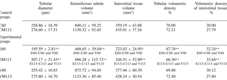
The tubular diameter (Table III) was largely diminished in both etoposide-experimental groups (E60 and EM113) in comparison to the correspon-dent sham-control and vehicle-experimen-tal groups.
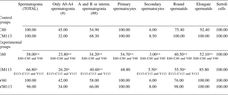
The frequencies of the tubular sections containing the various types of germinal lineage cells are shown in Table IV. In a hundred sections exam-ined per testis, the frequencies of all cell types of germinal lineage exhibited an ac-centuated reduction in the E60 group, when compared to both C60 and V60 groups.
In the mated etoposide-experimental group (EM113), various semi-niferous tubule sections exhibited similar qualitative histological characteristics such
TABLE III
TESTICULAR STEREOLOGY IN ALBINO RATS OF THE CONTROL AND EXPERIMENTAL GROUPS
Tubular Siminiferous tubule Interstitial tissue Tubular volumetric Volumetric density diameter volume volume density of interstitial tissue
Control (µm) (mm3) (mm3) % %
groups
C60 258.86 ± 16.39 840.11 ± 59.25 359.19 ± 43.88 70.00 30.00 CM113 276.60 ± 17.51 1130.32 ± 92.65 435.01 ± 37.26 72.21 27.79 Experimental
groups
E60 195.59 ± 2.81** 468.65 ± 39.04** 223.03 ± 24.95* 67.76** 32.24** E60<C60 and V60 E60<C60 and V60 E60<C60 and V60 E60<C60 and V60 E60>C60 and V60
EM113 207.17 ± 21.43** 486.28 ± 115.72** 246.51 ± 52.89** 66.36** 33.64** E113<C113 and V113 E113<C113 and V113 E113<C113 and V113 E113<C113 and V113 E113>C113 and V113
V60 255.42 ± 16.83 875.72 ± 94.65 377.46 ± 60.39 69.88 30.12 VM113 275.80 ± 16.70 1123.36 ± 85.40 428.24 ± 30.54 72.40 27.60
Values given as mean ± standard error *p<0.05 (significant).
**p<0.01 (highly significant).
Figure 1. Light micrograph of testicular section of a 60-day-old etoposide-treated rat. An altered seminiferous tubule showing low frequency of germinal lineage cells and many cells with pyknotic nuclei (arrows). PAS + H (x245).
Figure 2. Light micrograph of portion of se-miniferous tubule of a 60-day-old etoposide-treated rat exhibiting intraepithelial vacuola-tion (asterisks) and a multinucleated forma-tion of round spermatids (large arrow). Mar-gined condensations of chromatin are ob-served in some nuclei of this formation, sug-gesting apoptosis (small arrows). PAS + H (x615).
as those observed in the E60 group. How-ever, tubular sections showing low cell den-sity, intraepithelial vacuolation (Figure 5) and “Sertolization” were less frequent in the 113-day-old than in the 60-day-old eto-poside-treated experimental animals, al-though the frequencies of various cell types of germinal lineage were still low in rela-tion to both CM113 and VM113 groups (Table IV). Multinucleated formations of round spermatids (Figure 2) were very rare in the EM113 group and those of sperma-tocytes (Figure 4) were absent.
In addition, some folding of peritubular tissue was occasionally seen
in rats of the E60 group, while it was not observed in the animals of the EM113 group. The Leydig cells showed normal morphology under light microscope in both etoposide-experimental groups.
The Sertoli cells also exhibited nuclei with normal morphology under the light microscope and the fre-quency of the tubules containing these cells was also normal in both E60 and EM113 (Table IV).
The animals of the V60 and VM113 groups (Figure 6) displayed normal testicular histology, like the C60 and the C113 groups, respectively.
Body and testicular weights
Animals in the E60 group showed a highly significant reduc-tion of body weight in comparison to the correspondent C60 and V60 groups, while those of the EM113 group only exhibited a significant decrease in relation to the CM113 and VM113 groups (Table II). In addition, rats of both E60 and EM113 groups showed highly significant decreases of the absolute testicular weight in com-parison with the sham control and vehicle-experimental groups of the same age. Consequently, 113-day-old mated
etopo-TABLE IV
FREQUENCIES OF GERMINAL LINEAGE CELL TYPES IN ALBINO RATS OF THE EXPERIMENTAL AND CONTROL GROUPS (PERCENTAGE OF TUBULES WITH CELL TYPES)
Spermatogonia Only A0-A4 A and B or interm. Primary Secondary Round Elongate Sertoli (TOTAL) spermatogonia spermatogonia spermatocytes spermatocytes spermatids spermatids cells
(#) (##) Control
groups
C60 100.00 45.00 54.90 100.00 6.00 75.40 92.40 100.00 CM113 100.00 32.00 68.30 100.00 8.50 100.00 100.00 100.00 Experimental
groups
E60 58.00** 23.80** 34.20** 54.70** 3.00** 40.50** 52.10** 100.00
E60<C60 and V60 E60<C60 and V60 E60<C60 and V60 E60<C60 and V60 E60<C60 and V60 E60<C60 and V60 E60<C60 and V60
EM113 66.80* 26.20* 40.60** 68.80 5.50* 55.50* 85.80 100.00
E113<C113 and V113 E113<C113 and V113 E113<C113 and V113 E113<C113 and V113 E113<C113 and V113
V60 100.00 42.00 58.00 100.00 6.00 76.00 100.00 100.00 VM113 96.00 34.00 66.00 100.00 8.00 98.00 100.00 100.00
Values given as means
(#) percentage of tubules containing only type-A spermatogonia.
(##) percentage of tubules containing type-A and type-B or intermediate spermatogonia. *p<0.05 (significant).
**p<0.01 (highly significant).
Figure 4. Light micrograph of portion of seminiferous tubules of a 60-day-old etopo-side-treated rat showing a multinucleated formation of primary spermatocytes (arrow). PAS + H (x615).
Figure 5. Light micrograph of portion of se-miniferous tubule of a 113-day-old etoposide-treated rat exhibiting low frequency of germi-nal lineage cells, intratubular vacuolation (as-terisks) as well as sloughing cells and cellular debris in the cellular lumen. PAS+H (x310).
232 MAY 2002, VOL. 27 Nº 5
side-experimental rats showed a very sig-nificant reduction of relative testicular weight (g/100g bw), while 60-day-old eto-poside-experimental rats did not demon-strate any statistical difference in relation to the respective sham-control group and the vehicle experimental group of the same age (Table II). The results show that the differences of the body weight that were observed between the etoposide-treated rats and their respective control rats were more accentuated the younger the animals. On the contrary, there was no significant recuperation of the testis weight in older experimental rats.
Morphometric and stereological analysis of other parameters
Tables II and III contain data related to the testicular morphometry and stereology, obtained from the control and experimental groups. The values re-lated to the absolute volume of testicular components are shown in Table III and obtained from the data of each respective volume density shown in the same Table and from the total testicular volume ex-hibited in Table II. The majority of the morphometric and stereological param-eters showed a highly significant decrease in both E60 and EM113 groups in com-parison with the respective C60 and CM113 groups and the V60 and VM113 groups (Tables II, III). Exceptions were observed in relation to the volume of in-terstitial tissue, which only diminished significantly in the E60 group (Table III) as well as in relation to the testicular relative weight (Table II), which did not exhibit significant alterations in this
group. On the other hand, the volume density of interstitial tissue showed a highly significant increase in the E60 and EM113 groups, in comparison with the sham-control and vehicle-experimental groups of the same age. No significant morphometric and stereological testicular alterations occurred in the rats of the V60 and VM113 groups, in comparison to re-spective sham-control groups of the same age (C60 and CM113).
Serum testosterone level, reproductive performance and mortality of offspring
The statistical analysis did not show significant alterations on the serum testosterone levels in the rats of E60, V60, EM113 and VM113 experi-mental groups in comparison with the re-spective C60 and CM113 sham-control groups (Table V).
No significant alterations of the serum testosterone level were ob-served in the animals of etoposide-experi-mental groups in comparison to vehicle-experimental groups of the same age.
The data related to the reproductive performance of the rats of the EM113 group showed a highly sig-nificant decrease of the offspring occur-rence obtained from the mating of these rats in comparison to CM113 and VM113 groups. Consequently, the fertility index of the etoposide-experimental group was very low (Table V).
The newborn rats exhib-ited small size, low body weight (Table V) and cyanosis. There was a 100% mor-tality of the offspring in a seven-day pe-riod.
Discussion
Patients in reproductive age can be exposed to several side effects when chemotherapeutic agents are admin-istered for cancer treatment. Since cells of spermatogenic lineage are especially vulnerable because they are constantly under mitosis or meiosis, damage of the testis can occur and, consequently, a de-crease or loss of fertility.
The large and efficient activity of the etoposide in anticancer therapy has been reported (Relling et al., 1992), including its utilization in the treatment of refractory malignant neo-plasm such as tumor of testicular stroma cells (Stewart et al., 1993). Etoposide acts inhibiting the topoisomerase II, an ATP-dependent nuclear-enzyme that regu-lates DNA topology by transiently break-ing and rejoinbreak-ing double-stranded DNA (Wozniak and Ross, 1983). In mammals, this derivate of epipodophyllotoxyn inter-feres with the action of the topoisomerase II during the process of DNA replication and transcription. Thus, its chemothera-peutic efficacy is correlated with its abil-ity to stabilize the covalent DNA-topoi-somerase complex and this is the most probable explanation for this cytotoxicity. However, the stabilization of the covalent complex is not sufficient to ensure cell death, but the activation by etoposide of specific enzymes and precursors of en-zymes catalyzes a biochemical cascade of events culminating with apoptosis (Kauf-mann, 1998). In normal tissues, the activ-ity of etoposide varies during the cell cycle (Heck and Earnshaw, 1986) and in the S-phase this drug is very toxic (Ma-raschin et al., 1990). The long-term ad-ministration of low-daily-dose etoposide has been discussed and suggested as a possible superior schedule in treatments of some neoplasms, provoking less my-elotoxicity than the standard schedule (Greco and Hainswort, 1994). The former schedule was utilized in this report to ex-amine its toxicity amplitude on the pro-cess of spermatogenesis.
There is general agree-ment that various antineoplastic drugs ex-ert a prejudicial effect on spermatogonia provoking the death of these cells (Meist-rich et al., 1982; Russell and Russell, 1991). Besides, differentiated spermatogo-nia are more sensitive to chemotherapeu-tic agents than spermatogonia, which are reserve stem cells (Lu and Meistrich, 1979). The former include types B and intermediate, as well as the renewing stem cell categories that consist of four successive generations of A-type sper-matogonia (A1-A4; Lu and Meistrich, TABLE V
SERUM TESTOSTERONE LEVEL AND REPRODUCTIVE PERFORMANCE IN ALBINO RATS OF THE EXPERIMENTAL AND CONTROL GROUPS
Serum Total Total Index Weight testosterone number of number of of of the Control level newborn mated fertility offspring groups (ng/dl) rats females (g)
C60 197.00 ± 42.43 no mating no mating no mating -CM113 191.60 ± 69.82 194 20 9.70 6.22 Experimental
groups
E60 190.60 ± 101.09 no mating no mating no mating -EM113 274.40 ± 101.20 35** 20 1.75** 4.14**
E113<C113 and V113 E113<C113 and V113 E113<C113and V113
V60 253.40 ± 94.88 no mating no mating no mating -VM113 220.00 ± 74.86 203 20 10.15 6.30
1979; Dym and Clermont, 1987). Our re-sults showed a significant decrease of tu-bular sections containing the various ele-ments of the seminiferous epithelium in the etoposide-treated rats. This probably occurred because the testis is a highly prolific tissue, like the bone marrow, with fast cellular renewal and, for this reason, it probably presents a large quantity of topoisomerase and becomes an easy tar-get for the etoposide. Highly significant decrease of the frequencies of tubular sections containing the various types of spermatogonia, of primary and secondary spermatocytes, as well as of round and elongate spermatids were observed in eto-poside-treated 60-day-old rat testes. On the other hand, 113-day-old etoposide-treated rats showed only significant dimi-nution of the total number of tubular sec-tions containing spermatogonia as well as secondary spermatocytes and round sper-matids. Moreover, no statistically signifi-cant alterations of frequencies of tubular sections with primary spermatocytes and elongate spermatids occurred in these animals. The reserve stem cells (A0), which were less sensible to the drug, pro-liferated and populated the tubule again with renewing stem cells (A1-A4). Thus, an increase of tubules containing these types of stem cells was noted in the eto-poside-treated older rats. This may sig-nify that a light recuperation of the semi-niferous epithelium occurred since, on the contrary, in etoposide-treated younger rats the frequencies of these cellular types were highly decreased. Otherwise, mor-phometric and stereological testicular val-ues pertaining to the EM113 group did not return to normal level.
Etoposide inhibits pre-mitotic DNA synthesis more effectively at stages II–III and IV-VI of the seminiferous epithelium cycle in which DNA synthesis of late spermatogonia (intermediate and B-type spermatogonia) takes place (Parvinen, 1982; Hakovirta et al., 1992). The present results show that the frequencies of semin-iferous tubules presenting only A-type spermatogonia as well as A-type, B-type and intermediate spermatogonia were highly reduced in the E-60 group while they were respectively reduced and highly reduced in the EM113 group.
However, other pre-mi-totic peaks of DNA synthesis are also lo-cated at stages IX, XII, XIV and I, in-volving A1, A2, A3 and A4 spermatogo-nia, respectively (Hakovirta et al., 1992). This fact evinces data related to the evi-dent diminution of the frequency of the tubules containing only A-type sper-matogonia verified in this report.
On the other hand, DNA synthesis in pre-meiotic spermatocytes is
not as vulnerable to the etoposide action as pre-mitotic DNA synthesis is. This might be due to the fact that the role of topoisomerase II in pre-mitotic DNA syn-thesis, at determined stages of develop-ment, is more accentuated than in meiotic DNA synthesis. Moreover, in pre-meiotic DNA synthesis other topoiso-merases can be involved in the process (Hakovirta et al., 1992). Nevertheless, ac-cording to our results, etoposide adminis-tration caused a very accentuated diminu-tion of primary spermatocytes and of other more differentiated cells of germi-nal lineage in etoposide-treated 60-day-old animals. Probably, the phenomenon of depletion of maturation, which in-volves the progressive scarceness of sper-matocytes and spermatids after the de-struction of spermatogonia, was also an important reason for these decreases, act-ing together with the direct but minor ef-fect of etoposide on DNA pre-meiotic synthesis.
In addition to the quan-titative alterations in the components of seminiferous epithelium, qualitative mor-phological alterations such as intraepithe-lial vacuolation, presence of multinucle-ated cells from round spermatids and pri-mary spermatocytes, low cellularity of se-miniferous tubules, germinal lineage cells with pyknotic nuclei and tubular “Ser-tolization” were also observed. The oc-currence of multinucleated formations has been observed in testes of prepuberal normal rats (Miraglia and Hayashi, 1993), of elderly people (Holstein and Eckmann, 1986), in adverse conditions and patho-logical circumstances (Kaya and Har-rison, 1975; Hayashi and Cedenho, 1980; Martinova et al., 1989; Miraglia and Hayashi, 1993; Scott et al., 1996; Bo-telho Cabral et al., 1997; Sasso-Cerri et
al., 2001). However, in this work the
oc-currence of these formations from pri-mary spermatocytes was much rarer than from round spermatids. During the re-spective phases of the normal process of spermatogenesis, intercellular bridges connect groups of each specific cell type of the germinal lineage among them. These bridges remain intact until the later spermatids are released into the tubular lumen, to be, then, the free spermatozoa. Otherwise, multinucleated formations of round spermatids are groups of these cells that were unable to separate from each other during the spermiogenesis (Miraglia and Hayashi, 1993). This hap-pens because breakdown failures in the intercellular bridges take place (Holstein and Eckmann, 1986). Thus, these cells undergo karyokinesis without any cytoki-nesis and, then, coalesce themselves (Kaya and Harrison, 1975).
Multinucle-ated formations from round spermatids, suggesting apoptosis, were also relatively common in the 60-day-old etoposide-treated rats. Cancer chemotherapeutic drugs, including etoposide, are very po-tent inducers of apoptosis in male rat germ cells (Sjöblom et al., 1998). Death of these multinucleated formations may have occurred due to apoptosis, which is a way to eliminate damaged germ cells, as it is a controlled form of cell selection acting as a molecular control point regu-lating physiological processes, toxicities and diseases through cell deletion (Corcoran et al., 1994).
The presence of in-traepithelial vacuoles was probably a consequence of the death of germinal lin-eage cells resulting in large spaces be-tween contiguous Sertoli cells. Thus, these vacuoles were possibly extra-cellu-lar in relation to the Sertoli cell, due to the lack of elements of the seminiferous epithelium. Studies by transmission elec-tron microscope have shown that the vacuoles can also be aggregated within the seminiferous epithelium and arranged in sequential series along the course of the junctional complex between neighbor-ing Sertoli cells (Kerr et al., 1979; 1987). The results demonstrated that the etoposide had a negative action on the seminiferous epithelium; it fre-quently provoked a moderated tubular hipocellularity and sometimes an accentu-ated tubular atrophy, observed in 113 and 60-day-old etoposide-treated rats. Conse-quently, morphometric and stereological testicular alterations occurred in both groups. Lower and higher daily doses of etoposide than those utilized in this ex-periment can induce testicular and epid-idymal atrophies as well as a suppression of spermatogenesis with decrease of the total number of spermatozoa produced (Takahashi et al., 1986a; Kadota et al., 1989).
The evident diminution of germinal lineage cells observed had repercussions on the reproductive capac-ity of the 113-day-old etoposide-treated rats and provoked a decrease of their fer-tility index. In these cases, sterility and temporary infertility are probably related to the survival of testicular reserve stem cells as well as to the number of regener-ating stem cells and the kinetics of their differentiation into functional spermato-zoa.
234 MAY 2002, VOL. 27 Nº 5
The low fertility index observed in 113-day-old etoposide-treated male rats occurred due to deficient sper-matogenesis resulting in a decrease of functional spermatozoa. Damage and death of differentiated spermatogonia constituted the main cause. However, fetal mortality might also have contributed to the ob-served small progeny, as etoposide is a clastogenic (Maraschin et al., 1990; Mar-tin et al., 1999) and mutagenic drug (Sjöblom et al., 1994). It is possible that chromosome-type aberrations and frequent chromatid-type lesions occurred in cells of the spermatogenic lineage and probably neither occurred at random (Maraschin et
al., 1990). In the present study, the
off-spring from untreated multiparous normal female rats mated with etoposide-treated male rats died within 7 days after birth. High fetal mortality and many congenital malformations have been noted in the progeny from pregnant rats treated with etoposide (Takahashi et al., 1986b).
It has also been reported that anticancer agents can affect Leydig cells (Barcellona and Brinkley, 1973; Parvinen, 1979) and, consequently, ste-roidogenesis. Stereological and ultrastruc-tural studies about these subjects are be-ing carried out. Daily manipulation of rats has been cited as another factor that might modify the serum testosterone level (Grota, 1971). No statistically significant alterations of serum testosterone level were noted in the 60-day-old and 113-day-old rats treated with etoposide or ve-hicle for 30 consecutive days from 30 days of age.
In summary, the long-term administration of low daily doses of etoposide to rats from prepuberal phase in-duced accentuated testicular morphometric and stereological alterations in the adult phase. These morphological alterations were not specific and were similar to those caused by other anticancer agents. Prob-ably, differentiated spermatogonia (B-type, intermediate, A1-A4 types) were the most affected cells because they have high quantities of topoisomerase II, the main target of etoposide action. Previous reports have demonstrated that the forma-tion of a cleavable etoposide-topoisom-erase II-DNA complex triggers cytotoxic-ity but does not constitute the immediate cause of cellular death; on the other hand, there is a high degree of correlation between etoposide-induced sister chroma-tid exchanges and cytotoxicity (Chatterjee
et al., 1990). Sister chromatid exchanges
may also include non-homologous recom-bination in which unequal exchange of genetic material could lead to loss or gain of genetic sequences from progeny cells. This fact could lead to essential
gene inactivation, subsequent depletion of essential gene product and, finally, cellu-lar death (Berger et al., 1991). The fre-quencies of other more differentiated cells of germinal lineage also diminished, which might have been due to the cas-cade cellular depletion phenomenon since they derive from progressive differentia-tion of spermatogonia, but the direct ac-tion of etoposide on those cells should not be excluded. The rats showed restora-tion of body weight, and some light recu-peration of the seminiferous epithelium could be observed. Otherwise, their re-productive performance was very low. Thus, although a reduced fertility index has been noted when mating etoposide-treated rats with normal multiparous fe-males, it is also important to consider the high number of early deaths of cyanotic and little pups that occurred soon after birth. Also, it is possible that a percent-age of pre- or post-implantation embryo-nary loss could have happened since eto-poside is a clastogenic and mutagenic drug, deserving further investigation.
ACKNOWLEDGMENTS
This work was sup-ported by FAPESP. The authors thank José Gilberto Vieira, Ieda Varreschi and Ivonne F. Bianco of the Endocrinology Laboratory of UNIFESP for the testoster-one radioimmunoassay, Arilda M. Jardini for secretarial assistance and Isabel C.M. Westin for text revision.
REFERENCES
Agarwal K, Mukherjee A, Sen S (1994) Etopo-side (VP-16): citogenetic studies in mice.
Environ. Mol. Mutag. 23: 190-193.
Ahmad KN, Lennox B, Mack WS (1969) Esti-mation of the volume of Leydig cells in man. Lancet 30: 461-464.
Arnold AM (1979) Podophyllotoxin derivative VP 16-213. Cancer Chemother Pharmacol.
3: 71-80.
Barcellona WJ, Brinkley BR (1973) Effects of actinomycin D on spermatogenesis in the Chinese hamster. Biol. Reprod. 8: 335-349. Berger NA, Chatterjee S, Schmotzer JÁ, Helms
SA (1991) Etoposide (VP-16-213)-induced gene alterations: potential contribution to cell death. Proc Natl Acad Sci. 88: 8740-8743.
Botelho Cabral MG, Hayashi H, Miraglia SM (1997) Histomorphometry of sexually imma-ture albino rat testis after X ray-irradiation.
Interciencia 22: 71-80.
Calabresi P, Parks Jr RE (1985) Quimioterapia das doenças neoplásicas. In Gilman AG, Goodman LS, Rall TW, Murad F. As bases
farmacológicas da terapêutica. 7ed.
Guana-bara-Koogan. Rio de Janeiro. pp. 813-856. Chatterjee S, Trivedi D, Petzold SJ, Berger NA
(1990) Mechanism of epipodophyllotoxin-in-duced cell death in poly (adenosine diphos-phate-ribose) synthesis-deficient V79
Chi-nese hamster cell lines. Cancer Res, 50: 2713-2718.
Chen GL, Yang L, Rowe TC, Halligan BD, Tewey KM, Liu LF (1984) Nonintercalative antitumor drugs interfere with the breakage reunion reaction of mammalian DNA topoi-somerase II. J Biol Chem 259: 13560-13566. Corcoran GB, Fix L, Jones DP, Moslen MT, Nicolera P, Oberhammer FA, Buttyar R (1994) Contemporary issues in toxicology. Apoptosis: molecular control point in toxic-ity. Toxicol. Appl. Pharmacol. 128: 169-181. Dym M, Clermont Y (1987) Effects of x-rays on
type A spermatogonia in the rat. Anat. Rec.
157: 238.
Greco AF, Hainswort JD (1994) Prolonged ad-ministration of low-daily-dose etoposide: a superior dosing schedule? Cancer
Che-mother. Pharmacol. 34 (Suppl.): 101-104.
Grota LJ (1971) Effects of age and experience on plasma testosterone. Neuroendocrinology
8: 136-143.
Gundersen HJG, Bendtse TF, Korbo L (1988) Some new, simple and efficient stereological methods and their use in pathological re-search and diagnosis. APMIS 96: 379-394. Hakovirta H, Parvinen M, Lähdetie J (1992)
Ef-fects of etoposide on stage specific DNA synthesis during rat spermatogenesis. Mutat.
Res. 301: 189-193.
Hayashi H, Cedenho AP (1980) Fertilizing ca-pacity of the cryptorchid rat. J. Reprod. Fert.
59: 79-82.
Heck MMS, Earnshaw WC (1986) Topoi-somerase II: a specific marker for cell pro-liferation. J. Cell Biol. 103: 2569-2581. Hensle TW, Burbige KA, Shepard BR, Marbec
CC, Blanc WA, Wigger JH (1984) Chemo-therapy and its effect on testicular morphol-ogy in children. J. Urol. 131: 1142-1144. Holstein AF, Eckmann C (1986) Multinucleated
spermatocytes and spermatids in human se-miniferous tubules. Andrology 18: 5-16. Huckins C (1965) Duration of spermatogenesis
in pre and post puberal Wistar rats. Anat.
Rec. 151: 364.
Issel BF, Crooke ST (1979) Etoposide (VP-16-213). Cancer Treat. Rev. 6: 107-124. Joel SP, Slevin ML (1994) Schedule-dependent
topoisomerase II-inhibiting drugs. Cancer
Chemother. Pharmacol. 34: S84-S88.
Joel SP, Shah R, Slevin ML (1994) Etoposide dos-age and pharmacodynamics. Cancer
Chemo-ther. Pharmacol. 34: 69-75.
Kadota T, Chikazawa H, Takahashi N (1989) Toxicological study of etoposide (VP-16) in rats with special emphasis on testicular alter-ation. Toxicol. Lett. 45: 185-194.
Kaufmann SH (1998) Cell death induced by to-poisomerase-targeted drugs: more questions than answers. Biochim. Biophys. Acta. 1400: 195-211.
Kaya M, Harrison RG (1975) An analysis of the effect of ischaemia on testicular ultrastruc-ture. J. Pathol. 117: 105-117.
Kerr JB, Rich KA, Kretser DM (1979) Effects of experimental cryptorchidism on the ultra-structure and function of the Sertoli cell and peritubular tissue of the rat testis. Biol.
Reprod. 21: 823-838.
morphometric and hormonal assay study.
Cell Tissue Res. 249: 367-377.
Kobayashi K, Ratain MJ (1994) Pharmacody-namics and long-term toxicity of etoposide.
Cancer Chemother. Pharmacol. 34(Suppl.):
64-68.
Lox CD, Christian CD, Heine MW (1974) A simple radioimmunoassay for testosterone.
Am. J. Obst. Gynecol. 118: 114-118.
Lu CC, Meistrich ML (1979) Cytotoxic effects of chemotherapeutic drugs on mouse testis cells. Cancer Res. 39: 3575-3582.
Mandarim-de-Lacerda CA (1999) What is the in-terest of normal and pathological morpho-logical research to be quantitative? The ex-ample of the stereology. Braz. J. Morphol.
Sci.. 16: 131-139.
Maraschin J, Dutrillaux B, Aurias A (1990) Chromosome aberrations induced by etopo-side (VP-16) are not random. Int. J. Cancer
46: 808-812.
Martin RH, Ernst S, Rademaker A, Barclay L, Ko E, Summers N (1999) Analysis of sperm chromosome complements before, during, and after chemotherapy. Cancer Genet.
Cytogenet. 108: 133-136.
Martinova YS, Nikolova DB, Michova Z (1989) Early effect of the anticancer drug biocarbazin (DTIC synonym) on mice sper-matogenesis. Z. Mikrosk. Anat. Forsch. 103: 431-436.
Meistrich ML, Finch M, da Cunha M, Hacker U, Au WW (1982) Damaging effects of four-teen chemotherapeutic drugs on mouse testis cells. Cancer Res. 42: 122-131.
Minocha A, Long BH (1984) Inhibition of DNA catenation activity of type II topoisomerase by VP16-213 and VM26. Biochem. Biophys.
Res. Comm. 122: 165-170.
Miraglia SM, Hayashi H (1993) Histomorphome-try of immature rat testis after heating. J.
Morphol. 217: 65-74.
Myers SE, Schilsky RL (1992) Prospects for fer-tility after cancer chemotherapy. Sem. Oncol.
19: 597-604.
Parvinen LM (1979) Early effects of procarba-zine (N-Isopropyl-L-(2 Methylhydrazino)-p-Toluamide Hydrochloride) on rat spermato-genesis. Exper. Mol. Pathol. 30: 1-11. Parvinen LM (1982) Regulation of the
seminifer-ous epithelium. Endocr. Rev. 4: 404-417. Relling MV, Evans R, Dass C, Desiderio DM,
Nemec J (1992) Human cytochrome P450 metabolism of teniposide and etoposide. J.
Pharmacol. Exp. Ther. 261: 491-496.
Russell LD, Russell JA (1991) Short-term mor-phological response of the rat testis to ad-ministration of five chemotherapeutic agents.
Am. J. Anat. 192: 142-168.
Sasso-Cerri E, Giovanoni M, Hayashi H, Miraglia SM (2001) Morphological alter-ations and intratubular lipid inclusions as in-dicative of spermatogenic damage in cimeti-dine-treated rats. Arch. Androl. 46: 5-13. Scott CA, Desinan L, Maffezzini M, Simonato
A, Avelini C, Stefani S, Rizze V, Carmignani G, Beltrami CA (1996) Effects of cisplati-num and luteinizing hormone releasing hor-mone analogues on rat spermatogenesis. A morphological and flow cytometric study.
Anat. Quant. Cytol. Histol. 18: 361-373.
Sjöblom T, Parvinen M, Lähdetie J (1994) Germ-cell mutagenicity of etoposide: induction of meiotic micronuclei in cultured rat seminif-erous tubules. Mutat. Res. 323: 41-45. Sjöblom T, West A, Lähdetie J (1998) Apoptotic
response of spermatogenic cells to the germ cell mutagens etoposide, adriamycin, and diepoxybutane. Environ. Mol. Mutagen. 31: 133-148.
Stähelin H (1973) Activity of a new glycosidic lignun derivative (V.P. 16-213) related to podophyllotoxin in experimental tumours.
Eur. J. Cancer 9: 215.
Stewart AD, Stewart JD, Mai KT (1993) Active chemotherapy for metastatic stromal cell tu-mor of the testis. Urology 42: 732-734. Takahashi N, Kadota T, Kawano S (1986a)
Tox-icity studies of VP-16-213 (V). Intravenous three-month toxicity in rats. J. Toxicol. Sci.
11(Suppl.1): 123-161.
Takahashi N, Kai S, Kohmura H (1986b) Repro-duction studies of VP-16-213 (V). Intrave-nous administration to rats prior to and in the early stages of pregnancy. J. Toxicol.
Sci.;11 (Suppl. 1): 263-279.
Weibel ER (1963) Principles and methods for the morphometric study of the lung and the other organs. Lab. Invest. 12: 131-155. Wozniak AJ, Ross WE (1983) DNA damage as a