Universidade Católica Portuguesa
Faculdade de Engenharia
Reverse Engineering and Rapid Prototyping in Intervertebral Disc
Tissue Engineering
Sebastião Nicolau Dentinho van Uden
Dissertação para obtenção do Grau de Mestre em
Engenharia Biomédica: Especialização em Engenharia
Biomolecular, de Tecidos e de Órgãos
Orientador: Prof. Doutor Rui L. Reis
Co-Orientador: Doutor J. Miguel Oliveira
Júri
Prof. Doutor Manuel Barata Marques (Presidente)
Profª. Doutora Cecília Calado
Doutor Frederico Alves Ferreira
Doutor Joaquim Miguel Oliveira (Co-Orientador)
Junho de 2014
ii
É AUTORIZADA A REPRODUÇÃO INTEGRAL DESTA DISSERTAÇÃO APENAS PARA EFEITOS DE INVESTIGAÇÃO, MEDIANTE DECLARAÇÃO ESCRITA DO INTERESSADO, QUE A TAL SE COMPROMETE.
v
Resumo
A degeneração do disco intervertebral é considerada a maior causa de dor lombar, que por sua vez tem um impacto socioeconómico mundial na ordem dos 70 mil milhões de euros por ano. A Engenharia de Tecidos é uma área de investigação que está a crescer exponencialmente e que tem o potencial de desenvolver novos tratamentos, livres de rejeição imunológica, uma vez que são utilizadas células do próprio paciente. No entanto, é possível aumentar esse potencial de compatibilidade com o paciente combinando Engenharia Reversa com Prototipagem Rápida. Com isto visa-se o desenvolvimento de uma estrutura biodegradável e compatível tanto imunologicamente com estruturalmente, que vai sendo progressivamente substituída por novo tecido até se alcançar uma regeneração definitiva do disco intervertebral.
A prova-de-conceito preliminar desta estratégia terapêutica é reportada neste estudo através da utilização tanto de células, como a própria estrutura e morfologia, do disco intervertebral de coelho. A estrutura do anel fibroso foi replicada por Engenharia Reversa e Prototipagem Rápida em policaprolactona, enquanto que para servir como substituto do núcleo pulposo foram encapsuladas células num hidrogel de goma gelana metacrilada. A citotoxicidade, comportamento mecânico, morfologia superficial e porosidade da réplica do anel fibroso foram analisadas. Verificou-se que a porosidade é similar ao disco nativo e que o nível de biocompatibilidade está acima de 80%. As imagens de miscroscopia mostraram que as várias camadas da estrutura apresentam uma boa ligação após a solidificação do polímero. A adesão, proliferação e viabilidade celular na goma gelana metacrilada foram analisadas até 21 dias de cultura. Observou-se uma maior actividade metabólica nas células do núcleo pulposo do que na linha celular de fibroblastos, ambas encapsuladas em goma gelana metacrilada. Como trabalho futuro, pretende-se utilizar esta estratégia para tratar casos de degeneração do disco de uma forma efetiva em que o resultado final, após absorção da estrutura, é um disco intervertebral nativo biomecanicamente funcional.
Palavras-chave: Disco Intervertebral; Engenharia de Tecidos; Engenharia Reversa;
vii
Abstract
Intervertebral disc (IVD) degeneration disease (IDD) is considered the main cause for low back pain (LBP), which has a world socioeconomic burden of 70 billion euros a year. Tissue Engineering (TE) is an exponentially growing area due to its potential of finding patient-specific treatments in terms of immunological compatibility by using the patient’s own cells. Though, it is possible to increase TE patient-specificity by combining other technologies such as Reverse Engineering (RE) and Rapid Prototyping (RP). In this sense, it is possible to prepare a biodegradable scaffold that is both immunological and structurally compatible. This strategy has the potential to significantly increase implant integration and decrease immunological rejection, allowing the scaffold to be progressively replaced with newly synthesised tissue to ultimately regenerate the IVD into a fully functional anatomical motion segment.
Herein is reported a preliminary proof-of-concept for that strategy using rabbit IVD’s cells as well as morphology and structure. In this sense, the annulus fibrosus (AF) structure was replicated by RE and RP into a polycaprolactone scaffold, and the cells were encapsulated in methacrylated gellan gum (GG-MA) hydrogel as a nucleus pulposus (NP) substitute. The AF scaffold’s cytotoxicity, mechanical behaviour, porosity and superficial morphology were also analysed. It was observed a significant level of biocompatibility from the AF replica and a similar porosity in relation with the native IVD. Cell adhesion, proliferation and viability were assessed until 21 days of culture in GG-MA. The metabolic activity was higher in the NP cells than in the fibroblast cell line, both cultured in GG-MA. In the future, this novel strategy is envisaged to treat IDD, and remove LBP, by fully regenerating the intervertebral disc.
Keywords: Intervertebral Disc; Tissue Engineering; Reverse Engineering; Rapid Prototyping;
ix
Acknowledgments
I would like to express my special thanks of gratitude to the 3B’s Research Group and Prof. Doutor Rui L. Reis, who not only gave me the possibility of working in this stimulating group with state-of-the-art equipment, but also for allowing me to work under his supervision. It is truly a golden opportunity to have as supervisor the President of the Tissue Engineering and Regenerative Medicine International Society, which was recently appointed Commander of the Ordem Militar de Sant’Iago de Espada by the President of the Portuguese Republic, and won the Clemson’s prize among many other distinctions with no less credit. However, if there is a person that really gave me direct supervision during this project was Doutor J. Miguel Oliveira. He is my role model on how I want to be as a researcher, with collaborations, projects and prizes exponentially increasing, which will definitely make him one of the best Tissue Engineering researchers in the world. I never saw such a dedication and tireless working efficiency. I am extremely grateful to Doutora Joana Silva-Correia, who although having had a baby while I was doing my thesis managed to teach me encapsulation and culturing techniques needed for me to accomplish this work as well as proofreading my work with great detailed attention. I look at her on how to express my data in a transparent and flawless manner, with extreme ethics. I thank Doutor Vitor Correlo for helping me work with the Istron Mechanical Testing System and the Twin-screw Extruder. I would like to thank Profª. Doutora Cecília Leão for being so nice to help me find a thesis in Universidade do Minho, and in granting me the possibility to make my first contact with this thesis.
I would like to acknowledge Profª. Doutora Cecília Calado for all the support during my studies in Faculdade de Engenharia da Universidade Católica. And also together with Prof. Doutor Manuel Barata Marques for having prepared and created this really interesting and dynamic graduation, which now I am able to see how important were all those courses that at the beginning I could not understand their relevance. If I went back to the end of the highschool, I would chose this exact graduation in this Faculty again. On an entirely different subject, I appreciate all the patience and time spent while I was continually bothering both with academic association problems in my duty as vice-president of the academic association. To Prof. Pedro Simões I acknowledge my first contact with Tissue Engineering and Immunology. These were the only two courses that Prof. Pedro taught me, however I found his classes so interesting that when I was searching for a thesis my main choices were only between these two courses/fields.
I thank all people working in 3B’s Research Group that helped me in my work, and most of all for the after/off work time that we spent. I met a lot of foreign people in 3B’s that
x
shared their life stories and views, which come from completely different backgrounds and from all parts of the world.
I would like to acknowledge in a special way Daniela Pacheco for sharing with me her love for science research. I thank her for helping me to maintain focus in finishing the thesis at times that seemed this thesis would never end, which were several. When I started at 3B’s I was supposed to study the state-of-the-art regarding the subject of this thesis, however, sometimes I did not want to sit on my desk reading papers, but instead to start working in the lab, she kindly showed me around and helped me gain practical experience in the lab. She proofread my work in situations that I was exhausted and could not read another sentence. Daniela made possible to come work afterhours, on weekends and holydays when I needed and she could just go enjoy her day off instead, always with the attitude “if there is work to be done, (let’s) do it!”. To her I express my full gratitude.
I thank my colleagues Daniel Nunes, Elisete Duarte, Patrícia Bacelar, Patrícia Silva, and Pedro Júdice for the teamwork and sense of community. As the only MSc students in our year, we were only six, but we had a far greater determination not only to achieve good grades but most of all for learning. There were times that we surrounded the Professor, which was giving the class, with questions and more questions and we only let go when we got the answer and made sure that everyone of us understood. Therefore, I am grateful for this small community feeling that made me learn the courses I had with full long-lasting understanding of the given subjects, which in the end truly helped me in the thesis.
I would like to thank Materialise NV© for allowing me to use a trial version of their software, without which it would have been impossible to make this work.
Last but definitely not least, I would like to thank my mother and my sister Catarina for all the tasty homemade food that always felt like I was eating at home. However, if this thesis was possible to happen in every sense, from getting to start it until enabling to finish it, was my father. He was the one that gave me the chance to go to Vienna, to the World TERMIS, in 2012 to find Prof. Rui Reis to ask him for this thesis. He gave me the best toy an Engineer can have, a 3D printer, which was the most important equipment I needed for this thesis, without it I could not have prepared the scaffolds. I thank my father for contributing indirectly in my decision to study/follow the “Bio” world that alongside with my innate attraction towards Engineering made me chose to study Biomedical Engineering. However, the last push to study Biomedical Engineering was given by Profª. Doutora Isabel Spencer-Martins that in 2005 opened my eyes for the potentialities of this field for the future, and that I would regret not being part of it. If I never studied Biomedical Engineering this thesis would not have been made, for that reason I am truly thankful.
xiii
Table of Contents
I. General Introduction: Advanced Regenerative Strategies for
Treatment of the Intervertebral Disc degeneration ...1
1.
Low Back Pain: Socioeconomic Impact in the World and its Main Cause ... 1
2.
Spine: Anatomy and Function ... 5
3.
Intervertebral Disc Degeneration ... 9
3.1. Pathophysiology ... 9
3.1.1. Annular Tears ... 10
3.1.2. Disc Prolapse ... 11
3.1.3. End Plate Damage and Schmorl’s Nodes ... 11
3.1.4. Internal Disc Disruption ... 12
3.2. Biological and Molecular Changes... 12
4.
Treatment Strategies for Intervertebral Disc Repair/Regeneration ... 17
4.1. Repair Strategy ... 18
4.1.1. Discectomy/Arthrodesis ... 18
4.1.2. Replacement ... 19
4.2. Gene Therapy ... 21
4.3. Tissue Engineering and Regenerative Medicine Strategies Applied to the
Regeneration of Intervertebral Disc ... 24
4.3.1. Nucleus Pulposus ... 25
4.3.1.1. Matrices: Biomaterials/Scaffolds ... 26
4.3.1.2. Cells ... 30
4.3.1.3. Combined therapy: Cell-laden scaffolds ... 33
4.3.2. Annulus Fibrosus... 35
4.3.2.1. Matrices: Biomaterials/Scaffolds ... 36
4.3.2.2. Cells ... 39
xiv
5.
Future Advanced Strategies for Patient-specific Tissue Engineering: Reverse
Engineering and Rapid Prototyping ... 43
5.1. Reverse Engineering ... 43
5.2. Finite Element Method ... 45
5.3. Rapid Prototyping ... 46
5.3.1. Stereolithography ... 47
5.3.2. Fused Deposition Modelling ... 48
5.3.3. Selective Laser Sintering ... 49
5.4. Reverse Engineering and Rapid Prototyping Technologies Applied to Tissue
Engineering ... 51
5.5. Authors’ Considerations on Tissue Engineering the Intervertebral Disc Using
Bioprinting Technology ... 53
6.
Final Remarks and Future Trends ... 57
II. Patient-Specific Tissue Engineered Total Disc Replacement ... 61
1.
Hypothesis ... 61
2.
Materials and Methods ... 63
2.1. Spine Segments Extraction and Discriminated Intervertebral Disc Cells
Isolation ... 63
2.2. Replicating the Annulus Fibrosus: Scaffold Preparation ... 64
2.2.1. Reverse Engineering of Rabbit Intervertebral Disc... 64
2.2.1.1. Micro-Computed Tomography of Rabbit Intervertebral Discs: Acquisition ... 64
2.2.1.2. Conversion and Processing of Raw 2D Images into 2D Stack Images ... 65
2.2.1.3. Three Dimensional Modelling... 66
2.2.2. Rapid Prototyping of the Rabbit Intervertebral Disc’s 3D Model... 67
2.3. Patient-Specific Annulus Fibrosus Scaffolds Assessment ... 67
2.3.1. Micro-Computed Tomography Analysis of the Replicas ... 67
2.3.2. Scanning Electron Microscopy ... 68
2.3.3. Mechanical Testing ... 68
xv
2.3.4.1. MTS assay ... 69
2.4. Nucleus Pulposus Cell Construct Preparation ... 70
2.4.1. Methacrylated Gellan Gum Synthesis ... 70
2.4.2. Preparation of Ionic Methacrylated Gellan Gum Hydrogel Discs ... 70
2.4.3. In Vitro Culture Studies of Encapsulated Nucleus Pulposus Cells ... 71
2.4.3.1. In vitro cellular encapsulation ... 71
2.4.3.2. Live/Dead Viability and Adhesion Assay ... 71
2.4.3.3. DNA quantification ... 72
2.4.3.4. MTS Assay ... 73
3.
Results ... 75
3.1. Reverse Engineering and Rapid Prototyping the Intervertebral Disc ... 75
3.1.1. Scaffold Preparation ... 75
3.1.2. 3D Analysis ... 79
3.1.2. Mechanical Tests ... 81
3.1.3. Cytotoxicity Assessment ... 82
3.2. In vitro assessment of nucleus pulpous cells viability, adhesion and
proliferation ... 83
4.
Discussion ... 89
5.
Concluding Remarks and Future work ... 95
xvii
List of Figures
I. General Introduction: Advanced Regenerative Strategies for
Intervertebral Disc Degeneration Treatment
Figure 2.1. IVD Anatomy... 6
Figure 2.2. IVD’s biomechanics general scheme ... 7
Figure 3.1. Annular tear in hernia stage ... 10
Figure 3.2. Classification of annular tears ... 11
Figure 3.3. Scheme of a prolapsed disc ... 11
Figure 3.4. NP protrusions (Schmorl’s nodes) into the CEPs. ... 12
Figure 3.5. Scheme of the cascade of events associated with IDD. ... 14
Figure 4.1. Gene delivery strategy available, in vivo and ex vivo ... 23
Figure 4.2. Scheme of a Tissue Engineering strategy applied to the intervertebral disc. ... 25
Figure 4.3. Methacrylated gellan gum discs ... 27
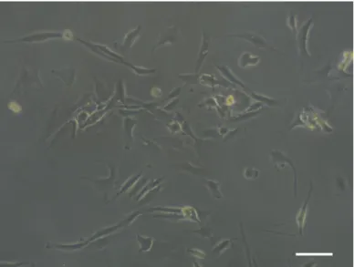
Figure 4.4. Micrographs of human NPCs after 3 days in culture ... 31
Figure 4.5. Micrograph of methacrylated gellan gum disc with encapsulated NPCs... 34
Figure 4.6. µCT top view of the AF and the NP ... 36
Figure 4.7. Photograph of a three-dimensional printed rabbit IVD replica ... 37
Figure 5.1. Fused Deposition Modelling 3D printer from Makerbot™. ... 47
Figure 5.2. Stereolithography process explained in the form of a scheme. ... 48
Figure 5.3. Fused Deposition Modelling process ... 49
Figure 5.4. Schematic representation of Selective Laser Sintering process. ... 50
Figure 5.5. Schematic representation of RE and RP allied to an IVD TE strategy ... 52
II. Patient-Specific Tissue Engineered Total Disc Replacement
Figure 3.1. Results of intervertebral disc reverse engineering and rapid prototyping ... 75Figure 3.2. Virtual representation of IVD replica models ... 77
Figure 3.3. SEM images from the lateral sides of the annulus fibrosus replica ... 78
Figure 3.4. Apparatus for culturing the IVD scaffold in pressurized conditions ... 79
Figure 3.5. Average pore size within each printed model and rabbit intervertebral disc. ... 80
Figure 3.6. Mechanical assessment results regarding the scaffold’s elastic deformation ... 81
Figure 3.7. Compressive Young’s Modulus of the scaffolds in dry and hydrated state. ... 82
Figure 3.8. Compressive stress at 15% specimen deformation. ... 82
Figure 3.9. Cytotoxicity assay results cells in culture with the scaffold extracts ... 83
xviii
Figure 3.11. DNA content of rabbit nucleus pulposus cells seeded within GG-MA. ... 85 Figure 3.12. MTS assay results of cells encapsulated in GG-MA ... 86
xix
List of Tables
I. General Introduction: Advanced Regenerative Strategies for
Intervertebral Disc Degeneration Treatment
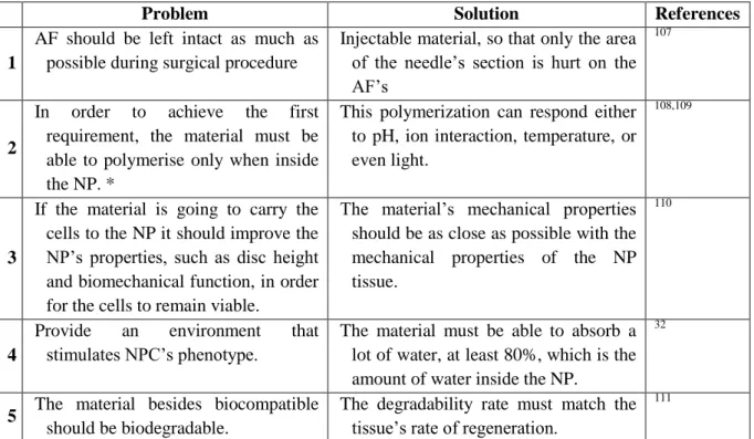
Table 4.1. Hydrogel requirements as NPCs carrier ... 27
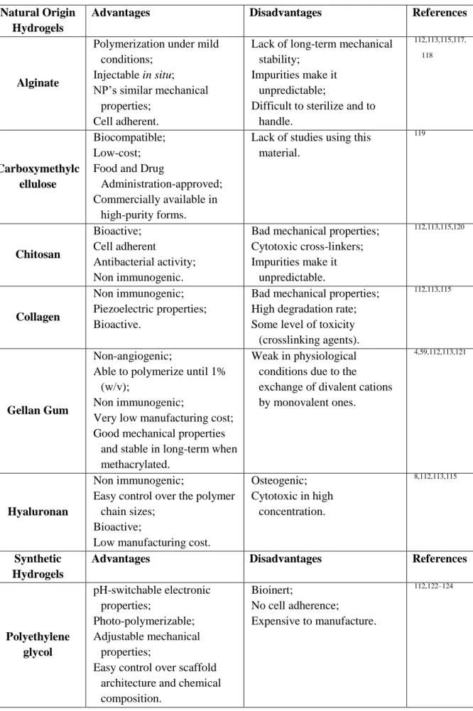
Table 4.2. Natural and synthetic origin hydrogels used in IVD TE strategies ... 29
Table 5.1. Solid-free based TE versus solid-scaffold based TE ... 54
II. Patient-Specific Tissue Engineered Total Disc Replacement
xxi
List of Acronyms
µCT - Micro-Computed Tomography 2D - Two-dimensional 3D - Three-dimensional AF - Annulus FibrosusAFCs - Annulus Fibrosus Cells ATB - Antibiotic Solution BMG - Bone Matrix Gelatin C - Cervical
CAD - Computer-aided Design CEP - Cartilaginous Endplates CMC - Carboxymethylcellulose CO2 - Carbon Dioxide
CT - Computed Tomography
DMEM:F12 - Dulbecco’s modified Eagle’s medium and nutrient mixture F12 DNA - Deoxyribonucleic acid
ECM - Extracellular Matrix FBS - Fetal Bovine serum f - Matemathical Function
FDM - Fused Deposition Modelling FEM - Finite Element Method GAG - Glycosaminoglycan GFs - Growth Factors
xxii
GG - Gellan Gum
GG-MA - Methacrylated-Gellan Gum GMA - Glycidyl Methacrylate
h - Compressed height of the NP h0 - Uncompressed height of the IVD HA - Hyaluronic Acid
IDD - Intervertebral Disc Degeneration Disease IL - Interleukin
IVD - Intervertebral Disc k - Permeability
L - Lumbar
L929 - Lung Fibroblasts Cell Line LA - Low Acyl
LBP - Low back pain
MMPs - Matrix Metalloproteinases MRI - Magnetic Resonance Imaging MSCs - Mesenchymal Stem Cells
MTS - 3-(4,5-dimethylthiazol-2-yl)-5-(3-carboxymethoxyphenyl)-2-(4-sulfophenyl)-2H-tetrazolium
MW - Molecular Weight
mRNA - Messenger ribonucleic acid NCs - Notochordal Cells
NP - Nucleus Pulposus
xxiii
O2 - Oxygen
OD - Optical Density
PBS - Phosphate Buffered Saline PCL - Polycaprolactone
PEEK - Polyetheretherketone PEG - Polyethylene Glycol PG - Proteoglycan
PGA - Polyglycolic Acid PLA - Polylactic Acid
PLGA - Polylactic-co-glycolic Acid PLLA - Poly (L-lactic Acid)
PPCLM - Poly(polycaprolactone triol malate) PVA - Polyvinyl Alcohol
PVP - Polyvinyl Pyrrolidine RE - Reverse Engineering RM - Regenerative Medicine RNA - ribonucleic acid RP - Rapid Prototyping S - Sacral
SEM - Scanning Electron Microscopy SLA - Stereolithography
SLS - Selective Laser Sintering T - Thoracic
xxiv
TDR - Total Disc Replacement TE - Tissue Engineering
TERM - Tissue Engineering and Regenerative Medicine TGF - Transforming Growth Factor
UV - Ultra Violet λ - Stretch Ratio
1
I. General Introduction: Advanced Regenerative Strategies for
Treatment of the Intervertebral Disc degeneration
1. Impact of Low Back Pain and the Promise of Advanced Therapies
Low back pain (LBP) is a major issue in our society these days, mainly in terms of socioeconomic impact and quality of life. Intervertebral disc (IVD) degeneration disease (IDD) is believed to be the main cause for LBP1,2. Tissue engineering (TE) and regenerative medicine (RM; TERM) are an application of multidisciplinary tools by researchers, engineers, and physicians to construct biological substitutes that can mimic tissues for diagnostic and research, with the final purpose to regenerate diseased and injured tissues3.
TERM is a field where a therapeutic strategy for IDD can be found, the doubt is not if it will work, but instead, how and when it will work. Indeed, many therapeutic strategies for IDD have been proposed within these two fields4–6, and although all of these have the same purpose, they use different ways to get there. Some research groups use cultured cells directly implanted in the native tissue, whether it is with nucleus pulposus (NP) cells (NPCs)7, annulus fibrosus (AF) cells (AFCs) and/or with stem cells8–11.
The aim of the cell therapy strategies is to increase IVD’s cell number levels and, therefore, the extracellular matrix (ECM) production. Another treatment option relies on biomolecules’ injection12
, which is intended to reduce the anabolic processes and increase the catabolic processes to restore ECM levels. However, there are also other approaches that can be taken for severe cases of IDD, regarding biomechanical dysfunction or lack of proper ECM content in the IVD, using third generation biomaterials that can mimic the native tissue’s physical and biochemical conditions13
. These three strategies are the main ways within TERM fields to stop and revert IVD’s degeneration process. They can be chosen according with the IVD’s stage of degeneration and type of malfunction, but probably, as many research groups have been suggesting, the most adequate approach, at least for moderate and severe cases of IDD, where IVD’s biomechanics is about to or is already compromised, would be a combination of two or all of these main regenerative strategies (e.g., a combination of scaffold with cells and bioactive molecules)14,15.
Bearing in mind that the world is in a difficult economic crisis and that getting enough funding for science research, as in other areas, is harder; a question must be asked - Is
2
researching an IVD’s regeneration strategy economically worthwhile? To answer that question, it must be taken into account the size of the disease in terms of prevalence in society and its socioeconomic impact, which is statistically masked as LBP. So the dependence between LBP and IVD degeneration should be analysed. In fact, the IVD degeneration is thought to be the primary cause of LBP, causing compression of the spinal nerves and adjacent vertebrae1,2. By the age of 50, 97% of the population show signs of IVD degeneration16, which is a time bomb for LBP. The incidence of LBP increases with age,
creating a relationship between age related IVD degeneration and the frequency of LBP17. Having in account the increasing lifetime average in first world countries, incidence of LBP is increasing with it. Considering the fact that modern lifestyle tends to fulfil some LBP’s risk factors, such as a large number of hours in a seated position, high levels of psychological distress, low levels of physical activity, obesity or job dissatisfaction, and the relation between IDD and LBP shows the problem is far from solved. Other risk factors for LBP are poor knowledge about self-state of health, previous back pain, pain below the knee, depression, fear avoidance behaviours, exposure to intense vibrations and smoking.
The most favourable way to analyse the economic importance/relevance of exploiting a regenerative strategy for IDD, is to check how much money is spent directly or indirectly (money not won on labour or which is spent in consequences due to LBP, e.g., impairment and/or unemployment benefits) every year on treatments for LBP, and consider the percentage of LBP that is related to disc degeneration cases. Most LBP cases resolve rapidly (approximately 80-90% until the twelfth week with LBP symptom, acute LBP cases), but the remaining cases are the ones (from the twelfth week of LBP symptom forward is considered chronic LBP), which incur most of the treatment challenges and healthcare costs. In the United States, LBP is: the first cause of impairment in people younger than 45 years, the second cause to visit the physician, the third cause for surgical procedures and the fifth cause of admission in the hospital18. In the United Kingdom, LBP accounts for 13% of the certificated sickness leave, with estimated indirect costs to the country of 9 billion euros per year (in today’s euro currency, X-Rates), and an annual direct cost to the National Health Service of 900 M€ (in today’s euro currency, X-Rates)19
. Resulting in 70 billion euros (in today’s euro currency, X-Rates) in annual costs to alleviate and treat this pain1
and it is rising.
This subject takes us to another question – Are there any good treatments for LBP? The current treatments for LBP are therapies addressed for LBP, and not for its cause, which means, that these treatments are focused on treating only the symptoms, and do not solve the actual problem, e.g., IDD. The current treatments addressed to LBP, which largely include IDD’s cases too, can be divided into pharmacological and non-pharmacological therapies16
3
The pharmacological treatment options include anti-inflammatories, muscle relaxants, antidepressants20, analgesics and opioids, and injection therapy (drug’s injection). Unfortunately, most of these treatment approaches have the danger of promoting addiction. On the other hand, the current treatments for chronic LBP are exercise, multidisciplinary therapy as in physical and psychological training, massage (classical/Swedish muscle massage)8, acupuncture21, behavioural therapy, back school, spinal manipulation22, electromyographic biofeedback, lumbar supports, traction and transcutaneous electrical nerve stimulation. These are all treatments applied nowadays for patients with IDD, which are diagnosed with LBP, and contribute for $90 billion annual costs applied directly in LBP patients’, money that could be spent in good and efficient treatments, which do not exist still. None of these treatments have shown already to be effective, sometimes it works, but it seems to depend on the cause of LBP, e.g., if a patient suffers from stress a muscle relaxant can be enough to solve the condition.
Herein, it is intended to address the basic anatomy and biomechanics of the spine and IVD, as well as the degeneration process of the IVD. It also overviews the state of the art regarding the repair strategies, gene therapy and TERM strategies developed to repair and regenerate the degenerated IVD. Other advanced strategies that have been exploited in new TE approaches are also discussed, which employs the use of reverse engineering (RE) and rapid prototyping (RP) technologies in order to prepare a patient-specific TE-total disc replacement (TE-TDR) implant for middle to severe cases of IDD, e.g., cases in which the AF is already compromised.
5
2. Spine: Anatomy and Function
The spine is composed of 33 vertebrae (9 are fused together in the sacrum and coccyx regions), most of them interspersed with IVDs, these being the biomechanical pivot of upper body motion. This allows the spine to be in the upper right position, bend and be submitted to torsion at the same time as it protects the spinal cord from trauma23,24. The spine provides strength and flexibility allowing the body to move in multiple spatial planes. The vertebrae composing the spine are numbered according to their spinal area location: 7 cervical (C1 to C7), 12 thoracic (T1 to T12), 5 lumbar (L1 to L5), 5 sacral (S1 to S5) and 4 coccygeal (fused vertebrae)25.
Cervical area’s main function is to support the head weight, which accounts for 8% of total bodyweight26. Thoracic area’s range of motion is very limited and its main function is to protect the chest’s internal organs by supporting the thoracic cage. The lumbar area is responsible for bearing the upper body’s weight; for that reason, it has the biggest vertebrae in the entire spine. Vertebrae from both sacral and coccygeal areas are, each one, fused together.
The spine has a total of 24 IVDs, with approximately 4 cm in diameter and 7 to 10 mm thick (in the lumbar area), which account for one third of its height, and are the main responsible for its flexibility. It also allows a variety of movements, namely in three different planes: lateral bending, axial rotation and flexion-extension. When these motions are accompanied with heavy lifting, forces up to 17 kN can be created in the lumbar area27. In fact, this tissue is under high pressure when the body is in a vertical position, especially the lumbar area’s discs, and even more when the body is in a seated position28. This indicates that the modern lifestyle, regarding its tendency for higher number of hours in which a person is seated, can be one more risk factor for chronic LBP and IVD degeneration.
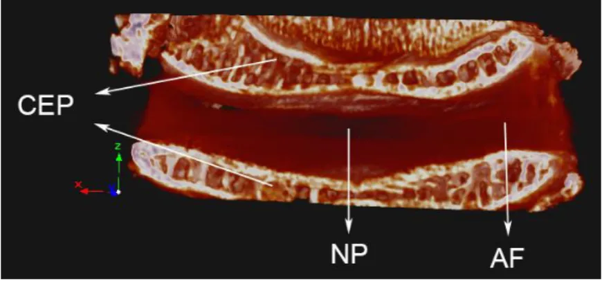
The IVD is a complex structure composed of three different, although interdependent, types of tissue: (1) NP, (2) AF and (3) the cartilaginous endplates (CEP) (Figure 2.1)1 located on both top and down IVD extremities which vertically delimit the AF and the NP. The NP is the gelatinous core of the IVD, and is contained by the strong and elastic AF.
6
Figure 2.1. Micro-computed tomography three-dimensional reconstruction of a rabbit IVD,
presenting the different components, namely: NP in the middle, AF around the NP and both top and down CEPs (Micro- computed tomography parameters: pixel size – 13.18µm, source 89kV / 112µA).
The NP’s hydrogel-like consistency is due to its proteoglycan (PG) and water content, held loosely by a random network of collagen type II and elastin fibres. This structure has a high water content due to its high PG predominance in28–31 which is about 80% and 65% (in dry weight), respectively at childhood, and drastically decreases with age3233. PGs are highly hydrophilic molecules, allowing them to adsorb large quantities of water, making NP’s ECM to swell, giving the NP the classic hydrogel-like morphology. This morphology has viscoelastic properties, which is ideal for the NP function, and also ideal for the IVD’s and spine’s biomechanics.
The AF structure is responsible for contain the NP, and avoid its extrusion followed by collapse, as NP is under high pressure and its consistency does not allow weight bearing on its own. The AF surrounds the NP with 10 to 25, extremely organized and highly fibrotic, annular elastic strips called lamellas28,31. Although collagen type II is also present in the AF as it is in the NP, the proportion is much smaller, on the other hand the presence of collagen type I is highly predominant30. The collagen ratio of type I/type II increases from the NP’s centre until the AF’s outer periphery. Actually, the AF varies so much radially that some research groups, including ours27 and Cassinelli et al., say the IVD is composed by four structures: CEP, NP, inner AF and outer AF17.
The outer AF is the peripheral layer of the IVD (in the transverse plane), and it is highly dense and organized17,27. The collagen fibres make a 60º angle with vertical axes, in the same lamella they are parallel to each other, but alternated from adjacent fibres. All together, the lamellas make a diamond mesh-like force field to contain the NP. This configuration allows the outer AF to contain large forces coming from the compression of the NP by the spine that are not contained by the inner AF30,31,34.
7
The inner AF is a less organized structure than the outer AF. It is somehow a combination between the outer AF and the NP both biochemically and biologically, although more similar with the outer AF. On a healthy IVD the inner AF has a clear and smooth appearance.
CEP divides both NP and AF from the adjacent vertebrae by covering the cortical bone’s surface. Vertebrae are connected to the CEPs through calcium structures, in addition the collagen fibres present in AF cross the border, tying the IVD to the vertebral bodies at its rim29. The CEP tissue is cartilaginous, and resembles articular cartilage in many properties. Studies have demonstrated that this tissue is the weakest of the three tissues in terms of mechanical properties30.
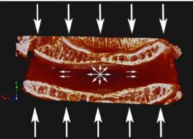
The IVD’s mechanism works by using the best properties of each of its three main types of tissue: CEP, stiffness; NP, uniaxial into hydrostatic pressure converter; AF, elastic resilience. This characteristics work on a force transfer cascade which begins with the spine being mechanically requested, resulting in its compression35–38. The uniaxial compression driven by this force is transferred through the CEPs to the NP, which reacts hydrostatically due to its fluid properties. As a result the AF ends up being pushed from within homogeneously, which reacts by containing it (Figure 2.2).
Figure 2.2.IVD’s biomechanics general scheme, when the spine is mechanically requested.
(Micro-computed tomography of rabbit IVD. Acquisition parameters: pixel size – 13.18µm, source 89kV / 112µA).
The assembled combination of both AF’s and NP’s mechanical properties make the IVD as it was composed of only one viscoelastic material ruled by two limit situations of
8
material mechanics: elastic solid and viscous liquid. As so, it is possible to consider, that the IVD has both elastic solid and viscous liquid properties.
9
3. Intervertebral Disc Degeneration (IDD)
The IVD suffers a great range of changes along the life of an individual - from the molecular phenotype expression, to cell type, to tissues’ (CEPs, AF and NP) morphology. Alongside with these changes, called aging process of the IVD, can be an underlying progressing process of IDD. This disease can be triggered by an acute overloading (e.g., lifting a heavy object). With aging, the needed threshold loading, to initiate the disease, progressively decreases. The degeneration of the disc converts the mild changes of aging IVD into serious conditions, e.g., water loss, ECM production activity and phenotype, among several other changes at the biomolecular level. Ultimately, these changes lead to severe morphological changes, expressed in the form of pathologies.
The IVD biomechanical functioning relies on a balance between the three tissues that compose it. The same way the pressure goes along the CEPs to the NP, further to the AF, as aforementioned; the water flow follows the same pattern. In young discs, the only existent vascularisation is located inside the CEPs, which provide hydration for the whole disc39. The water is absorbed due to the osmotic pressure created by the biochemical components that compose most of the NP. Therefore, during loading cycles where compression forces affect the disc, the NP is strongly squeezed and the water molecules “detach” from the PGs, flowing away through the AF tissue.
As previously mentioned, the PGs are extremely hydrophilic molecules that are responsible for the 80% water composition of the NP in young IVDs32. A combination of spine overloading with aging leads to NP’s ECM remodeling unbalance, loss of hydration, IVD height decrease, abnormal force distribution, and, ultimately, leads to the appearance of IDD morphological signs40.
3.1. Pathophysiology
Thompson et al. proposed a grading scale for the anatomical changes that happen along the IDD development41. With the evolution of the IDD, there are a number of pathophysiologic events that can develop with it, such as: annular tears, disc prolapse, end plate damage and Schmorl’s nodes, internal disc disruption, discogenic pain, disc narrowing, radial bulging, and vertebral osteophytes42, which are addressed below.
10 3.1.1. Annular Tears
The annular tears (Figure 3.1) are protrusions of NP tissue into the AF. As the NP’s material characteristics change and the AF tissue’s elastic properties weakens, with the help of an acute overloading recurrence on the spine, the NP is extruded through the AF and therefore creating an annular tear. There are three types of annular tears: circumferential tears, peripheral tears, and radial fissures. Circumferential tears may evolve from acute overloading in older IVDs; the peripheral rim tears can be associated with trauma or with bony outgrowths, and are more frequent in the AF’s anterior side; radial fissures are related with IDD and may appear on both posterior or posterolateral sides43.
Figure 3.1. Annular tear in hernia stage.
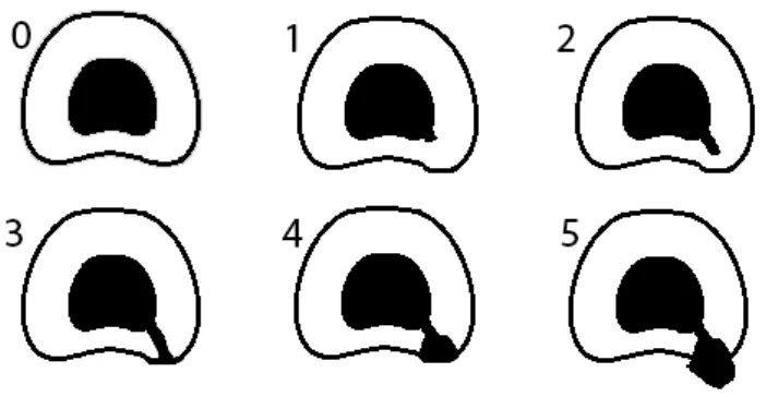
Annular tears can be classified as contained or herniated, the last being when the NP is extruding out of the AF. From contained to herniated NP there are five levels of severity for the annular tears progression, which is the gold standard for the computed tomography (CT) classification of annular tears (Figure 3.2). Grade 0 refers to a normal health IVD; grade 1 is when the NP extrudes until the inner one-third of the AF; in grade 2 the NP leakage progressed until two-thirds of the AF; when the NP finally completes the three-thirds of extrusion through the AF, but no more than that, is on the grade 3 of annular tear classification; after the NP extruding beyond the AF outer border line the annular tear is on the fourth grade; the last stage of the annular tear progression grading scale (grade 5) is when the extrusion of the NP reaches the epidural space, that is when it may press the spinal nerve or one nerve ending, creating LBP44.
11
Figure 3.2. Classification of annular tears first purposed by Sach et al. in 1990, which has been
modified by Bogduk et al. in 1992 and further modified by Schellhas et al. in 1996.
3.1.2. Disc Prolapse
When the NP is totally extruded throughout the AF there is a disc prolapse. In fact, the AF does not have any significant compressive properties, it is an elastic tissue and it acts like a rubber band when compressed, i.e., when it is submitted to traction it responds increasing the reaction force the more it is stretched, however when compressed the middle part runs away towards one of the sides. Therefore, when there is no NP to convert the received force into a hydrostatic pressure, the uniaxial compressive force is not contained and comes down on the AF, which bends easily and it prolapses the disc. The difference between a disc herniation and a stage 5 annular tear is the quantity of NP content outside of place, if it is total then a disc prolapse (Figure 3.3) is about to or already did happen45.
Figure 3.3. Scheme of a prolapsed disc, emphasized in red.
3.1.3. End Plate Damage and Schmorl’s Nodes
Another typical IVD’s pathology is CEP damage and the appearance of Schmorl’s nodes. The CEPs are the weakest tissue of the three composing the IVD, in terms of mechanical properties30. In aged IVDs, when the CEPs already undergone a severe trabecular
12
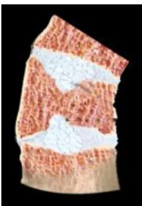
microdamage, the tissue can suffer protrusion from the NP. The NP invades the vertebral bodies (Figure 3.4) and it ceases to be under pressure, since the force presses the AF instead, which cannot bear it and bends42.
Figure 3.4. NP protrusions (Schmorl’s nodes) into the CEPs due to trabecular microdamage driven by
NP biochemical imbalance.
3.1.4. Internal Disc Disruption
Finally, the loss of water inside the NP makes its volume decrease in almost 1:1 proportional ratio, since as much as 80% of the NP is water. The decrease of NP’s volume has a threshold, when this level is crossed the AF tissue seizes to be pushed by the NP. Since the NP’s height is smaller than the AF, the CEPs start to compress the AF instead of the NP. The AF is not a hard tissue, thus it cannot hold compression in a functional way and internal disc disruption occurs, dividing the AF into two layers that bend to different sides, in and out46. Due to AF organisational morphology, which is divided in radial lamellas, tends to break apart between 2 of the 15 to 25 lamella stripes that compose the whole AF. This event creates a gap inside the AF, which significantly decreases the tissues mechanical performance, thus contributing to the progression of degeneration47.
3.2. Biological and Molecular Changes
Biological and molecular changes underlie the morphological signs of IDD. Regarding biological changes, the cells present in degenerating IVDs suffer changes in several domains. Zhao et al.48 made an extensive review on the cellular changes that happen during IDD. The
13
authors claim that there are cell changes in: type, density, death, proliferation, senescence, and phenotype. All these changes have an impact on molecular synthesis, which leads to an unbalanced ECM remodeling, and ultimately to loss of hydration that should be about 80%30. The IVD is composed of different types of cells in its different parts. The CEP as in resemblance to the hyaline cartilage is composed by chondrocytes; the outer AF is populated by elongated fibroblastic-like cells; the inner AF as more rounded chondrocyte-like cells. The cell population in CEP and AF does not change much with aging, however, it does vary greatly in the NP48. The CEP and the whole AF as most of the spinal structures are derived from the mesoderm germ layer, but not the NP49. This tissue on the other hand, has its origin on the endoderm germ layer that is also the notochordal cell’s origin. These cells are largely present in young discs, but disappear by the end of the first decade of life. The notochordal cells (NCs) are gradually replaced by chondrocyte-like cells which are believed to migrate from the inner AF and/or from the CEPs29.
There is some controversy around cell density changes. Zhao et al.48 believe the cell density has its turnovers in parallel with the change of cell type in the NP. As NCs start to disappear, the cell density drops creating a positive feedback for chondrocyte-like cells migration and proliferation into the NP tissue, coming from the already mentioned tissues. These chondrocyte-like cells increase density along IDD. Bae et al. do not agree though, they claim that the degenerated disc’s cell density is lower than that found in healthy discs50
. But both hypotheses have strong arguments, as degeneration process progresses the CEPs start to calcify and IVD’s vascular access starts to become scarce affecting the cells’ nutritional route and cellular waste removal, causing increased cell death (Figure 3.5). However, with IDD progression, ingrowth of blood vessels accompanied by nerve growth occurs into the AF and even into the NP (in advanced IDD)51. These changes increase nutritional accessibility and waste removal rate, which can increase as well the cell density in the NP. But with nutritional availability also oxygen concentration increases inside the IVD, even more than in healthy IVDs. NPCs phenotype and activity is stimulated by hypoxia, ultimately the excessive availability to oxygen leads to normoxia, which tends to make NPCs senescent52,53.
14
Figure 3.5. Scheme of the cascade of events associated with the morphological signs of IDD.
The cellular change, potentially most responsible for the morphological transformations observed due to IDD, is the NPC phenotype. Cell expression has direct influence in the anabolic-catabolic ECM pathway balance, as IDD progresses the catabolism increases in relation to anabolism, i.e., the matrix is more degraded than it is produced. In a normal IVD, the AF is largely composed of collagen type I whereas the NP is composed of collagen type II and aggrecan, which is a type of PG. But other molecules are present in the NP, such as: collagen type I, III, V, VI, IX and XI, biglycans, decorin and fibromodulin (other types of PGs), and fibronectin30. In healthy IVDs there is also production of catabolic
15
molecules with increased expression while IDD progresses. These molecules are matrix metalloprotainases (MMPs) and aggrecanases, which break down the ECM. There are several types of MMPs expressed in the NP: type 1, 2, 3, 7, 8 and 13. In addition to that, there is also an increased cytokine production, such as: interleukin (IL)-1α, IL-1β and tumour necrosis factor-α. These molecules also promote the MMPs synthesis, having a devastating effect on the ECM34. Zhao et al. summarized a table of the biochemical changes caused by the IDD, which complements this brief description of ECM remodeling mediation48.
The biomolecular changes throughout the IVD decrease the concentration of hydrophilic molecules in the NP. This has a strong effect on the IVD’s permeability that when affected can change the IVD’s biomechanics. NP´s hydraulic permeability greatly depends on the magnitude of the compression force made on the disc. Heneghan et al.54 defined a mathematical formula explaining this phenomenon, which is given by equation 1.
𝑘(𝜆) = 2.05 × 10⁻¹⁵(𝜆 − 0.2)1.13𝑒−0.01(𝜆2−1) (Equation 1)
Where, k, is the named permeability and, λ, is the stretch ratio, i.e., the ratio between the compressed sample’s height and the original height, on the apparatus used by this research group. In which, an IVD is submitted to a compression strain and the flow inlet and outlet are measured. This experiment besides giving a relation between permeability and compression, also shows that this relation does not follow a linear tendency but, instead follows an exponential tendency (λ = h / h0, as h0 is the uncompressed height of the IVD and
is always higher than h, which is the compressed height of the NP; therefore, λ is always between 0 and 1). Until a certain level of NP compression magnitude (λ ≤ 0.2), the tissue is not permeable, i.e., when k = 0 or is an imaginary value. But this formula can only be applied when the NP is healthy, since when the degeneration starts the PG number decreases inside the NP and its permeability increases with it, i.e., the water retention decreases and the whole IVD decreases its mechanical performance. This process is a cycle, with loss of biomechanical properties the typical loading starts to become an overloading, which also is a factor to progress the degeneration state that will in the end reduce even more the mechanical capacity, and the cycle goes on in a recessive spiral.
17
4. Treatment Strategies for Intervertebral Disc Repair/Regeneration
As previously stated, current treatments for LBP can cause addiction and only treat the symptoms. These treatments only work in specific situations, like in acute IVD traumas or in the beginning of the IDD’s appearance. They mainly work by taking pressure out of the IVD (e.g., muscle relaxants soften muscle strain over the IVD) thereby allowing the natural regeneration mechanism to solve the issue by itself. However, most of these treatments merely neutralize or reduce the patient’s pain, while the IDD is still progressing40
.
In more severe cases of IDD, the natural regeneration system cannot cope anymore, either by itself or with strain releasing help (such as current treatments for LBP, e.g., massage). In that case, only three strategies have the potential to remove pain completely, which are repair55, gene therapy56 and TERM strategies57. The first intends to solve IDD with artificial implants, whereas the second and third aim at the total regeneration of a patient’s IVD, removing all previous signs of degeneration.
Even though gene therapy is still far away from clinical application, it promises great results in the future, not only in IVD regeneration, but also in several different kinds of diseases e.g., cystic fibrosis58. The question of which one of these strategies is the best, cannot be answered yet, mainly due to the fact that there is still no clinically-approved IVD regeneration strategy available, despite promising potential in the currently researched treatment options59. Now, patients can only choose repair strategies as an alternative to current treatments in order to treat IDD.
In this section, it will be discussed the several repair approaches. Starting with the discectomy procedure, with its objective to remove the LBP generated by extruded NP material that compresses the peripheral spinal nerve or the spinal cord. This section gives a resume of some repair device strategies that not only remove LBP, but prevent it from coming back due to re-herniation after discectomy. Furthermore, the spinal fusion procedure is briefly explained, and how it treats IDD by removing the source of pain, preventing any possibility of it coming back. Well, at least in that intervertebral space, since herein it is also discussed how this procedure can stimulate adjacent IVDs to degenerate due to biomechanical imbalance through the spine after spinal fusion.
Moreover, TERM strategies to treat IDD in the NP are described and what pathways some research groups are taking to achieve that goal. This can be made whether by finding potential ways of differentiating stem cells into NPCs or by culturing isolated NPCs with growth factors (GFs) or serum-free chemically defined medium to make them metabolically
18
active. Then, which materials have been giving more interesting results to carry NPCs in the degenerated NP. Furthermore, it will be discussed which strategies are being followed nowadays to regenerate the AF, since without a good support, whether native or scaffold, for the NP, this will herniate on the first loading cycle. But to find the right material to mimic the complex mechanical properties of the AF is not an easy task, since it is a very efficiently organized tissue. And the synthesis of this ECM organization must, as well, be stimulated and timed with the scaffold rate of degradation.
4.1. Repair Strategy
4.1.1. Discectomy/Arthrodesis
Surgical methods for degenerative lumbar conditions include discectomy, arthrodesis or a combination of both. Discectomy is the surgical removal of NP fragments following herniation that compresses the spinal nerve. This compression distribution on the affected nerve causes pain, sensory changes, or weakness60,61. Discectomy is successful in relieving the radicular pain caused by the herniated disc. However, this procedure alone is unable to restore the nucleus to its original load sharing capacity, which controversially affects long-term benefits and re-herniation rates. Moreover, discectomy may accelerate the progression of disc degeneration by damaging the AF, which in turn will lead to a decrease in NP pressure, decreasing the disc height, impairing the disc’s ability to rehydrate, and increasing the AF stresses and strains61. Besides the anatomical problems, the removal of the degenerated or damaged disc tissue typically provokes negative biomechanical changes1,60. Furthermore, it is unknown whether the effect of discectomy depends on the degenerative state of the disc61.
Due to the disadvantages and limited success of discectomy procedures in general, arthrodesis was developed as an alternative method within clinical treatments. Arthrodesis, also known as spinal fusion, has been practiced since the beginning of the 20th century. Spinal fusion involves the use of bone tissue, traditionally derived from autografts, allografts, as well as the application of demineralized bone matrix, ceramics, and more recently bone morphogenetic proteins to bridge two or more vertebrae62. This procedure aims at stabilizing the moving segment, and slowing the progression of disc degeneration and relative pathological motion between vertebrae. Arthrodesis is based on the hypothesis that the mechanical and environmental changes will relieve the pain63,64. Even though spinal fusion is
19
a common procedure, its efficacy in treating discogenic LBP has been resulted in conflicting results65. Long-term consequences such as adjacent segment disease have increased concerns for the use of spinal fusion1. Furthermore, several changes have been observed such as dehydration, disc space narrowing, osteophyte formation, and progressive deformity at levels adjacent to a fused spinal segment66.
4.1.2. Replacement
In an effort to improve results of fusion and to decrease the incidence of adjacent IVD degeneration, TDR techniques have been introduced and studied extensively.
Artificial disc replacement (nucleus and annulus) technology was first considered in the early 1950’s to produce an implant that could mimic, to some extent, the function of the normal IVD (maintain the mobility of the intervertebral motion segment and restoring natural disc function)62,67. Moreover, implants for disc replacement should be biocompatible, durable, and easily implantable68. There are generally two types of disc arthroplasty devices, which are nucleus replacement or TDR devices, with the latter being more frequently used1.
Nucleus substitutes are aimed at restoring disc height and returning annular fibres to their natural length. Adding to the appeal is the minimally invasive nature of this treatment method69. However, despite minimal invasion, a passage through the annulus for the prosthesis has to be created. Nevertheless, this approach allows the rehabilitation of the normal load distribution among the nucleus, the annulus and the facet joints, as well as promoting the healing of the annulus and thwart degeneration by themselves.
In general, the substitute should provide resistance to pressure with position change recreating the disc “bellows” effect60
. A wide range of materials has been tried in order to replace the nucleus of the IVD, including: polymethylmethacrylate, polyvinyl alcohol (PVA)/polyvinyl pyrrolidine (PVP) copolymer, polycarbonate urethane, albumin, silicon, and stainless steel62. The most well-known device is the prosthetic disc nucleus, which is comprised of two sections: the first is made up of a non-degradable hydrogel pellet (polyacrylamide) and the second, which surrounds the hydrogel, is a polymer mesh or jacket composed of polyethylene70. However, in advanced stages of degeneration, these devices cannot be applied and TDR becomes again the most favourable approach.
TDR aims at restoring the physiological kinematics of the IVD, such as resisting wear and relieving pain, while avoiding instability and protecting the adjacent discs and facet
20
joints from undue degeneration62. Three artificial disc options have been proposed for TDR: metallic, non-metallic, or a combination of both materials27. SB-Charité®, a metal-polyethylene-metal construct was the first total disc arthroplasty, which is still in use today following only minor changes to the original design. Pro-disc® is another prosthesis that has been widely used71.
Each artificial disc is composed of two or three components, which include two endplates and an articulating mechanism with either a metal-on-metal or metal-on-polymer surface. In order to keep the disc in place and provide stability within the host vertebral body, devices feature different designs - teeth-like compounds fixed into the vertebral bone; a porous coated surface onto the endplates, which promotes the growth of fibrous tissue around the device; or implant securing with screws into the recipient vertebral body27,72. Even though theoretically appealing, there are several challenges with current TDR strategies and also there is insufficient data to assess the performance of IVD arthroplasty adequately. Despite being in use for at least the last 20 years, there are some concerns regarding the safety and efficacy of these methods73. Consequently, patients may require revision surgery, which may be very dangerous due to the adjacent great vessels and the nerve plexus. Another solution, posterior fusion, requires the removal of the disc prosthesis followed by spinal fusion to immobilize the affected tissue, which again is very risky and dangerous1,73.
Regarding the obvious downfalls of spinal fusion, the development of dynamic, or semi rigid-, constructs for lumbar spine instrumentation has emerged as an alternative option. This method is based on a load-sharing device, allowing for fusion without excessive rigidity, which, if disregarded, may lead to adjacent segment complications74. Some dynamic constructs have also been used without fusion75. Several authors have reported that posterior dynamic instrumentation, compared to rigid instrumentation, increases the amount of load transmitted through the anterior column and the interbody bone graft, which will avoid stress-shielding phenomena. Consequently, this may favour osteogenesis and enhance interbody fusion in accordance with Wolff’s Law, which states that bone will adapt to the load it is placed under through piezoelectric phenomena74.
Systems for spinal fusion can be described as pedicle screw–based constructs that are semi-rigid, or allow constrained motion in compression or flexion and extension. Pedicle screw-based systems are classically divided into semi-rigid rod systems and tension band-based posterior systems used in non-fusion technology74. The rigidity of these constructs depends on the material and design of the rods, which are connecting the pedicle screws. Although, solid stainless steel and titanium are commonly used in spinal fusion constructs76, the semi-rigid constructs often include polyetheretherketone (PEEK) rods77, nitinol rods78,
21
especially cut rods (e.g., Accuflex), articulated rods, and polyethylene terephthalate cords (Dynesys)75,79.
Unlike the aforementioned prostheses that are fixed to the vertebrae by the use of pedicle screws, there are “floating devices”, which are interspinous implants. These implants have the advantage of reducing the risk of implant loosening during motion80.
During the last decade, spinal cage implantation has gained a lot of attention. This approach enhances spinal fusion and stability in cervical spine surgery, ensuring, at the same time, an adequate increase in the height and helping to correct cervical kyphosis, i.e., a curvature in the cervical area of the spine81. PEEK cages have recently been used in cervical surgery, since they provide both strength and stiffness in the intervertebral space. A major advantage of these implants is their radio transparency and magnetic resonance imaging (MRI) compatibility, which are traditionally used in the visualization of the spinal cord and the root. Since the polymer is radiolucent, visualization in the aforementioned methods can be performed without the generation of implant artefacts in the resulting images81.
Finally, intradiscal electrothermal therapy is a percutaneous technique reported by Saal et al., and it is another option for the treatment of discogenic LBP82. A navigable electrothermal catheter is inserted inside the posterior annulus, which delivers heat. The proposed mechanism of action of this technique is collagen modulation, cauterization of granulation tissue, deactivation of inflammatory agents and possibly annular denervation. The heat induces the retraction of the annular collagen (collagen fibrils shrink at temperatures greater than 60ºC), therefore coagulating inflammatory tissue and nerve endings in the periphery of the disc’s posterior side82
. This heating method has been shown to produce temperatures sufficient to cause nerve fibre death as well as collagen denaturation83. Moreover, there are no biomechanical modifications and destabilizations after applying this procedure.
4.2. Gene Therapy
A potential therapeutic strategy next to the classic LBP management treatments and repair strategies is the gene therapy. This possibility was first thought as a way of treating chronic diseases, targeting the problem on its core by modifying, adding or disabling a gene or a cocktail of genes. Several problems occurred, when it was first tested; unfortunately the
22
idea gained a bad reputation and gone into a research standby for a decade84. Recently, the number of groups researching gene therapy started rising again, being nowadays a possibility of stopping and reverting degeneration of the IVD.
The first question when applying this strategy for IVD regeneration is – what should change on a molecular level, to stop and revert IVD’s degeneration? As it has been explained above, the malfunction in the NP’s ECM production and maintenance is one of the main reasons for the IVD to fail biomechanically. So, the strategy must be focused on balancing the catabolic-anabolic equilibrium into the ECM production side. For that, two things can be done, alone or together, increase the ECM anabolism and decrease the catabolic pathway. This can be done by transfecting the right genes into the NPC population, genes that codify potential therapeutic GFs and cytokines such as: transforming GF (TGF)-β1, TGF-β3, insulin like GF-1, osteogenic 1, IL-1, bone morphogenic 2, latent membrane protein-1, SOX9, among others85. Transfecting a combination of these genes would increase ECM production, but the reverse strategy can also show equally positive results, by silencing targeted catabolic proteins using RNA interference, such as proteolytic enzymes (e.g., MMPs and disintegrin and metalloproteinase with thrombospondin motifs). This RNA suppresses the overall production of the targeted gene by using a small interfering RNA, which binds specifically to the gene’s mRNA sequence, leading to a suppressed translation or increased mRNA’s degradation86,87
.
After choosing the proper cocktail of genes to be delivered, another question may be posed - how does the gene reach its final destination? With few exceptions naked DNA alone is not a feasible way to deliver it to target the cells’ nucleus; a vector is needed that can be viral or non-viral. Non-viral vectors are systems, which do not have viral origin, such as liposomes88, DNA-ligand complexes89, gene gun90 and microbubble enhanced ultrasound91. However, the non-viral vectors give the host a transient gene expression that is not suitable for the treatment of chronic diseases. A longer lasting strategy must be applied, for that, viral vectors are the most favourable since they use viral’s natural way of infecting the host and integrating DNA into the target cells’ genome, e.g., metabolism senescent cells like the ones present inside the IVD in order to enhance their activity. Viral vectors, on the other hand, bring the risk of alarming the immune system that is a huge issue in high-vascularised tissues, which is not the case as the IVD is the most avascular tissue in the body. Viral vectors utilized for gene therapy applications include adenovirus92, adeno-associated virus93, herpes simplex virus94, lentivirus and retrovirus95.
In addition to the selection of the appropriate gene and vector, another important consideration with gene therapy applications is the delivery strategy. There are two main
23
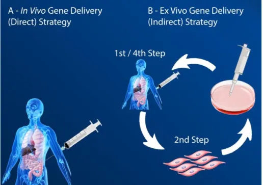
strategies for gene delivery as proposed by Nishida et al96, in vivo and ex vivo (Figure 4.1). The first strategy takes fewer steps than the second, it involves the direct transfer of the vector-gene complex into the target tissue within the living host. The ex vivo strategy involves a more complex approach. First the cells are isolated from the host; after the culture is transfected with the desired DNA material; the cells, which were successfully genetically modified, are then implanted into the desired tissue. This strategy seems to be much safer, since while cells remain in vitro they can be assessed before implantation in order to control what goes inside the patient’s organism; so those cells that had a bad reaction to the transfection can be removed, and only the desired cells are implanted97. Also, the in vitro culture required in ex vivo strategies may change cells characteristics in a way that they cannot survive in the harsh environment observed in a degenerated NP (low oxygen, low pH, poor nutrition)98. Mimicking the IVD’s conditions is needed in order to reproduce in vitro, as much as possible, the IVD’s cell environment, and probably the only possible strategy is to proceed with the culture in a bioreactor. On the other hand, the in vivo strategy has its disadvantages as well, using this approach the viral vectors are injected with an unknown concentration in comparison with the cells present in the target tissue. This relation between the number of viral particles and target cells is called multiplicity of infection, which in high values is extremely cytotoxic52.
Figure 4.1. Gene delivery strategy available, A. in vivo – which implants the vector-gene complex
within the living host – and B. ex vivo – which involves isolation of the host target cells (1st and 2nd steps), transfection (3rd step) and implantation (4th step).