Universidade de Lisboa
Faculdade de Medicina de Lisboa
Modulatory role of adenosine upon
GABAergic transmission:
consequences for excitability control
Diogo Miguel Santos Rombo
Doutoramento no Ramo de Ciências Biomédicas
Especialidade em Neurociências
Faculdade de Medicina de Lisboa
Modulatory role of adenosine upon
GABAergic transmission:
consequences for excitability control
Diogo Miguel Santos Rombo
Tese orientada pela Professora Doutora Ana Maria
Sebastião
Doutoramento no Ramo de Ciências Biomédicas
Especialidade em Neurociências
Júri: Prof. Doutor. J. Melo Cristino (Presidente), Faculdade de Medicina da Universidade de Lisboa; Prof. Doutor Alfonso Araque, University of Minnesota, USA; Prof. Doutora. Ana Luísa Carvalho, Faculdade de Ciências e Tecnologia da Universidade de Coimbra; Prof. Doutor. Joaquim Alexandre Ribeiro, Prof. Doutora. Ana Maria Sebastião, Prof. Doutor. Alexandre de Mendonça e Prof. Doutora. Raquel B. Dias, Faculdade de Medicina da Universidade de Lisboa.
ii
A impressão desta dissertação foi aprovada pelo
Conselho Científico da Faculdade de Medicina de
iii
Todas as opiniões expressas nesta publicação são da exclusiva responsabilidade do seu autor, não cabendo qualquer responsabilidade à Faculdade de Medicina de Lisboa pelos conteúdos apresentados.
All opinions expressed in this document are of the sole responsibility of its author and Faculdade de Medicina de Lisboa is not liable in any way for its content.
v
O trabalho experimental constante da presente tese foi realizado no Instituto de Farmacologia e Neurociências, Faculdade de Medicina de Lisboa e Unidade de Neurociências, Instituto de Medicina Molecular, sob orientação da Professora Doutora Ana Maria Ferreira de Sousa Sebastião e no Department of Pharmacology, University of Oxford, Oxford, Reino Unido, sob a supervisão do Doutor Karri Lämsä.
The experimental work described in this thesis was performed at the Instituto de Farmacologia e Neurociências, Faculdade de Medicina de Lisboa e Unidade de Neurociências, Instituto de Medicina Molecular, under the orientation of Professor Ana Maria Sebastião and at the Department of Pharmacology, University of Oxford, Oxford, United Kingdom, under the supervision of Doctor Karri Lämsä.
vii
ix
Publications
The scientific content of this thesis was included in the publication of the following original articles:
- Rombo DM, Dias RB, Duarte ST, Ribeiro JA, Lamsa KP, Sebastião AM (2014). Adenosine A1 receptors suppress tonic GABAA receptor currents in hippocampal pyramidal cells and in a defined subpopulation of interneurons. Cerebral Cortex. (Epub ahead of print).
- Rombo DM, Newton K, Nissen W, Badurek S, Horn J, Minichiello L, Jefferys J, Sebastiao AM, Lamsa K (2015). Synaptic mechanims of adenosine A2A receptor mediated hyperexcitability in the hippocampus. Hippocampus 25, 566-80.
Other publications closely related to the content of this thesis: - Dias RB, Rombo DM, Ribeiro JA, Henley JM, Sebastião AM (2013). Adenosine: setting the stage for plasticity. Trends Neurosci 36, 248-57.
- Sebastião AM, Rombo DM, Ribeiro JA. (2015). Adenosine Receptor Modulation of GABAergic Transmission. In Adenosine Signaling Mechanisms: Pharmacology, Functions and Therapeutic Aspects., eds. Vickram Ramkumar, Roberto Paes de Carvalho. New York: Nova Science Publishers
x Other publications from the author:
- Diógenes MJ*, Dias RB*, Rombo DM*, Vicente Miranda H, Maiolino F, Guerreiro P, Näsström T, Franquelim HG, Oliveira LM, Castanho MA, Lannfelt L, Bergström J, Ingelsson M, Quintas A, Sebastião AM, Lopes LV, Outeiro TF (2012). Extracellular alpha-synuclein oligomers modulate synaptic transmission and impair LTP via NMDA-receptor activation. J Neurosci 32, 11750-62. *Co-fist authors.
- Dias RB, Rombo DM, Ribeiro JA, Sebastião AM (2013). Ischemia-induced synaptic plasticity drives sustained expression of calcium-permeable AMPA receptors in the hippocampus. Neuropharmacol 65, 114-22.
- Félix-Oliveira A, Dias RB, Colino-Oliveira M, Rombo DM, Sebastião AM (2014). Homeostatic plasticity induced by brief activity deprivation enhances long-term potentiation in the mature rat hippocampus. J Neurophysiol 112, 3012-22.
- Santos AR, Mele M, Vaz SH, Kellermayer B, Grimaldi M, Colino-Oliveira M, Rombo DM, Comprido D, Sebastião AM, Duarte CB (2015). Differential role of the proteasome in the early and late phases of BDNF-induced facilitation of LTP. J Neurosci 35, 3319-29.
- Fernandes TG, Duarte ST, Ghazvini M, Gaspar C, Santos DC, Porteira AR, Rodrigues GM, Haupt S, Rombo DM, Armstrong J, Sebastião AM, Gribnau J, Garcia-Cazorla À, Brüstle O, Henrique D, Cabral JM, Diogo MM (2015). Neural commitment of human pluripotent stem cells under defined conditions recapitulates
xi
neural development and generates patient-specific neural cells. Biotechnol J (Epub ahead of print).
xiii
Table of contents
Publications ... ix
Table of contents ... xiii
Figure index ... xvii
Table index ... xxii
List of abbreviations ... xxiii
Resumo ... xxx
Abstract ...xxxiv
1 Introduction ... 1
1.1 The hippocampal formation ...4
1.1.1 Excitatory glutamatergic connections in CA1 region ...9
1.1.2 Hippocampal interneurons ... 12
1.1.2.1 Anatomical classification ... 12
1.1.2.2 Neurochemical classification... 14
1.1.2.3 Functional classification ... 15
1.2 GABA and GABA receptors ... 16
1.2.1 GABAA receptors ... 18
1.2.2 Phasic receptor activation ... 23
1.2.3 Tonic receptor activation ... 24
1.2.4 Functional role of phasic and tonic transmission ... 28
1.3 Neuromodulation ... 30
1.3.1 Adenosine ... 31
xiv
1.3.1.2 Modulation of hippocampal GABA transmission .... 43
2 Aim ... 45
3 Techniques ... 47
3.1 Patch-clamp recordings ... 47
3.2 Field recordings ... 57
3.3 Optogenetics ... 59
4 Material and Methods ... 63
4.1 Animals ... 63
4.2 Hippocampal slice preparation ... 64
4.3 Chemicals ... 66
4.4 Electrophysiological recordings ... 70
4.4.1 Patch-clamp recordings ... 73
4.4.1.1 Muscimol-evoked postsynaptic currents ... 75
4.4.1.2 Electrical-evoked inhibitory postsynaptic currents .. 76
4.4.1.3 Miniature inhibitory postsynaptic currents ... 77
4.4.1.4 Tonic inhibitory currents ... 77
4.4.1.5 Electrical-evoked excitatory postsynaptic currents . 79 4.4.2 Optogenetic recordings ... 79
4.4.2.1 Light-evoked EPSCs/disynaptic IPSCs ... 81
4.4.2.2 Light-evoked IPSCs ... 82
4.4.3 Firing patterns ... 83
4.4.4 Field recordings ... 84
4.4.5 Spontaneous epileptiform discharges ... 86
xv
4.6 Morphologic and immunohistochemical analysis ... 93
4.6.1 Tissue fixation and re-sectioning ... 93
4.6.2 Cell reconstructions ... 94
4.6.3 Immunohistochemistry ... 95
4.7 Immunoblot assay ... 97
4.8 Statistical analysis ... 98
5 Results ... 99
5.1 Adenosine A1R suppresses tonic GABAAR currents in hippocampal pyramidal cells and in a defined subpopulation of interneurons ... 99
5.1.1 Summary ... 100
5.1.2 Rational ... 101
5.1.3 Adenosine A1R inhibits agonist-evoked GABAA R-mediated currents in CA1 pyramidal cells... 102
5.1.4 Phasic GABAAR-mediated currents are not affected by adenosine A1R in CA1 pyramidal cells ... 108
5.1.5 Adenosine A1R suppresses tonic GABAergic currents in CA1 pyramidal cells ... 112
5.1.6 Adenosine A1R-mediated effect on GABAA currents is PKA/PKC-dependent ... 116
5.1.7 Adenosine A1R suppresses tonic GABAAR currents in a specific subpopulation of hippocampal interneurons ... 121
5.1.8 Discussion ... 131
5.2 Synaptic mechanisms of adenosine A2AR-mediated hyperexcitability in the hippocampus ... 143
xvi
5.2.2 Rational ... 145
5.2.3 Adenosine A2AR facilitates glutamatergic synapses and amplifies CA1 pyramidal cell input-output transformation ... 146
5.2.4 Adenosine A2AR increases excitation and suppresses feedforward inhibition to pyramidal cells ... 153
5.2.5 Adenosine A2AR facilitates glutamatergic Schaffer collateral synapses selectively to pyramidal cells ... 157
5.2.6 Adenosine A2AR enhances GABAergic inhibition in the CA1 area selectively between interneurons ... 161
5.2.7 Endogenous adenosine promotes synchronous pyramidal cell discharge via A2ARs in hippocampal slices ... 170
5.2.8 Modulation of spontaneous epileptiform pyramidal cell discharge by adenosine A2AR ... 173
5.2.9 Discussion ... 178
6 General Discussion and Conclusions ... 183
7 Future Perspectives ... 189
8 Acknowledgements ... 195
9 References ... 203
xvii
Figure index
Figure 1.1. The human hippocampus compared with a seahorse
... 4
Figure 1.2. Illustration of the neuronal circuitry of the rodent hippocampus ... 6
Figure 1.3. Hippocampal operations performed by distinct populations of CA1 interneurons ... 11
Figure 1.4. Neuronal inhibition mediated by GABAAR ... 20
Figure 1.5. Phasic and tonic activation of GABAARs ... 26
Figure 1.6. Adenosine modulation sites... 32
Figure 1.7. Schematic representation of adenosine metabolism and receptors ... 33
Figure 1.8. Adenosine receptors and classical signaling pathways ... 38
Figure 2.1. Schematic representation of the context and main targets of this study. ... 46
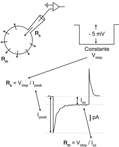
Figure 3.1. Oscilloscope traces obtained in response to constant test pulses for establishment oh whole-cell recording ... 50
Figure 3.2. The voltage-clamp technique ... 52
Figure 3.3. Whole-cell voltage-clamp recordings ... 55
Figure 3.4. Method for approximate series resistance and membrane resistance calculation ... 56
xviii
Figure 3.5. Schematic representation of a field excitatory postsynaptic potential (fEPSP) recorded in stratum radiatum of hippocampal CA1 region ... 58 Figure 3.6. Cell specific targeting of adeno-associated virus (AAV2/5:ChR2-eYFP) into transgenic Cre-recombinase mice .. 61 Figure 5.1. Local agonist (muscimol)-evoked GABAA currents in pyramidal cells... 103 Figure 5.2. Adenosine A1R suppresses muscimol-PSC in
pyramidal cells. ... 104 Figure 5.3. Adenosine A1R antagonist facilitates recovery of muscimol.PSC after agonist action. ... 105 Figure 5.4. Endogenous activation of A1R suppress muscimol-PSCs ... 106 Figure 5.5. A1R-mediated suppression of muscimol-PSC is independent of glutamatergic transmission and neuronal firing ... 107 Figure 5.6. Pharmacology on A1R-mediated suppression of muscimol-PSCs ... 108 Figure 5.7. Adenosine A1R agonist fails to suppress electrical-evoked IPSCs ... 109 Figure 5.8. Spontaneous inhibitory activity is not affected by A1R activation ... 111 Figure 5.9. Recording and measurement of tonic inhibitory
currents ... 114 Figure 5.10. Tonic-ICs are suppressed by A1R activation ... 115
xix
Figure 5.11. PKA and PKC are involved in A1R-mediated
suppression of muscimol-PSCs ... 117 Figure 5.12. PKC activity is downstream PKA activity to suppress muscimol-PSCs ... 118 Figure 5.13. Adenosine A1R decreases GABAAR δ-subunit immunoreactivity ... 120 Figure 5.14. Schematic representation of the signaling cascade involved in A1R-mediated suppression of GABAAR ... 121 Figure 5.15. Hippocampal interneurons are affected differently by A1R activation ... 123 Figure 5.16. Characterization of interneurons by their firing pattern ... 124 Figure 5.17. A1R activation suppresses muscimol-PSCs in GABAergic interneurons expressing axonal CB1R, but not in CB1-immunonegative interneurons. ... 126 Figure 5.18. Tonic GABAAR currents in CB1R-immunoposivite interneurons are inhibited by adenosine A1R activation ... 128 Figure 5.19. Adenosine A1R suppresses tonic-ICs recorded in the presence of endogenous concentrations of GABA ... 129 Figure 5.20. Phasic synaptic IPSCs in interneurons are not suppressed by adenosine A1R ... 130 Figure 5.21. Schematic representation of the A1R-mediated actions upon GABAergic transmission into CA1 hippocampal pyramidal cells and interneurons. ... 140
xx
Figure 5.22. Activation of adenosine A2AR facilitates
glutamatergic transmission in hippocampal Schaffer collaterals ... 148 Figure 5.23. Activation of adenosine A2AR amplifies CA1
pyramidal cell input-output function ... 152 Figure 5.24. Schematic of light-evoked EPSCs/disynaptic IPSCs ... 153 Figure 5.25. Adenosine A2A receptor facilitates excitatory
Schaffer collateral synapses and suppresses feed-forward
GABAergic inhibitory input to CA1 pyramidal cells ... 154 Figure 5.26. Effect of CGS21680 on EPSC and disynaptic IPSC charge in all experiments. ... 156 Figure 5.27. Adenosine A2AR facilitates glutamatergic synapses to pyramidal cells ... 158 Figure 5.28. Adenosine A2AR does not affect synapses to two major feed-forward GABAergic inhibitory interneuron populations expressing either PV or CCK ... 160 Figure 5.29. Adenosine A2AR agonist facilitates IPSCs elicited from GABAergic PV-positive cells to various inhibitory
interneurons ... 163 Figure 5.30. Adenosine A2AR fails to modulate IPSCs from PV-positive GABAergic synapses to identified pyramidal cells... 164 Figure 5.31. The CGS21680-induced IPSC facilitation in
interneurons is associated with reduced paired-pulse ratio (PPR) ... 166
xxi
Figure 5.32. Optogenetic-evoked IPSC facilitation by CGS21680 occurs in various different postsynaptic interneuron types ... 167 Figure 5.33. The IPSCs elicited from CCK-positive interneurons are not modulated by the A2AR agonist... 168 Figure 5.34. Optogenetically-evoked IPSCs from CCK-positive interneurons are inhibited by CB1R activation ... 170 Figure 5.35. Facilitation of hippocampal pyramidal cell discharge through A2ARs activated by high-frequency electrical stimulation. ... 172 Figure 5.36. Modulation of spontaneous epileptiform pyramidal cell discharge by A2AR antagonist. ... 175 Figure 5.37. Modulation of spontaneous epileptiform pyramidal cell discharge by A2AR agonist. ... 177 Figure 6.1 Schematic with the main achievements of the work presented in this thesis. ... 184
xxii
Table index
Table 1.1. Adenosine Receptors in CNS ... 37 Table 4.1 Solutions for preparation, storage and recording of hippocampal slices ... 66 Table 4.2. Pharmacological tolls ... 67 Table 4.3 Intracellular solutions ... 71 Table 4.4 Schematic of all experimental designs performed in electrophysiological recordings ... 88 Table 4.5 Primary and seconday antibodies ... 96 Table 5.1. Baseline-normalised slope values of CGS21680 (agonist) effect alone or in the presence of SCH58261
xxiii
List of abbreviations
5-HT3R – 5-hydroxytryptamin (serotonin) tupe 3 receptor A1R – A1 receptor
A2AR – A2A receptor A2BR – A2B receptor A3R – A3 receptor AA – arachidonic acid
AAV2/5 – adeno-associated vírus serotype 2 or 5 AAC – axo-axonic cell
ABC – ATP-binding cassete transporter AC – adenylate cyclase
ACC - associational commissural connection aCSF – artificial cerebrospinal fluid
ADA – adenosine deaminase ADP – adenosine 5’-diphosphate AK – adenosine kinase
AM-251 -
N-(Piperidin-1-yl)-5-(4-iodophenyl)-1-(2,4-dichlorophenyl)-4-methyl-1H-pyrazole-3-carboxamide AMP – adenosine 5’-monophosphate
AMPA - α-amino-3-hydroxy-5-methyl-4-isoxazolepropionic acid AMPAR – AMPA receptor
ATP – adenosine 5’-triphosphate BC – basket cell
BDNF – brain derived neurotrophic factor BSC – bistratified cell
BSNP – burst-spiking non-pyramidal cell CA – cornu ammonis
xxiv
CAM – calcium/calmodulin-dependent protein
CAMK – calcium/calmodulin-dependent protein kinase cAMP – cyclic adenosine 5’-monophosphate
CB – cannabinoid
CB1R – cannabinoid type 1 receptor CB2R – cannabinoid type 2 receptor CCK – cholecystokinin
CGP55845 - (2S)-3-[[(1S)-1-(3,4-Dichlorophenyl)ethyl] amino-2-hydroxypropyl] (phenylmethyl) phosphinic acid hydrochloride CGRP - calcitonin gene-related peptide
CGS21680 - 4-[2-[[6-Amino-9-(N-
ethyl-β-D-ribofuranuronamidosyl)-9H-purin-2-yl] amino] ethyl] benzenepropanoic acid hydrochloride
ChR2 – channelrhodopsin-2 Cl- - chloride ion
CNQX - 6-cyano-7-nitroquinoxaline-2,3-dione disodium salt CNS – central nervous system
CPA - N6-cyclopentyladenosine
CREB – cAMP response element binding protein D2R – dopamine type 2 receptor
DAG - diacylglycerol DG – dentate gyrus
DIO – doble-floxed inverted open reading frame DIC-IR – differential interference contrast-infrared dIPSC – disynaptic inhibitory postsynaptic current
DL-AP5 - DL-2-Amino-5-phosphonopentanoic acid sodium salt DMSO – dimethyl sulfoxide
DPCPX - 1,3-dipropyl-8-cyclopentylxanthine DR – dopamine receptor
xxv DTT - dithiothreitol
EC – entorhinal cortex eCB – endocannabinoid
ECL – enhanced chemiluminescence detection method ECl – equilibrium potential for chloride ion
EDTA – ethylenediamine tetra-acetic acid EGABA – equilibrium potential for GABA
EHCO3 – equilibrium potential for bicarbonate ion ENa – equilibrium potential for sodium ion
ENT – equilibrative nucleoside transporter EPSC – excitatory postsynaptic current EPSP – excitatory postsynaptic potential eYFP – enhanced yellow fluorescent protein fEPSP – field excitatory postsynaptic potential FSI – fast-spiking interneuron
GABA - gamma-aminobutyric acid GABAAR – GABA type A receptor GABACR – GABA type C receptor GABABR – GABA type B receptor GAD - glutamic acid decarboxylase
GAPDH – glyceraldehyde-3-phosphate dehydrogenase Ginput – membrane input conductance
GAT – GABA transporter GAT-1 – GABA transporter 1 GAT-3 – GABA transporter 3
GF109203x - 2-[1-(3-Dimethyl aminopropyl)indol-3-yl]-3-(indol- 3-yl) maleimide
GIRK – G-protein dependent inwardly rectifying potassium channel
xxvi Glu - glutamate
GPCR – G-protein coupled receptor
H-89 - N-[2-[[3-(4-Bromophenyl)-2-propenyl] amino]ethyl]-5-isoquinoline sulfonamide dihydrochloride
HCO3- - bicarbonate ion
HFS – high frequency stimulation I – current
IN - interneuron Ipeak – current peak ISS – steady-state current IP3 – inositol 1,4,5-triphosphate
IPSC – inhibitory postsynaptic current IPSP – inhibitory postsynaptic potential IS-I - interneuron-selective interneuron K+ - potassium ion
KA - kainate
KCC2 – potassium-chloride co-transporter 2 kDa – kilo Dalton
KN-62 - 4-[(2S)-2-[(5-isoquinolinylsulfonyl) methylamino]-3-oxo-3-(4-phenyl-1-piperazinyl) propyl] phenyl isoquinoline sulfonic acid ester
LAC – Local axon collateral
MAPK – mitogen-activated protein kinase
MCPG - (RS)-α-Methyl-4-carboxyphenylglycine disodium salt MF – mossy fibers
mGluR – metabotropic glutamate receptor
mIPSC – miniature inhibitory postsynaptic current
muscimol-PSC – muscimol-evoked postsynaptic current Na+ - sodium ion
xxvii nAChR – nicotinic acetylcholine receptor
NBQX - 2,3-Dioxo-6-nitro-1,2,3,4-tetrahydrobenzo[f] quinoxaline-7-sulfonamide disodium salt
NF-kB – nuclear factor-κB NHS – normal horse serum
NKCC1 – sodium-potassium-2chloride co-transporter 1 NMDA - N-methyl-D-aspartate
NMDAR – NMDA receptor NPY - neuropeptide Y
NR-RSNP – non-rebounding-regular spiking non-pyramidal cell NTPDase - ecto-nucleoside triphosphate diphosphohydrolase NT5 – cytosolic 5’-nucleotidase
NT5E – ecto-5’-nucleotidase
O-LM - oriens-lacunosum moleculare PB – phosphate buffer
PC – pyramidal cell
PCl – permeability for chloride ion PDD - Phorbol 12,13-didecanoate PDE - phosphodiesterase
PHCO3 – permeability for bicarbonate ion PI3K – phosphatidylinositol 3-kinase
PIP3 – phosphatidylinositol-4,5-biphosphate PiTX – picrotoxin
PKA – protein kinase A PKB/AKT – protein kinase B PKC – protein kinase C PLC – phospholipase C PP – perforant path PPR – paired-pulse ratio
xxviii PV – parvalbumin PVDF – polyvinylidene fluoride QX-314 - N-(2,6-Dimethylphenyl carbamoylmethyl) triethylammonium bromide R – resistance
R-RSNP – rebounding-regular skipink non-pyramidal cell RMP – resting membrane potential
Rm – membrane resistance
Rp-cAMPs - R)-Adenosine, cyclic 3',5'-(hydrogen phosphorothioate) triethylammonium
Rs – series resistance Rseal – seal resistance
RSNP – regular-spiking non-pyramidal cell s.l-m. – stratum lacunosum-moleculare s.o. - stratum oriens
s.p. – stratum pyramidale s.r. – stratum radiatum
SAH – S-adenosyl-L-homocysteine
SAHH – S-adenosyl-L-homocysteine hydrolase SC - schaffer collaterals
SCA - schaffer-collateral associated interneuron
SCH58261 - 2-(2-Furanyl)-7-(2-phenylethyl)-7H-pyrazolo[4,3-e][1,2,4]triazolo[1,5-c]pyrimidin-5-amine
SDS – sodium dodecyl sulfate SEM – standard error of the mean
SFK-89976A - 1-(4,4-Diphenyl-3-butenyl)-3-piperidinecarboxylic acid hydrochloride
SNAP5114 - 1-[2-[tris(4-methoxyphenyl) methoxy]ethyl]-(S)-3-piperidinecarboxylic acid
xxix SOM – somatostatin
SR-95531 – gabazine (2-(3-Carboxypropyl)-3-amino-6-(4 methoxyphenyl) pyridazinium bromide)
Sub – subiculum
TAP - temporoammonic pathway TBS – tris-buffered saline
TPS-Tx – tris-buffered saline with 0.3% Triton-X-100 Tonic-IC – tonic inhibitory current
TTX - tetrodotoxin V – voltage / volts
VDCC – voltage-dependent calcium channel Vh – holding voltage
VIP - vasoactive intestinal polypeptide Vm – membrane potential
Vstep – voltage-clamp step
WIN 55,212-2 -
(R)-(+)-[2,3-Dihydro-5-methyl-3-(4-morpholinylmethyl) pyrrolo[1,2,3-de]-1,4-benzoxazin-6-yl]-1-naphthalenyl methanone mesylate
xxx
Resumo
A transmissão glutamatérgica no hipocampo é continuamente controlada por neurónios inibitórios, denominados interneurónios, que libertam o neurotransmissor ácido gama-aminobutírico (GABA). Estas células apresentam uma grande diversidade anatómica, fisiológica e bioquímica, estando descritos mais de vinte e um tipos diferentes de interneurónios no hipocampo. Estes são capazes de comunicar quer com células principais excitatórias (denominadas células piramidais), quer com outros interneurónios inibitórios, com resultados diferentes para a excitabilidade do sistema. A inibição de células piramidais leva a uma diminuição direta da sua excitabilidade; ao passo que a inibição de outros interneurónios pode resultar na desinibição das células principais e consequente aumento da excitabilidade. Desta grande variedade de interneurónios, destacam-se duas grandes classes que correspondem às duas populações de interneurónios mais importantes e abundantes no hipocampo - os neurónios que expressam colecistocinina (CCK) e os neurónios que expressam parvalbumina (PV). As funções de cada uma destas populações no hipocampo são únicas e complementares no controlo da atividade das redes neuronais. Desta forma, um controlo rigoroso destes circuitos inibitórios é de extrema importância na regulação das funções do hipocampo. A adenosina é um neuromodulador ubíquo do sistema nervoso central que atua através de dois grandes tipos de recetores de alta afinidade – os recetores A1 (A1R) e os recetores A2A (A2AR). Os primeiros têm ações principalmente inibitórias da excitabilidade neuronal, e portanto estão normalmente
xxxi
associados a funções neuroprotetoras, enquanto os segundos atuam no sentido de aumentar a excitabilidade no hipocampo e induzir excitotoxicidade. Enquanto que a função da adenosina no controlo da transmissão excitatória glutamatérgica tem vindo a ser caracterizada há várias décadas, o papel da adenosina na modulação da transmissão inibitória tem sido muito menos explorada.
O trabalho apresentado nesta tese tem como objetivo a caracterização das ações dos A1Rs (Capítulo 5.1, p99) e dos A2ARs (Capítulo 5.2, p143) na comunicação neuronal inibitória no hipocampo bem como tentar perceber quais as consequências que uma possível modulação a este nível tem na excitabilidade das células piramidais e no desenvolvimento de atividade do tipo epiléptica.
Para responder a estas questões foi planeado e executado um trabalho experimental que envolveu o registo da atividade elétrica neuronal no hipocampo de ratos e ratinhos através de técnicas eletrofisiológicas ex vivo (nomeadamente registos extracelulares e registos de patch-clamp).
Relativamente às ações dos A1Rs, foi demonstrado que apenas um tipo de respostas inibitórias, denominadas por respostas tónicas, são afetadas pela ativação dos A1Rs, levando à sua diminuição. Este tipo de resposta tónica tem caraterísticas lentas e prolongadas no tempo e é mediada principalmente por recetores ionotrópicos do GABA do tipo A (GABAAR) que estão localizados em porções peri- e extrasináticas dos neurónios. Pelo contrário, as respostas habitualmente rápidas e concertadas no tempo, denominadas por respostas fásicas, e que são mediadas por recetores localizados nas sinapses, não parecem ser afetadas
xxxii
pela ativação dos A1Rs. Curiosamente, estas ações ocorrem seletivamente em neurónios excitatórios piramidais e numa subpopulação de interneurónios que expressam o neuropéptido CCK. O efeito dos A1Rs na diminuição das respostas tónicas está associado a uma cascata de sinalização intracelular que envolve as proteínas cinase A (PKA) e C (PKC) e é acompanhado pela diminuição da expressão de GABAARs que contêm a subunidade δ, habitualmente implicada nas respostas tónicas.
Neste trabalho foi também demonstrado que a adenosina, através dos A2ARs, também influencia a transmissão inibitória no hipocampo. De facto, os efeitos da ativação dos A2ARs levam a um aumento da excitabilidade das células piramidais, que pode ser explicado pela ação destes recetores em dois locais: (1) a ativação dos A2ARs aumentam diretamente as respostas glutamatérgicas sobre as células piramidais; (2) simultaneamente, os A2ARs vão desinibir as células principais através de um mecanismo que envolve o aumento da libertação de GABA dos terminais sinápticos de neurónios que expressam PV e que contactam com outros neurónios inibitórios. Estas ações moduladoras têm implicações importantes em modelos de hiperexcitabilidade neuronal induzida pelo aumento das concentrações extracelulares de potássio, na medida em que a ativação ou inibição dos A2ARs leva a um exacerbação ou diminuição, respetivamente, desta hiperatividade neuronal sincronizada.
No seu conjunto, os resultados apresentados nesta tese revelam, pela primeira vez, o envolvimento dos recetores de adenosina na modulação da transmissão neuronal inibitória no hipocampo. Estes resultados poderão abrir novas e promissoras perspetivas
xxxiii
relativamente ao envolvimento da adenosina no controlo das funções do hipocampo em condições fisiológicas e patológicas.
xxxiv
Abstract
Glutamatergic principal cell excitability in the hippocampus is regulated by local circuit neurons that release the inhibitory neurotransmitter gamma-aminobutyric acid (GABA). These GABAergic interneurons exhibit vast structural, physiological and biochemical diversity, innervating both excitatory principal cells and other inhibitory interneurons. In the hippocampus, two classes of interneurons, the cholecystokinin (CCK)- and parvalbumin (PV)-containing neurons, are the most significant and abundant cell type displaying unique and complementary functions in the control of principal cells output. Hence a tuned modulation of inhibitory circuits is of great importance in the control of network hippocampal function. Adenosine, acting through high affinity A1 receptor (A1R) and A2A receptor (A2AR), is a well-recognized endogenous modulator of glutamatergic principal cells excitability. Actions mediated by A1Rs are long-known to decrease hippocampal excitability with neuroprotective effects while actions through A2ARs are associated with increased neuronal excitability and excitotoxicity. However, the role of adenosine to modulate inhibitory transmission is much less known.
This work aimed to evaluate and characterize the involvement of A1Rs (Chapter 5.1, p99) and A2ARs (Chapter 5.2, p143) on inhibitory neuronal communication in CA1 hippocampus and its impact on principal cells excitability and in the control of epileptiform discharges.
These main goals were achieved by performing ex vivo electrophysiology recordings (field and patch-clamp recordings) from rat and mice hippocampus.
xxxv
Regarding A1R-actions, it was found that tonic - mediated by GABA receptor type A (GABAAR) localized peri- and extrasynaptically - but not phasic - mediated by GABAARs located at synapses - inhibitory transmission in pyramidal cells and CCK-positive interneurons were diminished after A1R activation. The effect was dependent on a signaling cascade involving both protein kinase A (PKA) and protein kinase C (PKC) and was accompanied by decreased GABAAR δ-subunit expression. On the other hand, it was also found that A2AR-mediated increase in pyramidal cells excitability results from a direct increase of glutamatergic transmission in parallel with disinhibition of principal cells by a mechanism that involves increased GABA release from PV-positive cells to other interneurons. Also, A2AR activation or blockage respectively promotes or reduces synchronous pyramidal cell firing in hyperexcitable conditions induced by elevated extracellular potassium or following high-frequency electrical stimulation.
Together the results presented in this thesis show for the first time a direct involvement of adenosine receptors in the control of inhibitory network transmission in the hippocampus. This results open new promising perspectives for the involvement of adenosine in the control of physiological hippocampal operations and maladaptive conditions.
1
1 Introduction
The main goal of neuroscience is to “understand the biological mechanisms that account for mental activity” (Albright et al. 2000). This concept includes the understanding of how the complex neuronal circuits that are assembled during development allow individuals to perceive the world around them, how this perception is recalled from memory and how is translated into emotions, thinking and behavior. Historically, the first written record about the nervous system can be dated back to the 17th century BC, with the Edwin Smith Surgical Papyrus, an Ancient Egyptian medical text describing 48 case histories of trauma, with the first two cases being related to brain injuries (Gross 1987). This treatise shows already a vague recognition from Ancient Egyptians of the effect of brain trauma on the human body. Until the end of the 19th century, the history of neuroscience was made of a combination of breakthroughs and setbacks with great names of science, such as Hippocrates, Aristotle, Galen, Vesalius and Descartes. Most of the works were anatomical descriptions of brain and nerves, although several of its functions were already proposed. In fact, Hippocrates (in On The Sacred Disease, 400 BC) recognized already epilepsy as an abnormal functioning of the brain rather than a spiritual affliction and Galen (AD 129–199) considered the brain as the site of sensation and thought as well as the controller of movement.
Last century was incredibly enthusiastic for neuroscience, with many disciplines contributing for our current knowledge of brain’s structure and function. In anatomy, the microscopic era was
2
flourishing and the work made by the great Spanish anatomist Ramón y Cajal marked the beginning of modern neuroscience. Ramón y Cajal used Golgi’s technique of neuronal staining to visualize individual cells in the brain and demonstrate that each nerve cell with axons and dendrites is an individual unit (Ramón y Cajal 1911). This finding extended Hook’s cell theory (Hooke 1665) to the nervous system creating what is now known as the neuron doctrine (Gest 2004) - only completely confirmed with electron microscopy (Gray 1959a,b). In physiology, experimentation started with Galvani’s pioneering work on animal electricity (see Piccolino, 1998). Galvani was followed by many others that were driven to understand the electrical nature of neuronal signaling: Émile du Boi-Reymond differentiated nerve currents from muscle currents (du Bois-Reymond 1848); his student Julius Bernstein introduced the modern membrane theory of action potential (Bernstein 1902); later, Alan Hodgkin and Andrew Huxley, together with Bernard Katz, uncovered its ionic basis (Hodgkin & Huxley 1939, 1947, 1952a; Hodgkin et al. 1952). The next great step in electrophysiology was made by Neher and Sakmann who developed the “patch-clamp” technique (Neher & Sakmann 1976), revolutionizing the recordings of neuronal activity. Pharmacological sciences gave an enormous contribution to the understanding of nervous system. Here, is worth mentioning the work of John Langley, who introduced the concept of “receptive substance” or “receptors” as we now call it (Langley 1905); Otto Loewi, that studied the chemical nature of neuronal communication (Loewi 1921); the identification of many neurotransmitters, as acetylcholine (Dale & Dudley 1929), adrenaline and noradrenaline (von Euler 1946, 1948),
gamma-3
aminobutyric acid (GABA) (Awapara et al. 1950, Roberts & Frankel 1950, Udenfriend 1950) or glutamate (Curtis et al. 1959) occurring right after Loewi’s discoveries.
This brief historical perspective, although lacking many other important breakthroughs in the field, already shows the significance of multi-disciplinarity for the progress of neuroscience. In fact, neuroscience is one of the most inter-disciplinary areas of knowledge, influenced not only by anatomy, physiology and pharmacology, as already mentioned, but also with strong contributions from psychology, genetics, molecular biology, mathematics, computer science among many others. In the work described in this thesis, I used some of these approaches to understand how hippocampal inhibitory network is regulated and modulated by adenosine. Many of the neuromodulatory capabilities of adenosine in the hippocampus are long known by the scientific community (see Chapter 1.3.1, p31 for details). However, regardless the fact that adenosine is released by all brain cells and its receptors are ubiquitously distributed in neurons including GABA-releasing interneurons (Rivkees et al. 1995, Ochiishi et al. 1999), the study of its role in hippocampal inhibitory neurotransmission has been mostly neglected. There is also strong evidence for adenosine influence on neuronal plasticity (de Mendonça et al. 1997, Izumi & Zorumski 2008, Fontinha et al. 2009, Dias et al. 2012), meta-plasticity (Dias et al. 2013), hippocampal rhythms (Schulz et al. 2012) and neuronal excitotoxicity (de Mendonça et al. 2000), all phenomena leaning on GABAergic regulation. All these evidences denote that the study of the modulatory role of adenosine on hippocampal inhibitory system should not be delayed.
4 1.1 The hippocampal formation
The term hippocampus (derived from the Greek word hippos meaning "horse" and kampos meaning "sea monster") was first used by the anatomist Giulio Cesare Arantius, in 1587, after linking the shape of the hippocampus to the tropical fish seahorse (Figure 1.1).
Figure 1.1. The human hippocampus compared with a seahorse
Preparation of the human hippocampus dissected free (left) alongside with a specimen of Hippocampus leria (right). Not in scale. Preparation by László Seress in 1980.
The hippocampal formation is a specialized cortical structure located in the medial temporal lobe, in the floor of the inferior horn of the lateral ventricle. During late nineteenth and early twentieth centuries, this part of the brain has been proposed to be responsible for many functions ranging from olfaction (Ferrier 1886, Jackson & Beevor 1890, Penfield & Erickson 1941), emotion (Papez 1995) and attention control (Jung & Kornmüller 1938, Green & Arduini 1954). Today it is largely accepted as
5
mostly involved in memory acquisition, spatial learning and navigation (Stark 2007).
The hippocampal formation is a group of distinct but related brain regions that together comprise one functional system. These regions include the dentate gyrus (DG), hippocampus proper, subiculum, presubiculum, parasubiculum, and entorhinal cortex (EC), which are linked, one to the next, by a largely unidirectional neuronal pathway (Amaral & Witter 1989) (Figure 1.2). Often, as in this thesis, the word hippocampus is used to refer to a structure comprising the hippocampus proper and DG.
The hippocampus proper can be further divided into three major subregions identified by the neuroanatomist Rafael Lorente de Nó (Lorente de Nó 1934) that comprise the Cornu Ammonis (CA) fields (CA1, CA2 and CA3). Early neuroanatomical studies together with electrophysiological recordings identified a powerful excitatory feedforward glutamatergic circuit known as the trisynaptic circuit (Andersen et al. 1971) [EC → DG (synapse 1); DG → CA3 (synapse 2); CA3 → CA1 (synapse 3); see Figure 1.2B].
6
Figure 1.2. Illustration of the neuronal circuitry of the rodent hippocampus
(A) Original drawing by Ramón y Cajal of the rodent hippocampus, processed with Golgi and Weigert staining. Schematic in (B) shows the flow of information from the Entorhinal Cortex (EC) to Dentate Gyrus (DG) and CA3 pyramidal neurons via Perforant Path (PP) and to CA1 pyramidal neurons through Temporoammonic pathway (TAP) and from DG to CA3 neurons via the mossy fibers (MF). From CA3 region, cells project to CA1 pyramidal neurons via Schaffer Collateral Pathway (SC) which than project to Subiculum (Sub) and back to EC forming a uni-directional loop. (C) Magnification of CA1 region in (A) showing the different strata contained in a cross section of the hippocampus and the projection of basal and apical dendrites of pyramidal cells. The drawing in (A) and (C) is adapted from Ramón y Cajal 1911.
The first synaptic connections to form the intrinsic hippocampal circuit are axons from layer II of the EC. These will form the major hippocampal input pathway called the perforant path (PP) and project, among other destinations, to granule cells of DG (Steward 1976). From these cells, the information flows unidirectionally
CA3 EC DG CA1 Sub A EC II EC III DG CA3 CA1 Sub PP MF SC EC deep B Hippocampal sulcus CA1 alveus s. oriens s. pyramidale s. radiatum s. lacunosum-moleculare C Basal dendrites Cell soma Apical dendrites PC Layers Distal dendrites TAP
7
through mossy fibers (MF) to CA3 pyramidal cells forming the second hippocampal synapse (Claiborne et al. 1986). The third connection in the trisynaptic loop brings the information from the CA3 cells via Schaffer collaterals (SC) to the CA1 pyramidal cells. Adding to this major trisynaptic loop, shorter monosynaptic pathways also occur. Thus, we can find monosynaptic connections from layer II of the EC directly to CA3 neurons through PP (Steward 1976), and from layer III of the EC to CA1 pyramidal cells through temporoammonic pathway (TAP) (Amaral 1993). At CA3 region, the information is further processed through auto-association fibers that connect CA3 pyramidal cells with one another (Schaffer 1892, Le Duigou et al. 2014). This recurrent network activity can also be observed in DG where granule cells excite mossy cells, another type of cell in DG (Scharfman & Schwartzkroin 1988), that project back to granule cells (Hetherington et al. 1994, Jackson & Scharfman 1996). The CA1 field of the hippocampus projects monosynaptically (Nakashiba et al. 2008) or disynaptically via subiculum pyramidal cells to deep layers of the EC. The monosynaptic pathway was suggested to be relatively weaker compared to the disynaptic one (Swanson et al. 1978, Amaral & Witter 1989). These connections close the hippocampal excitatory unidirectional loop (Figure 1.2B).
The detailed anatomical knowledge of hippocampal circuitry described above has been of great value to comprehend the functional contribution of each subregion for memory formation and navigation (Lisman 1999, van Strien et al. 2009). Indeed, the EC was found to work as an input-output structure that maintains information flow from and towards the cortex (Naber et al. 1997). Moreover, EC also integrates generic and contextual information
8
before entering the hippocampus (Selden et al. 1991, Mayeaux & Johnston 2004, Sargolini et al. 2006). The processed contextual patterns reach the DG where they are separated and contrasts are recognized and amplified (Bakker et al. 2010). At the CA3 field, the recurrent connections will work as an auto-associative network and have been proposed as essential for reconstructing already encoded patterns and retrieving previous experiences (Hasselmo et al. 1995, Nakazawa et al. 2002, Rolls 2007). Finally, the CA1 field operates as a match/mismatch decoder, switching from encoding new information arriving from direct EC inputs or feedforwarding retrieved information from CA3 inputs (Duncan et al. 2012). Importantly, the existence of place cells in CA1/CA3 fields (O’Keefe & Dostrovsky 1971, O’Keefe & Conway 1978) and grid cells in EC (Fyhn et al. 2004, Hafting et al. 2005) also confer to the hippocampus a fundamental role in navigation processes. Cells at the CA2 subregion (located between CA3 and CA1) have been subject of substantial controversy due to their less distinct anatomy. However, recent studies have begun to stablish a unique connectivity and physiology for these cells (Jones & McHugh 2011).
Hippocampal subregions are structured in a lamellar organization. Each lamella is called stratum and the CA1 field is composed of five clearly defined strata (Figure 1.2C). The most superficial layer is the stratum alveus that is virtually devoid of cell bodies but contains the bulk of axons from CA1 pyramidal cells; next to alveus is the stratum oriens, a layer that contains the cell bodies of GABAergic interneurons as well as collaterals from CA3 principal cells and basal dendrites of CA1 pyramidal neurons; the stratum pyramidale corresponds to a thin layer containing
9
neuronal cell bodies of principal pyramidal cells (making up 90% of total neurons in CA1 region) and disperse interneurons; the stratum radiatum is the largest CA1 layer, containing not only sparse interneuron cell bodies but mostly the SC fibers from CA3 cells that terminate in CA1 pyramidal cell dendrites; finally, the stratum lacunosum-moleculare is adjacent to the hippocampal fissure (sulcus) and contains the distal and apical dendritic ramifications of pyramidal cells together with fibers from TAP (EC → CA1) (Figure 1.2C).
1.1.1 Excitatory glutamatergic connections in CA1 region
Excitatory connective inputs into CA1 neurons can arise mainly from four different pathways (Figure 1.3): (1) SC fibers projecting from CA3 pyramidal cells. These will target both basal and apical dendrites of CA1 pyramidal neurons and interneurons from all CA1 layers (Ishizuka et al. 1990, Li et al. 1994). (2) Local axon collaterals (LAC) of CA1 pyramidal cells synapsing with CA1 pyramidal basal dendrites and stratum oriens interneurons (Deuchars & Thomson 1996). (3) TAP inputs from EC layer III that will predominantly target distal apical dendrites of principal cells and interneurons. (4) Associational Commissural connections (ACC) that project from contralateral CA3 region hippocampus to CA1 cells (Blackstad 1956, Fricke & Cowan 1978). These fibers are termed commissural fibers since they cross from one hemisphere of the brain to the other. These synapses (contralateral) differ from SC fibers (ipsilateral) in many molecular, anatomical and functional properties (Shinohara et al. 2008, Kohl et al. 2011) (Figure 1.3).
10
There are also two other less explored inputs to CA1 hippocampus from thalamic nucleus reuniens targeting distal dendritic tuffs (Dolleman-Van Der Weel & Witter 1996) and from amygdala terminating in stratum oriens (Pikkarainen et al. 1999).
As mentioned before, excitatory fibers project not only to principal glutamatergic cells but also to CA1 interneurons, resulting in feedforward and feedback inhibitory operations (Figure 1.3B). The direct recruitment of interneurons from afferent pathways originates feedforward inhibition and enforces the temporal fidelity of pyramidal cells discharges (Pouille & Scanziani 2001). Local CA1 pyramidal cell projections to interneurons results in feedback recurrent inhibition that sequentially recruits somatic-targeting or dendritic-targeting inhibitory circuits which synergistically restrain principal cell activity (Pouille & Scanziani 2004, Somogyi & Klausberger 2005).
11
Figure 1.3. Hippocampal operations performed by distinct populations of CA1 interneurons
(A) Schematic representation of a coronal slice of the hippocampus highlighting the CA1 region. Orientation of the slice corresponds to orientation of schematic circuits represented in (B) and (C). Schematic in (B) shows a simplistic representation of forms of feedback and feedforward operations performed by interneurons. It is also shown interneurons that selectively innervate other interneurons disinhibiting principal cells. (C) Principal subtypes of interneurons in hippocampal CA1 area and their laminar distribution. The main glutamatergic inputs to CA1 region are indicated on the left. For (B) and (C), thick lines coming out from the soma correspond to neuronal dendrites; thin lines terminating in circles correspond to axonal projections; PC: pyramidal cell (black); I: interneuron (red); BC / AAC: Basket cell/Axo-axonic cell (blue); O-LM: oriens-lacunosum moleculare cell (yellow); BSC/SCA: bistratified cell/schaffer-collateral associated interneuron (green); IS-I: interneuron-selective interneuron (orange); ACC: associational commissural connection; LAC: Local axon collateral; TAP: temporoammonic pathway; SC: schaffer collaterals fibers; sub: subiculum; s. l-m: stratum lacunosum-moleculare; s. rad: stratum radiatum; s. pyr:
stratum pyramidale; s. ori: stratum oriens. (Somogyi & Klausberger 2005).
A CA1 O-LM BC/ AAC BSC/ SCA IS-I PC TAP SC/ ACC LAC s. l-m s. rad s. pyr s. ori B C Feedforward Feedback Disinhibition PC I I I TAP/SC/ACC to sub
12 1.1.2 Hippocampal interneurons
Contrary to what happens to pyramidal cells, GABAergic interneurons in the cortex are very diverse, which has hindered a satisfactory consensus in its classification (DeFelipe et al. 2013). This diversity is manifested in many aspects of their phenotype, such as their distinct anatomical, neurochemical and physiological features (Ascoli et al. 2008). These different characteristics confer to interneurons distinct roles in controlling pyramidal cell excitability and the overall hippocampal activity. The CA1 region, given its well-organized laminar structure and well-characterized oscillatory activity patterns is the most studied cortical structure with respect to interneuron diversity and function (Somogyi & Klausberger 2005).
1.1.2.1 Anatomical classification
From the earliest work of Ramon y Cajal (Ramón y Cajal 1911) and later from the work of Janos Szentágothai (Szentágothai 1975) it was hypothesized that different neuronal shapes could have distinct roles in cortical functions. Extensive morphological studies allow us today to discriminate more than twenty different types of interneurons (Somogyi & Klausberger 2005). The analysis of anatomical characteristics of interneurons provides intuitive insights about its contributions to network operations. In fact, the dendritic arborization and axonal projections of basket cells (BC) (Freund & Buzsáki 1996) and axo-axonic cells (AAC) (Szentágothai & Arbib 1974, Somogyi et al. 1983) places them in optimal position to contribute to both feedforward and feedback
13
network processes and to play a major role in controlling pyramidal cells final integration and output (Miles et al. 1996, Pouille & Scanziani 2001). BC axonal projections target the soma and proximal dendrites of pyramidal cells and AAC project selectively to axon initial segments of pyramidal cells (Figure 1.3C, Blue). Other neurons that are driven in feedback and feedforward manner are bistratified cells (BSC) (Buhl et al. 1994) and schaffer-collateral associated interneurons (SCA) (Vida et al. 1998). With some exceptions, these cells receive inputs from SC and ACC fibers and span their axons to the entire width of stratum radiatum and stratum oriens (Figure 1.3C, Green).
Although the majority of interneurons work in a feedback– feedforward dichotomy, there are GABAergic neurons exclusively operating feedback inhibition. These include oriens-lacunosum moleculare (O-LM) cells (Lacaille et al. 1987, McBain et al. 1994). The O-LM GABAergic interneurons receive most glutamatergic inputs from CA1 pyramidal cells (Blasco-Ibáñez & Freund 1995) and innervate the distal dendrites of the same pyramidal cells (Maccaferri et al. 2000) (Figure 1.3C, Yellow). There is another group of interneurons that selectively target other inhibitory cells, and are hence called interneuron-selective interneurons (IS-I) (Acsády et al. 1996, Gulyás et al. 1996). The IS-I are particularly relevant in synchronizing interneuron outputs and disinhibitory actions (inhibition of inhibitory cells culminating in increased excitability of principal cells) (Freund & Buzsáki 1996) (Figure 1.3B and Figure 1.3C, orange). It is noteworthy that interneurons such as BC, AAC or O-LM cells can also synapse with other interneurons at different layers of the hippocampus and also contribute to disinhibitory phenomena.
14
Other types of interneurons also occur in CA1 region such as neurogliaform cells, lacunosum moleculare neurons, trilaminar cells or back projecting cells (Somogyi & Klausberger 2005).
1.1.2.2 Neurochemical classification
Despite the usefulness of anatomical characterization, this is not always sufficient criteria to distinguish different types of interneurons. Also, the role of an interneuron is not only influenced by its morphology but also strongly shaped by its biochemical properties. The first evidence for biochemical differences in neurons that were translated in completely different functional outputs came from the distinction between glutamate and GABA-releasing neurons (Storm-Mathisen et al. 1983). However, some years earlier, Roberts’ group had already described the GABA-synthesizing enzyme, glutamic acid decarboxylase (GAD), in neurons from cerebellum, spinal cord, substantia nigra and olfactory bulb (Saito et al. 1974, McLaughlin et al. 1975, Ribak et al. 1976, 1977), clearly identifying inhibitory cells. Many markers were later found to distinguish different types of interneurons which include peptides [e.g. somatostatin (SOM), cholecystokinin (CCK), neuropeptide Y (NPY) and vasoactive intestinal polypeptide (VIP)] or calcium-binding proteins [e.g. calbindin, parvalbumin (PV) and calretinin] (Somogyi & Klausberger 2005). For example, there are morphologically identified BC that can be further sub-divided into two groups based on their neurochemical content: one expressing the calcium-binding protein PV and the other containing the peptide CCK. These two BC differ markedly in their functional characteristics (Bartos & Elgueta 2012). The PV
15
BC are associated with fast, stable and time-controlled inhibition onto their target cells (Kraushaar & Jonas 2000, Bartos et al. 2002, Hefft & Jonas 2005, Doischer et al. 2008) and CCK BC are known to generate asynchronous, fluctuating and less timed inhibitory outputs (Hefft & Jonas 2005, Daw et al. 2009, Ali & Todorova 2010).
On the other hand, different types of morphological identified interneurons may express the same neurochemical marker. For example, PV can be found in four anatomical-identified interneurons (AAC, BC, BSC and O-LM cells) and CCK can be found in three types of neurons (BC, SCA and lacunosum moleculare neurons) (Somogyi & Klausberger 2005).
These examples show that a combination of anatomical and neurochemical evaluation is required to unambiguously distinguish interneurons operating in the hippocampus.
1.1.2.3 Functional classification
The morphological and neurochemical approaches have been combined with a physiological characterization of interneurons. These characteristics include, among others, passive and subthreshold properties of neurons, action potential measurements and firing pattern (Ascoli et al. 2008). The knowledge of the electrophysiological characteristics of a particular neuronal population is important to understand its role in circuit activity and computation. As an example, CCK-positive BC and PV-positive BC largely differ in their intrinsic functional properties. The first show slow and accommodating trains of action potentials when depolarized by suprathreshold current
16
injection (Lee et al. 2011) while PV cells show a high frequency and non-accommodating discharge pattern (Doischer et al. 2008). The fast time constants of PV-positive neurons make them temporally precise followers of pyramidal cell input and the less accurate CCK-positive BCs are better suited to integrate feedforward and feedback inputs (Klausberger et al. 2005, Glickfeld & Scanziani 2006, Freund & Katona 2007). However, we should bear in mind that although some of these features correlate well with anatomical and biochemical characteristics, others do not.
1.2 GABA and GABA receptors
Since the early 1950’s that the amino acid GABA was found to be present in the mammalian brain (Awapara et al. 1950, Roberts & Frankel 1950, Udenfriend 1950). However, GABA was not readily acknowledged as a natural transmitter (Elliott & Van Gelder 1958, Hayashi 1958, Curtis 1959) and only in 1967, with the work of Krnjević and Schwartz on cerebral cortical neurons, GABA was unequivocal accepted as a neurotransmitter of the central nervous system (CNS) (Krnjević & Schwartz 1967) (Roberts 1986, Martin & Olsen 2000, Bowery & Smart 2006). Today, GABA is considered the main inhibitory neurotransmitter in the adult brain, being primary released by around 20% of brain neurons (Beaulieu et al. 1992, Somogyi et al. 1998). These GABA-releasing neurons are characterized by the presence of GAD, the enzyme which catalyzes the decarboxylation of glutamate to GABA (Roberts & Kuriyama 1968) beeing considered as the principal marker of GABA-releasing interneurons.
17
When first described in neurons, GABA was shown to produce inhibitory hyperpolarizing responses (Krnjević & Schwartz 1967) that were blocked by bicuculline (Curtis et al. 1970). These actions were later found to be mediated by the chloride (Cl−) permeable ionotropic receptor called GABAA receptor (GABAAR) (Schofield et al. 1987). However, attempts to identify GABA receptors on peripheral nerve terminals revealed that GABA application led to a reduction of noradrenaline release in the rat heart, an effect that was not blocked by bicuculline and was mimicked by baclofen (Bowery et al. 1980). These actions were later found to be mediated by a new GABA receptor called GABAB receptor (GABABR) (Bowery et al. 1981, Hill & Bowery 1981, Kerr & Ong 1995). This GABABR does not increase Cl− flux like GABAAR, but is coupled via second messengers (Hill 1985) to potassium (K+) channels at the postsynaptic site and to calcium (Ca2+) channels at presynaptic terminals. The former produces the late inhibitory postsynaptic potential characteristic of a GABA response (Newberry & Nicoll 1985) and the later mainly decreases transmitter release (Dunlap & Fischbach 1981). A third type of GABA receptor, mostly localized in subpopulations of retinal neurons (Feigenspan et al. 1993, Qian & Dowling 1993), that is bicuculine- and baclofen-insensitive was identified (Johnston et al. 1975) and named GABAC receptor (GABACR) (Drew et al. 1984, Bormann & Feigenspan 1995). This receptor was, however, later included in the GABAAR class, on the recommendations of IUPHAR Nomenclature Committee (Barnard et al. 1998).
18 1.2.1 GABAA receptors
The GABAAR is a member of the “cys-loop” superfamily of ligand-gated ion channels to which nicotinic acetylcholine receptor (nAChR), glycine receptor and serotonin (hydroxytryptamine) 5-HT3 receptor also belong (Unwin 1989, Barnard et al. 1998). All of these receptors are heteromeric pentamers composed of five subunits arranged around a central pore. When the ligand binds to the receptor it triggers a conformational change in the channel protein that results in the flow of ions through the transmembrane pore that will depend on the electrochemical gradient of the particular permeant ion. GABAAR is permeable to Cl− and bicarbonate (HCO3−) ions (Bormann et al. 1987, Kaila 1994). The net flow response that results from the increasing membrane permeability to Cl− and HCO3− caused by GABAAR activation will depend on the distribution of these two ions across the membrane and on the membrane potential of the cell. In most mature neurons of the CNS the expression of the K+ - Cl− co-transporter 2 (KCC2) (Payne et al. 2003, Rivera et al. 2005), a Cl− extruder , will result in a Cl− equilibrium potential (ECl) that is more negative than the resting membrane potential (RMP) of the neuron (Thompson & Gähwiler 1989a, Rivera et al. 1999). On the other hand, the equilibrium potential for HCO3− (EHCO3) is more positive then the RMP (Roos & Boron 1981, Chesler 1990), but the GABAAR permeability to HCO3− is about fivefold less than that to Cl− ions (Bormann et al. 1987, Kaila 1994). Thereby, GABAAR activation in these conditions will lead to the net entry of anions (outward current) that results in a hyperpolarizing inhibitory postsynaptic potential (IPSP).
19
The GABAAR action is, therefore, considered “inhibitory” for two main reasons (Figure 1.4): (1) there is a general increase in membrane input conductance that shunts the ability of excitatory potentials to depolarize the membrane (Figure 1.4A); (2) the Cl— -mediated hyperpolarization of the membrane will summate to any eventual depolarizing signal arriving to the neuron that reduces the probability of the cell to fire an action potential (Figure 1.4B) (see Kuffler 1960; McCormick 1989).
20
Figure 1.4. Neuronal inhibition mediated by GABAAR
The inhibitory action mediated by GABAARs results from a combination of two main
effects: ∆Vm = I Ginput Ohm’s Law: > Ginput (GABAAR activation) Cl− = I (excitatory current) Glu < ∆Vm (decreased depolarization)
Shunting effect
A
ERMP GABA Inhibition Summation Excitation PCl > 5 * PHCO3 HCO3− [Cl−]i Cl− K+ Cl− K+ Na+ GABAAR KCC2 NKCC1 in out [Cl−] o EGABA = RT F ln PCl [Cl—]o + PHCO3 [HCO3—]o PCl [Cl—]i + PHCO3 [HCO3—]iHyperpolarization effect
B
21
(A) Shunting effect, corresponds to an increase in membrane input conductance (Ginput) due to activation of GABAARs. According to Ohm’s law, GABAAR-mediated
increase in chloride permeability will lead to an overall increase in input conductance. This increased Ginput will necessarily decrease membrane depolarization induced by
any excitatory glutamatergic current (I) arriving to the neuron. The shunting effect does not result in a direct hyperpolarization of the neuron but it limits any changes in glutamate-induced membrane depolarization.
(B) Hyperpolarizing effect, contrary to the shunting effect, corresponds to a direct hyperpolarizing action of GABAARs. The GABAARs are primary permeable to chloride
ions and, in a less extent, to bicarbonate ions (PCl is 5 times bigger than PHCO3). The
expression of chloride transporters (KKC2 and NKCC1) in the adult brain results in low concentration of chloride inside the cell compared to outside. Considering the relative permeability of GABAARs to chloride and bicarbonate and the concentration
of the ions inside and outside the cell, the Goldmann equation calculates the equilibrium potential for GABA (EGABA) in physiological conditions more negative than
the resting membrane potential (RMP). When an inhibitory input arrives to the neuron, the RMP will get more negative, towards EGABA, hyperpolarizing the cell. The inhibitory
potential will propagate to the soma and summate to any excitatory potential arriving simultaneously to the neuron and restrain neuronal excitability. F: Faraday’s constant (≈9.6 x 10⁴ J/mol*V); I: current; R: ideal gas constant, (≈8.3 J/K*mol; T: temperature
(37°C = 310 K); Vm: membrane potential.
In immature and developing neurons, however, the activation of GABAAR can lead to membrane depolarization and, in some cases, firing of action potential (Ben-Ari et al. 1989, Brickley et al. 1996, Chen et al. 1996, Owens et al. 1996, 1999; Dammerman et al. 2000, Gao & van den Pol 2001, Wang et al. 2001). This results from a higher intracellular concentration of Cl− due to early developmental expression of Na+ - K+ - 2Cl− co-transporter 1 (NKCC1) (Delpire 2000) pumping Cl− inside the cell, and lack of expression of KCC2 (Rivera et al. 1999) involved in extruding Cl− from the neuron. This intracellular accumulation of Cl− in immature neurons leads to depolarized ECl compared to the resting membrane potential and excitatory actions of GABA during development. Also, neuronal activity, such as epileptiform discharges, can transiently change the reversal potential for GABA and turn GABAAR currents into depolarizing and excitatory (Alger & Nicoll 1982, Huguenard & Alger 1986, Perreault & Avoli 1988, 1992; Thompson & Gähwiler 1989b, Michelson & Wong
22
1991, Grover et al. 1993, Staley et al. 1995, Kaila et al. 1997). The shift in GABAAR response polarity results from an increased and prolonged receptor conductance that dissipates Cl− (Thompson et al. 1988, Thompson & Gähwiler 1989a) and HCO3− (Kaila & Voipio 1987, Grover et al. 1993, Staley et al. 1995) gradient towards an equilibrium potential of GABAAR more positive then the RMS, explaining the depolarizing responses of GABA (Kaila 1994). As mentioned before, the GABAAR is a heteropentameric glycoprotein of about 275kDa and composed of five subunits (Olsen & Tobin 1990). To date, there are seven subunit families described and some of them have multiple subtypes making a total of 19 different subunit isoforms: α1-6, β1-3, γ1-3, δ, ε, π, ρ1-3, and θ (Schofield et al. 1987, Macdonald & Olsen 1994, Mehta & Ticku 1999). In addition, further structural complexity exists due to alternative splicing of subunits such as γ2 subunit (Whiting et al. 1990, Kofuji et al. 1991). Within a subunit family there is about 70% sequence homology that drops to around 30% homology in between families (Schofield et al. 1987, Olsen & Tobin 1990, DeLorey & Olsen 1992). Despite the multiplicity of receptor subunits, there is a limited number of GABAAR subunit combinations in vivo (Olsen & Sieghart 2008). Current evidence shows that most GABAAR subtypes are formed from two copies of a single α, two copies of a single β, and one copy of another subunit, such as γ, δ, ε, π or θ (McKernan & Whiting 1996). The ρ subunit contribute to the assembly of GABACR (Cutting et al. 1991).
The physiological significance of the structural heterogeneity of GABAAR may lie on the provision of functional diversity such as channel kinetics, affinity for GABA, rate of desensitization and
23
susceptibility for transient chemical modification (e.g. phosphorylation) (Macdonald & Olsen 1994). Also, given the differential subunit expression throughout brain regions, different GABAAR subunit compositions also distributes differently between cell-types and subcellular locations, where they can mediate distinct forms of GABAAR inhibition (phasic vs tonic inhibition) (Farrant & Nusser 2005, Glykys & Mody 2007a).
1.2.2 Phasic receptor activation
Phasic GABAAR-mediated synaptic transmission allows a fast and precisely-timed communication between GABAergic presynaptic terminal and the postsynaptic target. With the arrival of an action potential at the interneuron axonal terminal, a pool of GABA-containing vesicles is synchronously released to the synaptic cleft in a calcium-dependent manner. This will transiently increase local GABA concentration up to about 1.5 to 3.0 mM that lasts between 10-100 ms (Mody et al. 1994, Nusser et al. 2001, Mozrzymas et al. 2003). Released GABA is rapidly removed from the synapse either by high affinity GABA transporters in presynaptic nerve terminals and surrounding astrocytes or, in a less extend, by passive diffusion (Iversen & Neal 1968, Conti et al. 2004). Ten to a few hundred GABAARs clustered opposite to the releasing site are activated (Edwards et al. 1990, Mody et al. 1994, Nusser et al. 1997), producing an inhibitory postsynaptic current (IPSC). The kinetics of this inhibitory synaptic response will mainly depend on the properties and number of receptors and by the magnitude and duration of the GABA transient. Each GABAAR will transit between a closed state, a brief open state, a desensitized state (a closed