DETECTION OF Pasteurellaceae IN LABORATORY MICE BY FECAL PCR
Thesis presented to Escola Superior de Biotecnologia of the Universidade Católica Portuguesa to fulfill the requirements of Master of Science degree in Microbiology
by
Tânia Sofia Aguiar Ribeiro
under the supervision of Isabel Fidalgo-Carvalho
Marta Vaz Mendes
Resumo
A utilização de animais de laboratório microbiologicamente definidos na investigação biomédica tem-se tornado prática comum nas últimas décadas. A uniformização microbiológica, com base na realização de testes de rotina aos animais em intervalos regulares, tem contribuído para a política dos 3Rs (Refinement, Reduction e Replacement). Permite a redução do número de animais utilizados na medida em que diminui a variação dentro e entre grupos experimentais, e desempenha um papel importante no refinamento do estado de saúde dos animais melhorando assim o seu bem-estar. Adicionalmente, tem reduzido os riscos para a saúde humana devido a zoonoses. O estatuto sanitário SPF (Specific Pathogen Free) foi desenvolvido para garantir a ausência de patogénicos específicos em animais de laboratório enquanto o estatuto sanitário SOPF (Specific Opportunistic and Pathogen Free) garante a ausência dos principais agentes oportunistas para além dos patogénicos específicos. Estas definições têm base numa detalhada lista de exclusão de agentes susceptíveis de infectar roedores e confundir resultados de experiências com animais, tal como os membros da família Pasteurellaceae. Entre estes, a Pasteurella pneumotropica é considerada o patogénico oportunista mais frequente e é geralmente isolada do tracto respiratório superior, pulmões, tracto genital e gastrointestinal. A patogenicidade deste organismo em ratos e murganhos imunocompetentes é considerada baixa, mas em animais imunodeprimidos pode levar ao desenvolvimento de pneumonia, conjuntivite e infecção dos tractos respiratório e genital. Contudo, graças à estrutura taxonómica irresoluta da família Pasteurellaceae e à ocorrência de outros taxa para além da P. pneumotropica, a FELASA recomenda a monitorização de roedores SPF para todos os membros desta família. As técnicas de diagnóstico disponíveis para a detecção de Pasteurellaceae incluem métodos bacteriológicos e caracterização imunológica e bioquímica. Estes procedimentos são morosos e por vezes produzem resultados indeterminados dada a diversidade fenotípica desta família bacteriana. Ensaios de PCR com base na sequência do gene 16S rRNA foram recentemente descritos como alternativas para a detecção de Pasteurellaceae. No entanto, os protocolos utilizados baseiam-se em métodos de amostragem invasivos que requerem o sacrifício dos animais. Neste estudo desenvolvemos um ensaio de PCR simples, não invasivo e específico para a detecção de Pasteurellaceae usando DNA isolado de fezes de murganhos. Discutimos ainda o impacto desta técnica não invasiva na avaliação da prevalência de Pasteurellaceae em roedores de laboratório.
Abstract
The use of microbiologically defined laboratory animals in biomedical research has become standard practice in the last few decades. Microbiological standardization, based upon routine testing of the animals at regular intervals, has contributed to the 3Rs policy (Refinement, Reduction and Replacement). It allows the reduction of the number of animals used as it decreases the variation within and between test groups, and it plays an important role in the refinement of the overall health of laboratory animals thus improving their welfare. Additionally, it has reduced human health risks due to zoonotic diseases. Specific pathogen free (SPF) health status was developed to guarantee the absence of specific pathogens in laboratory animals whereas Specific Opportunistic and Pathogen-Free (SOPF) health status guarantees the absence of the major interfering opportunistic agents in addition to the specific pathogens. These negative definitions are based on a detailed exclusion list of agents which are likely to infect laboratory rodents and confound results from animal experiments, such as members of the Pasteurellaceae family. Among these, Pasteurella pneumotropica is considered to be the most frequently occurring opportunistic pathogen in laboratory rodents and it is usually isolated from the upper respiratory tract, lungs, genital and gastrointestinal tracts. The pathogenicity of this organism in immunocompetent laboratory mice and rats is regarded as low, but in immunodeficient animals it may lead to pneumonia, conjunctivitis, and respiratory and genital tract infections. However, due to the unsettled taxonomic structure of the Pasteurellaceae family and the occurrence of taxa other than Pasteurella pneumotropica, FELASA recommends the monitoring of SPF rodents for all Pasteurellaceae. The available diagnostic techniques for Pasteurellaceae screening traditionally include bacteriological methods, immunological and biochemical characterization. These procedures are time-consuming and sometimes yield indeterminate results due to the phenotypical diversity of this bacterial family. PCR assays based on the 16S rRNA gene sequence have recently been reported as alternatives to biochemical and culture methods for Pasteurellaceae detection. However, the protocols used are based on invasive sampling methods that require the sacrifice of animals. In this study we developed a simple, non invasive and specific PCR assay to detect Pasteurellaceae by using DNA isolated from mice feces. Furthermore we discuss the impact of this non-invasive technique in assessing the prevalence of Pasteurellaceae on laboratory rodents.
Acknowledgements
À Doutora Isabel Carvalho, minha orientadora, por esta tese. Muito obrigada pela disponibilidade, pelas críticas, sugestões e correcções durante a orientação deste trabalho. E sobretudo pela amizade, pelo carinho, pela motivação e pela confiança. Estou verdadeiramente grata por tudo o que tens feito por mim. Muito, muito obrigada!
À Doutora Marta Vaz Mendes, que prontamente aceitou a co-orientação deste trabalho, por ter sempre as portas abertas para mim. Por toda a ajuda indispensável, pela disponibilidade e motivação constantes.
À Sílvia Pires, pela ajuda preciosa na execução do trabalho prático relacionado com clonagem e sequenciação.
À Paula Magalhães pelos conselhos para as condições de PCR, e ao Francisco Morinha pela ajuda nos géis e no tratamento das sequências. Aos dois, muito obrigada pela amizade, pela cumplicidade e pela motivação.
Aos meus amigos, sempre interessados e solícitos em ajudar-me, por perdoarem as minhas ausências ao longo destes meses de escrita. Obrigada por estarem sempre presentes.
À minha família, a grande força que me impulsiona em tudo o que faço. À minha mãe, a minha fonte de inspiração, e aos meus irmãos, os melhores do mundo! Obrigada pelo apoio incondicional e por estarem presentes em todos os momentos.
Index
Resumo………... ii
Abstract………... iii
Acknowledgements………. iv
Index………... v
List of Abbreviations………. vii
. 1. Introduction………... 1
1.1. Microbiological standardization and health monitoring of laboratory rodents…… 1
1 1.2. Pasteurellaceae……… 3 1.2.1. Taxonomy………... 3 1.2.2. General characteristics………... 4 1.3. Pasteurella pneumotropica………... 1.4. 5 1.3.1. Taxonomy………... 5 1.3.2. Organism description……….. 6 1.3.3. Hosts………... 6 1.3.4. Basic biology……….. 7
1.3.5. Clinical disease and pathogenesis………... 7
1.3.6. Treatment and elimination……….. 8
1.4. Prevalence of Pasteurellaceae in laboratory rodents………. 9
1.5. Effects of Pasteurellaceae on animal based research………. 9
1.6. Detection of Pasteurellaceae in laboratory rodents……... 10
1.6.1. Traditional methods………... 10
1.6.2. Molecular based methods………... 11
1.6.3. Sampling techniques……….. 12
1.7. Work objectives……….. 13
2. Materials and Methods………... 14
2.1. Animals………... 14
2.3. DNA isolation and quantification………... 15
2.4. Primers………... 16
2.4.1. Specificity of Bootz primer pair - in silico analysis………... 16
2.5. Polymerase Chain Reaction………... 18
2.5.1. Detection of PCR products………. 18
2.6. Cloning and sequencing of PCR products……….. 19
2.6.1. Sequencing analysis and multiple alignment…….……... 19
2.7. Comparison of the fecal PCR with standard culture methods………... 19
2.8. Prevalence of Pasteurellaceae in SOPF mice……….…… 20
3. Results and Discussion………. 21
3.1. The feces as a source of bacterial DNA…….………... 21
3.2. Specificity of Bootz primer pair - in silico analysis……… 22
3.3. Evaluation of the fecal PCR for Pasteurellaceae detection……… 24
3.3.1. Assay sensitivity………... 24
3.3.2. Detection of Pasteurellaceae in fecal samples……... 25
3.4. Sequencing analysis and multiple alignment………..…...…………... 26
3.5. Comparison of the fecal PCR with standard culture methods………... 29
3.6. Prevalence of Pasteurellaceae in SOPF mice….……… 30
3.6.1. Sex distribution of Pasteurellaceae infection………..….. 31
3.6.2. Age distribution of Pasteurellaceae infection………..…. 32
3.6.3. Breeding status and Pasteurellaceae infection………... 32
3.6.4. Impact of housing conditions in Pasteurellaceae prevalence……… 33
3.6.5. Impact of mice immunological status in Pasteurellaceae prevalence……… 33
4. Concluding remarks……… 34
5. Future Work………. 35
List of Abbreviations
ATCC: American Type Culture Collection
BLASTn: Nucleotide Basic Local Alignment Search Tool bp: base pairs
DNA: Deoxyribonucleic acid
ELISA: Enzyme Linked Immunosorbent Assay
EMBL-EBI: European Molecular Biology Laboratory - European Bioinformatics Institute FELASA: Federation of European Laboratory Animal Science Associations
GC: Guanine-Cytosine
HEPA: High-Efficiency Particulate Air
IBMC: Instituto de Biologia Molecular e Celular
ICSP: International Committee on Systematics of Prokaryotes IVC: Individually Ventilated Cage
LPSN: List of Prokaryotic Names Standing in Nomenclature NAD: Nicotinamide Adenine Dinucleotide
NCBI: National Centre for Biotechnology Information NCTC: National Collection of Type Cultures
PAH: Pasteurella-Actinobacillus-Haemophilus PCR: Polymerase Chain Reaction
RNA: Ribonucleic acid rRNA: Ribosomal RNA
RTX: Repeats in the structural ToXin
SOPF: Specific and Opportunistic Pathogen Free SPF: Specific Pathogen Free
1. Introduction
1.1. Microbiological standardization and health monitoring of laboratory rodents
The use of microbiologically defined laboratory animals in biomedical research has become standard practice in the last few decades. Several groups of microorganisms (viruses, mycoplasmas, bacteria, fungi and parasites) have been recognized as responsible for infections in rodents, most of which are latent and do not lead to overt clinical signs. Latent infections can challenge the validity of animal studies and lead to a decreased reproducibility. There are numerous examples of the influence of latent microorganisms on the inflammatory cascade, immune response, physiology and behavior of laboratory animals. Indeed, all infections, apparent or unapparent, are likely to affect animal health and/or welfare and consequently increase variability on the results of animal experiments resulting in an increase in animal use. Importantly, some of the microorganisms that affect laboratory animals can also infect humans making animal health monitoring programs crucial for occupational health and safety in animal based research.
Standardized animals are important prerequisites for reproducible animal experiments. Microbiological standardization aims to produce animals that meet pre-established requirements of microbiological quality and to aid in the maintenance of this quality during experiments (Nicklas et al., 2002; Nicklas, 2008). Furthermore, it allows the reduction of the number of animals used as it decreases the variation within and between test groups, and it plays a role in the refinement of the overall health of laboratory animals thus contributing to the 3Rs policy (Refinement, Replacement and Reduction). Additionally, it has reduced human health risks due to zoonotic diseases. Proper health monitoring is vitally important to the evaluation of the microbial status of laboratory rodent colonies and it is based on periodic routine assessment of resident animals via random screening or targeted testing of sentinel animals.
With the advent of barrier-facility production techniques, rodents free of unwanted microorganisms can be produced for use in biomedical research. Specific pathogen free (SPF) health status was developed to guarantee the absence of specific pathogens in laboratory animals whereas Specific Opportunistic and Pathogen-Free (SOPF) health status guarantees the absence of the major interfering opportunistic agents in addition to the specific pathogens.
These negative definitions are based on a detailed exclusion list and indicate that the rodents have been tested and found to be free of the pathogen and opportunistic agents designated in that exclusion list (Otto and Franklin, 2006).
To promote international harmonization, the Federation of European Laboratory Animal Science Associations (FELASA) has established an exclusion list (Figure 1) which is the most followed recommendation for rodents’ health monitoring in European and non European animal facilities. Pasteurellaceae are present in the FELASA exclusion list for SPF rodent colonies. Whether they should be a part of this list remains quite controversial, but due to the absence of overt clinical signs and to their high prevalence in laboratory rodent colonies there is general consensus in including these agents in health monitoring programs. According to the FELASA recommendations for health monitoring of mouse and rat colonies, the presence of Pasteurellaceae should be monitored quarterly (Nicklas et al., 2002).
1.2. Pasteurellaceae
1.2.1. Taxonomy
The name Pasteurellaceae was established by Pohl in 1979 and validly published in 1981. DNA hybridization studies revealed a close relationship between Haemophilus spp, Pasteurella spp and Actinobacillus spp, leading to the establishment of the family Pasteurellaceae which was located in the gamma division of the Proteobacteria phylum (Mannheim, 1984). In 1992, the phylogenetic relationships in representative strains of some Pasteurellaceae species were assessed by comparison of 16S rRNA gene sequences (Dewhirst et al., 1992, 1993). Since then, the taxonomic classification of these fastidious bacteria has been under constant revision as a consequence of the increasing phylogenetic data obtained by molecular methods. Sequencing of the housekeeping genes atpD, infB, rpoB and gyrB, and of the 16S rRNA gene has redefined the relationships among the various species of the family, providing new insights to delineate the phylogeny of the Pasteurellaceae family (Christensen et al., 2004, 2007; Korczak et al., 2004; Hayashimoto et al., 2005b, 2006, 2007b; Sasaki et al., 2006a, 2006b, 2009a). However, the correct taxonomic position of these agents has not yet been defined.
According to the most recent taxonomy from the Subcommittee on the Taxonomy of Pasteurellaceae of the International Committee on Systematics of Prokaryotes (ICSP; Christensen, 2009) and to the List of Prokaryotic names Standing in Nomenclature (LPSN; Euzéby, 2011), the following 15 different genera have been described to date: Actinobacillus, Aggregatibacter, Avibacterium, Basfia, Bibersteinia, Chelanobacter, Gallibacterium,
Haemophilus, Histophilus, Lonepinella, Mannheimia, Nicoletella, Pasteurella, Phocoenobacter and Volucribacter. Bergey’s Manual of Systematic Bacteriology subdivides the Pasteurellaceae into 21 phylogenetic clusters. Only five clusters are represented by named genera, namely Actinobacillus sensu stricto, Haemophilus sensu stricto, Pasteurella sensu stricto, Manheimia and Lonepinella. The remaining 16 clusters contain a wide variety of hitherto unnamed species and strains, among which are strains misclassified as Actinobacillus, Haemophilus and Pasteurella (Olsen et al., 2005).
1.2.2. General characteristics
Bacterial species belonging to the Pasteurellaceae family are Gram-negative, facultative anaerobic, coccoid- or rod shaped, non-spore-forming and nonmotile. The GC content of the DNA is 37-47% and the genome size range is between 1.7 and 2.6 Mb. They produce acid from glucose, usually without the production of gas. Nitrate reductase, oxidase, catalase and alkaline phosphatase tests are almost always positive, although the oxidase reaction may be weak or delayed. These species are routinely grown in vitro under aerobic conditions, but an atmosphere enriched with 5 to 10% carbon dioxide usually improves growth. The optimum growth temperature is 35-37ºC and most strains grow within 24 to 48 hours of incubation. They are easily cultured in blood agar but most species also grow well on chocolate agar. Certain species are dependent on growth factors such as X-factor (protoporphyrin, hemin) or V-factor (β-NAD) (Nicklas, 2007; Hayashimoto et al., 2007b, 2008; Dousse et al., 2008).
The species of the Pasteurellaceae family generally colonize the mucosal membranes of the respiratory, digestive and genital tracts of mammals, birds and reptiles. They may also be recovered from secretions. The family includes both pathogenic and non-pathogenic species for many animals and comprises mostly commensal species. However, several Pasteurellaceae are known as opportunistic secondary invaders, able to cause infections under predisposing circumstances, and some species are primary pathogens that cause severe disease in many animals (Steffen and Nicklas, 1999;Olsen et al, 2005).
Few molecular studies have been undertaken and many strains and species infecting laboratory rodents have not been sufficiently characterized. Although members of the genera Actinobacillus and Haemophilus have been reported in laboratory rodents (Csukas, 1975; Bisgaard, 1986), very little information has been published on these organisms. The genus Pasteurella includes the most well-known bacteria of this family found in mice (Mus
musculus) and rats (Rattus norvegicus). Among these, Pasteurella pneumotropica is considered to be the most frequently occurring opportunistic pathogen in contemporary conventional and barrier-maintained laboratory rodents (Nicklas, 2007; Pritchett-Corning et al., 2009).
1.3. Pasteurella pneumotropica
1.3.1. Taxonomy
Pasteurella pneumotropica was first described as a latent pneumotropic Pasteurella isolated from the lungs of laboratory mice (Jawetz, 1950). The biotype Heyl was proposed latter, based on the utilization of carbon sources and amino acids (Heyl, 1963), and P. pneumotropica was reclassified as P. pneumotropica biotypes Jawetz and Heyl. The third biotype, Henriksen, is not found in rodents and has been reclassified as P. dagmatis (Mutters et al., 1985). Subsequently, the detailed biochemical variations of isolates of Pasteurellaceae obtained from laboratory and wild rodents were compared with those of reference strains, including P. pneumotropica Heyl ATCC 12555 (FJ685623) and P. pneumotropica Jawetz NCTC 8141 (AY362924) (Boot and Bisgaard, 1995c). Further, a report showed that P. pneumotropica isolates could de differentiated by haemagglutination properties (Boot et al., 1993).
In bacterial taxonomy, P. pneumotropica is closely related to Actinobacillus muris and Haemophilus influenzaemurium and it is commonly referred as belonging to the Pasteurella-Actinobacillus-Haemophilus (PAH) group. However, these pathogenic agents have not yet been formally classified under genera since there is insufficient knowledge regarding the taxonomic classification of these bacteria. Although many wild-type strains of P. pneumotropica have been isolated mainly from laboratory rodents, and their biochemical properties have been aligned, their phenotypic and genotypic characteristics have diversified with the increase of new isolates. Furthermore, details regarding the phylogeny of wild type strains of P. pneumotropica have not yet been clarified (Hayashimoto et al., 2005b; Sasaki et al., 2006a, 2006b). Strains reported as P. pneumotropica are genetically diverse and Pasteurellaceae from hamsters and guinea pigs belong to other taxa than those from mice and rats (Olsen et al., 2005). A report suggests the existence of P. pneumotropica isolates that can infect both mice and rats, whilst other isolates can only infect one of these hosts (Nakawaga et
al., 1981). Other reports suggest the existence of a host-independent group of isolates in addition to the host-dependent group (Sasaki et al., 2006b, 2009a). Moreover, although P. pneumotropica is well known as a non-hemolytic bacterium, isolates from mice and rats showing β–hemolytic activity on blood agar were also reported (Sasaki et al., 2006a, 2006b). Various methods of detection and identification of P. pneumotropica in laboratory rodents have been proposed and developed in recent years, but the taxonomic differences in P. pneumotropica strains remain unelucidated (Sasaki et al., 2009a).
1.3.2. Organism description
P. pneumotropica is a Gram-negative, aerobic or facultative anaerobic, nonhemolytic, nonmotile and non-spore forming short rod or coccobacillus. On primary culture, the organism grows well on blood agar producing smooth, convex, light gray to yellow nonhemolytic colonies after 48 hours incubation at 37ºC. It produces acid but not gas from glucose. It is oxidase, urease and catalase positive (Steffen and Nicklas, 1999; Hayashimoto et al., 2005a; Nicklas et al., 2007).
There have been several opinions about the separation of the P. pneumotropica Jawetz and Heyl biotypes. However, reports in the literature on the biochemical and phenotypical characteristics of both biotypes are very inconsistent and contain conflicting information (Heyl, 1963; Kodjo et al., 1999; Nicklas, 2007).
Heyl and Jawetz biotypes are approximately 96% similar with respect to 16S rRNA gene Although there are data suggesting that the Heyl biotype is more pathogenic than Jawetz, correlations between a specific biotype and pathogenicity have not yet been firmly established (Hayashimoto et al., 2005a; Dole et al., 2010).
1.3.3. Hosts
P. pneumotropica is an opportunistic organism prevalent in many commercial and research colonies of rodents. It has been frequently isolated from both healthy and diseased laboratory rodents, notably from mice and rats, but also from hamsters and guinea pigs (Kunstyr and Hartmann, 1983; Nicklas et al, 2007). Surveys of conventional rodent colonies have revealed a very high incidence of asymptomatic infection with this organism.
Association of P. pneumotropica with disease in humans is exceedingly rare, but sporadic case-reports of human infections are found in literature, such as septicemia in a 72-year-old immunocompetent woman who was frequently scratched by cats and dogs (Frebourg et al., 2002) or peritonitis in a peritoneal dialysis 8-year old patient resulting from a contamination of the dialysis tube by a pet hamster bite (Campos et al., 2000). Human pasteurellosis are usually associated with animal contact, and are most often caused by dog and cat bites resulting in cellulitis and subcutaneous abscesses. The second most common site of infection or colonization is the respiratory tract. Human systemic infection is very rare and mostly occurs in patients with underlying disease as respiratory chronic disease (Gautier et al., 2005; Guillard et al., 2009, 2010).
1.3.4. Basic biology
In laboratory rodent colonies, P. pneumotropica is most likely transmitted by direct contact between infected animals, but indirect exposure to contaminated bedding or other fomites may also occur (Sharmann and Heller, 2001). The organism does not appear to be vertically transmitted and it is not secreted in the milk. In neonates, the transmission occurs through intravaginal infection at the parturition, and oro-nasal infection through the maternal feces and saliva (Mikazuki et al., 1994). P. pneumotropica is ubiquitous in many rodent colonies and it can be detected in most mucous membranes of their hosts. Main colonization sites are the upper respiratory tract, lungs and genital tracts but they are also frequently isolated from the intestinal tract, feces, urinary bladder, oral cavity, conjunctiva and skin (Needham et al., 1975; Ward et al. 1978; Mikazuki et al., 1994; Wang et al., 1996; Goelz et al., 1996). The pharyngolarynx is the primary colonization site of P. pneumotropica in mice (Mikazuki et al., 1994).
1.3.5. Clinical disease and pathogenesis
In contemporary mice colonies, P. pneumotropica infection is usually subclinical but may be associated with sporadic disease, including conjunctivitis, panophthalmitis, respiratory genital and intestinal tract infections, metritis, cystitis, dermatitis, abscesses and suppurative lesions (Needham and Cooper, 1975; Ward et al., 1978; Nicklas, 2007). These manifestations
occur most commonly in immunodeficient or genetically modified mouse strains, in which P. pneumotropica infection may lead to various serious diseases such as lethal pneumonia and sepsis. It is known that coinfection with Pneumocystis carinii and P. pneumotropica leads to fatal pneumonia in B cell-deficient mice (Macy et al., 2000). In mice lacking functional alleles at MHCII, Tlr4 and Nramp1 genes, experimental challenge with P. pneumotropica results in lung infections and lethal pneumonia (Chapes et al., 2001; Hart et al., 2003). Furthermore, orbital abscesses were caused by P. pneumotropica infection in Cd28-mutated mice (Artwohl et al., 2000). Historically, P. pneumotropica infection in laboratory rodents was associated with disease but only when concurrent infections with other pathogens were present, such as Sendai virus (Jakab and Dick, 1973; Carthew and Aldred, 1988), Kilham rat virus (Carthew and Gannon, 1981), Pneumocystis carinii (Macy et al., 2000) or Mycoplasma pulmonis (Brennan et al., 1969; Laubach et al., 1978). Reports of P. pneumotropica as the sole cause of disease in immunocompetent mice are scarce, even when a large proportion of the mice colony is colonized with the bacterium.
P. pneumotropica pathogenesis has remained unknown because the virulence factors involved in the pathogenicity have not yet been thoroughly identified and characterized (Kawamoto et al., 2007). The phenotypic characteristics related to the virulence of P. pneumotropica are hemagglutination and hemolysis (Sasaki et al., 2009b). RTX toxins are considered to be important virulence factors in Pasteurellaceae. These pore-forming protein toxins are produced by a broad range of pathogenic gram-negative bacteria. In vitro, they mostly exhibit a cytotoxic and often also a hemolytic activity. They are particularly widespread in species of the Pasteurellaceae family which cause infectious diseases, most frequently in animals but also in humans (Frey and Kuhnert, 2002). In two recent studies, three hemolysin-like proteins similar to RTX toxin were identified and characterized in P. pneumotropica (Sasaki et al., 2009b, 2011). However, details about their functions and cytotoxicity remain to be clarified. More recently, a study suggest that RTX toxins may have a determinant role in host specificity of pathogenic species of Pasteurellaceae (Frey, 2011).
1.3.6. Treatment and elimination
P. pneumotropica infections in laboratory rodents can be effectively treated with antibiotics. Enrofloxacin, a fluoroquinolone bactericidal antibiotic, has been shown to be effective in eliminating this organism from mice colonies when administered by oral route
(Goelz et al., 1996; Ueno et al., 2002; Matsumya et al., 2003; Sasaki et al., 2007). Rederivation techniques such as hysterectomy and embryo transfer are known to be effective in eliminating P. pneumotropica (Goelz et al., 1996; Macy et al., 2000). However, treatment and elimination procedures are time-consuming and require special facilities and equipment. Therefore, to prevent infection in laboratory rodents, it is necessary to periodically perform microbiological monitoring of the animals and maintain the rodent colonies in a clean environment.
1.4. Prevalence of Pasteurellaceae in laboratory rodents
Pasteurellaceae infections in laboratory rodents have a worldwide distribution. Most published reports mention only P. pneumotropica, but it has to be expected that all species are likely to be found in laboratory rodents in all parts of the world due to the extensive exchange of animals between research institutes. Historically, the number of rodent colonies infected with P. pneumotropica has been higher for conventional colonies but the agent has also been commonly found in barrier-maintained animals (Nakagawa et al., 1984). In a retrospective study from laboratory rodent colonies in France, more than 40% of mice and rat colonies were reported to be positive for P. pneumotropica (Zenner and Regnault; 1999/2000). Recently, in a microbiologic screen of 109.403 mice from North America and Europe, the prevalence of P. pneumotropica was found to be 12,9% (Pritchett-Corning et al., 2009). Even today, Pasteurellaceae are frequently tolerated as “normal flora” and eradication steps are not taken, which is one reason for the high prevalence rate in laboratory rodents colonies.
1.5. Effects of Pasteurellaceae on animal based research
Members of the Pasteurellaceae family are prevalent in laboratory rodent colonies and frequently colonize the upper respiratory tract and other mucosal surfaces of rodents. The species of widest distribution and of major concern is undoubtedly P. pneumotropica, but its role in rodents’ health monitoring remains quite controversial. Because clinical disease is infrequent in immunocompetent mice colonized with P. pneumotropica, this bacterium is often perceived as having little or no clinical relevance to most biomedical research studies (Ueno et al., 2002). However, infections with other agents, including mouse parvoviruses,
mouse rotavirus, and Helicobacter spp., can alter host physiology or biologic responses without causing clinical signs of illness (Baker, 1998). A recent study showed that experimental inoculation of immunocompetent mice with P. pneumotropica can induce perturbations in the transcription of inflammatory cytokines (Patten et al., 2010).
The most common viewpoint regarding P. pneumotropica is that it is a major opportunistic pathogen or co-pathogen capable of contributing to serious disease by acting synergistically with other infectious agents. As such, it has the potential to confound interpretation of data collected from infected animals. Knowing the full health status of experimental mice is therefore paramount to avoid unwanted experimental variables. Although most laboratories or institutions report the presence or absence of P. pneumotropica, FELASA recommends that all Pasteurellaceae should be listed in laboratory rodents’ health reports because the taxonomy of the Pasteurellaceae family is complex and at present incompletely resolved (Nicklas et al., 2002).
1.6. Detection of Pasteurellaceae in laboratory rodents
Pasteurellaceae infection in laboratory rodents can be monitored by standard microbiological culture and morphological examination, immunological and biochemical characterization, serologic assays and, more recently, molecular based diagnostic methods (Steffen and Nicklas, 1999; Nicklas et al., 2002; Hayashimoto et al., 2005a; Dousse et al., 2009).
1.6.1. Traditional methods
Although there is no standardized identification procedure of Pasteurellaceae in microbiological monitoring of laboratory rodents, isolation and identification using classical bacteriology techniques is still used as the “gold standard” in diagnostic laboratories. Several primary isolation media can be used although blood agar is common choice. However, the organisms are sometimes difficult to culture because they are present in small numbers or may be overgrown by other bacteria (Bootz et al., 1998). Furthermore, morphological examination is subjective and it has been shown repeatedly that different laboratories come to
different conclusions on the same strain of Pasteurellaceae (Hayashimoto et al., 2005a; Nicklas et al., 2002).
Commercial systems for biochemical characterization, including API® 20 NE and Vitek® 2 systems (BioMérieux), are commonly used for Pasteurellaceae identification, but failure of these commercial systems to satisfactorily identify microorganisms is of concern. A case of Pasteurella dagmatis misidentified as P. pneumotropica in the automated system Vitek® 2 was reported because the system database included very few Pasteurellaceae species (Guillard et al., 2009). Furthermore, the FELASA Working Group on Health Monitoring of Rodent and Rabbit Colonies indicated that commercial kits do not identify Pasteurellaceae properly (Nicklas et al., 2002).
Several ELISA assays have been described for the detection of Pasteurellaceae and P. pneumotropica antibodies in the sera of laboratory rodents (Manning et al., 1989; Boot et al., 1995a, 1995b; Boot and van den Berg, 2006). However, there is a high risk of false-positive reactions due to the complex antigenic structure of bacteria and to unpredictable cross-reactivities between different isolates. Moreover, owing to the diversity of Pasteurellaceae, several antigens are required to cover all the species in the family (Bootz et al., 1998; Nicklas et al., 2002).
Molecular based techniques have been reported as alternatives to biochemical, serologic and culture methods as these procedures are time-consuming and sometimes yield indeterminate results due to the phenotypical diversity of the Pasteurellaceae family.
1.6.2. Molecular based methods
A number of Polymerase Chain Reaction (PCR) assays have been described to identify and biotype cultured isolates of Pasteurellaceae from laboratory rodents or for their detection in clinical samples. Target sequences for primers are usually located on the 16S rRNA gene (Wang et al., 1996; Bootz et al., 1998; Weigler et al., 1998; Kodjo et al., 1999; Nozu et al., 1999; Hasegawa et al., 2003) but assays targeting the gyrB and the rpoB genes have also been described (Hayashimoto et al, 2007a; Dole et al., 2010).
16S rRNA qualifies as the most comprehensive single gene database that can be used to classify bacteria phylogenetically and has emerged as the most prominent target in microbial detection. Although much of the 16S rRNA gene is highly conserved among many bacterial families, portions of the gene are unique and can be used to speciate bacteria (Dole et al.,
2010). Wang et al. (1996) established their PCR based on the 16S rRNA gene sequence of the type strain of P. pneumotropica Jawetz. However, due to the genetic heterogeneity of P. pneumotropica, their primer sets do not detect P. pneumotropica Heyl or other Pasteurellaceae infecting or colonizing rodents. Nozu et al. (1999) reported a PCR with new primers designed also from P. pneumotropica 16S rRNA gene sequences. A number of reference and field strains were used to evaluate the PCR, and differences were shown between their results and those obtained by Wang and colleagues (1996). However, neither the sensitivity nor the specificity can be evaluated on the basis of the given data since P. pneumotropica was identified by API® 20 NE which is not suited for this purpose. Kodjo et al. (1999) developed a PCR which detects P. pneumotropica biotypes Jawetz and Heyl. Bootz et al. (1998) selected primers on the basis of 16S rRNA gene sequences of various rodent isolates representing different phenotypic groups of Pasteurellaceae together with all sequences from NCBI GenBank and EMBL-EBI databases available at that time. This PCR was established with the goal of detecting all Pasteurellaceae known to colonize mice and rats.
The different PCR primer sets targeting the 16S rRNA gene described to date differ in their ability to detect Pasteurellaceae infection in rodents. According to a recent study, primer pairs based on the 16S rRNA gene sequence described by Wang et al. (1996) and Kodjo et al. (1999) should be considered unsuitable to monitor rodents for Pasteurellaceae infection as they will detect only a fraction of the taxa from the bacterial family cultured from rodents. The primer pair developed by Bootz et al. (1998) revealed to detect significantly more Pasteurellaceae than both the other pairs (Boot et al., 2009).
The success of detection assays based on PCR has been largely due to its rapidity in comparison to many conventional diagnostic methods. Additionally, there is the enormous potential of DNA amplification assays with regard to sensitivity and specificity.
1.6.3. Sampling techniques
A variety of samples have been used for Pasteurellaceae screening in laboratory rodents, both with traditional and molecular based methods. However, the sampling techniques described are frequently invasive and require the sacrifice of animals. Usually samples are obtained from tissue specimens or swabs, including pharynx, trachea and lung samples (Bootz et al., 1998; Chapes et al., 2001), oropharyngeal swabs (Ueno et al., 2002), swabs from
conjunctiva, nasal cavity, pharyngolarynx, vagina, uterus and cecum (Nozu et al., 1999; Scharmann and Heller, 2001) or nasopharynx and reproductive tract specimens and swabs (Steffen and Nicklas, 1999; Kodjo et al., 1999). The use of oral swabs from live animals has been described as an alternative non-invasive sampling method (Patten et al., 2010). However, this method is technically demanding and it requires the restraint of animals causing a certain level of distress and discomfort. PCR assays using fecal pellets have been described for several rodent pathogens, including Helicobacter spp. (Beckwith et al., 1997), Clostridium piliforme (Furukawa et al., 2002) and Citrobacter rodentium (McKeel et al., 2002). Fecal samples are easy to obtain and provide a good source of bacterial DNA. Therefore, they can be used as a non-invasive means of rapidly screening large numbers of animals. Furthermore, this sampling method follows the 3Rs policy by refining animal welfare, and by reducing the number of animals used for health monitoring purposes.
1.7. Work Objectives
Recently, immunodeficient animals have become important for experimental biomedicine. Therefore, managing pathogens that do not significantly affect the health of immunocompetent animals but cause severe diseases in immunodeficient and genetically modified animals has become a standard practice. Preventing the propagation of P. pneumotropica and monitoring infections are important issues in the management of laboratory rodents and, therefore, this bacterium is included as a routine test item for the microbiological monitoring of laboratory rodents. But due to the unsettled taxonomic structure of the Pasteurellaceae family and the occurrence of taxa other than P. pneumotropica in rodents, FELASA recommends the monitoring of rodents for all Pasteurellaceae (Nicklas et al., 2002).
A simple, sensitive and non-invasive PCR assay to detect the presence of Pasteurellaceae would facilitate the detection of latent infections and would be especially valuable as a surveillance tool in large colonies of SPF rodents. Therefore, the purpose of this work was to,
- develop a novel fecal PCR assay for detection of Pasteurellaceae in laboratory mice; - compare this assay with traditional bacteriological methods;
- discuss the impact of this non-invasive technique in assessing the prevalence of Pasteurellaceae on laboratory rodents.
2. Materials and Methods
2.1. Animals
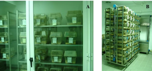
This investigation was carried out using mice from the IBMC’s Animal House internal health monitoring program. A total of 362 mice of 49 strains including knockout, transgenic and wild type mice of various genetic backgrounds, immune status, ages and either sex were examined. The animals used were breeders, experimental mice, stock mice or bedding sentinels. Except for 2 animals, kept in a non-barrier conventional room, all other 360 mice were kept in full-barrier rooms and their health status was considered to be SOPF for the agents listed in the FELASA recommendations for the monitoring of mice colonies (Nicklas et al., 2002). All animals were kept in rooms under positive pressure HEPA-filtered ventilation, at 22 ± 2ºC with a 12/12h light/dark cycle and 55 ± 10% relative humidity, housed in static polycarbonate top filtered cages on corn cob bedding, either in shelf racks (Figure 2A) or in Individually Ventilated Cages (IVCs; Figure 2B). Autoclaved diet (ref. 4RF25GLP, Mucedola) and reverse-osmosis filtered water were available ad libitum. Cages and bedding, feeders and water bottles were autoclaved and changed every week. All procedures were carried out in accordance with the national law Portaria nº 1005/92 (23rd of October) and the European Communities Council Directive 86/609/EEC.
Figure 2: Animals housed in static polycarbonate top filtered cages. A: Shelf racks. B: IVC.
2.2. Sampling
For DNA isolation, stool samples were obtained by taking individual mice out of their cage and collecting a fecal pellet directly into a sterile 1.5 ml reaction tube by holding the mouse by the base of the tail (Figure 3). More than 90% of mice excreted a fecal pellet within one minute. After collection, samples were immediately frozen at -20ºC and DNA isolation was performed within 24 hours. For culture purposes, oral swabs (Becton, Dickinson and Company) were obtained by restraining each mouse for about 2 minutes while swabbing the animal’s mouth. Swabs were placed in transport medium and kept at 4ºC.
Figure 3: Fecal sample collection.
2.3. DNA isolation and quantification
DNA from individual fecal pellets was isolated using a QIAamp DNA Stool Mini Kit (Qiagen) according to the manufacturer’s protocol for isolation of DNA from stool for pathogen detection. DNA concentration and purity were determined spectophotometrically by measuring the absorbance at 260 nm and 280 nmin a NanoDrop ND-1000 Spectrophotometer (NanoDrop Technologies). One unit of absorbance corresponds to 50 μg/ml for double-stranded DNA.
2.4. Primers
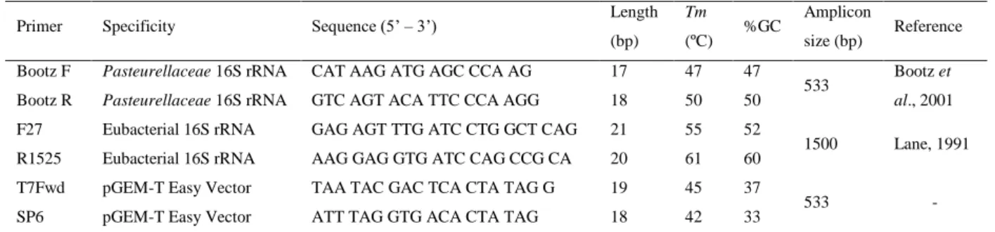
Primers used in this work are described in Table 1. Primer pair Bootz F / Bootz R has been previously described for the specific detection of Pasteurellaceae (Bootz et al., 1998) and amplifies a 533 bp fragment of the 16S rRNA gene. The prokaryotic broad-range F27 / R1525 primer pair, targeting also the 16S rRNA gene, was used as PCR positive control and for detection of putative false negative results. All primers were synthesized by IDT – Integrated DNA Technologies.
Table 1: Primers used to amplify regions of the 16S rRNA gene.
Primer Specificity Sequence (5’ – 3’) Length
(bp) Tm
(ºC) %GC
Amplicon
size (bp) Reference Bootz F Pasteurellaceae 16S rRNA CAT AAG ATG AGC CCA AG 17 47 47
533 Bootz et al., 2001 Bootz R Pasteurellaceae 16S rRNA GTC AGT ACA TTC CCA AGG 18 50 50
F27 Eubacterial 16S rRNA GAG AGT TTG ATC CTG GCT CAG 21 55 52
1500 Lane, 1991 R1525 Eubacterial 16S rRNA AAG GAG GTG ATC CAG CCG CA 20 61 60
T7Fwd pGEM-T Easy Vector TAA TAC GAC TCA CTA TAG G 19 45 37
533 -
SP6 pGEM-T Easy Vector ATT TAG GTG ACA CTA TAG 18 42 33
Tm, Melting temperature calculated by manufacturer; bp, base pairs
2.4.1. Specificity of Bootz primer pair - in silico analysis
The specificity of Bootz primer pair was analyzed in silico using the EMBL-EBI multiple sequence alignment program ClustalW2 (Chenna et al., 2003). An alignment of 16S rRNA gene sequences available at the NCBI Genbank database from Pasteurella pneumotropica (n=26), Haemophilus parainfluenzae (n=2), Haemophilus sp (n=2), Actinobacillus muris (n=2), Escherichia coli (n=6), Proteus mirabilis (n=2) and Enterococcus faecalis (n=1) was performed. The sequences of primers’ targets were then searched throughout the aligned sequences. The Pasteurellaceae sequences selected for the alignment were submitted to GenBank after the primers description (1998). The Genbank accession numbers and submission dates of the sequences used in the alignment are given in Table 2.
Table 2: Genbank accession numbers of 16S rRNA gene sequences used in the alignment.
Accession Organism Strain Submission date
M75083 Pasteurella pneumotropica Jawetz NCTC 8141 24-01-2000
AF012090 Pasteurella pneumotropica Heyl CNP 160 28-11-2001
AY362924 Pasteurella pneumotropica Jawetz NCTC 8141 28-07-2004
DQ875933 Pasteurella pneumotropica Heyl Q480011-V1 30-08-2006
FJ685629 Pasteurella pneumotropica Heyl T08711-V2 01-02-2010
FJ685626 Pasteurella pneumotropica Jawetz J426011 01-02-2010
FJ685623 Pasteurella pneumotropica Heyl ATCC 12555 01-02-2010
GU809188 Pasteurella pneumotropica Jawetz CR3 08-04-2011
GU809187 Pasteurella pneumotropica Jawetz CR51 08-04-2011
GU809186 Pasteurella pneumotropica Jawetz CR53 08-04-2011
GU809185 Pasteurella pneumotropica Jawetz CR19 08-04-2011
GU809184 Pasteurella pneumotropica Jawetz CR54 08-04-2011
GU809183 Pasteurella pneumotropica Jawetz CR17 08-04-2011
GU809182 Pasteurella pneumotropica Jawetz CR13 08-04-2011
GU809181 Pasteurella pneumotropica Heyl CR28 08-04-2011
GU809180 Pasteurella pneumotropica Heyl CR26 08-04-2011
GU809179 Pasteurella pneumotropica Heyl CR24 08-04-2011
GU809178 Pasteurella pneumotropica Heyl CR10 08-04-2011
GU809177 Pasteurella pneumotropica Heyl CR5 08-04-2011
GU809176 Pasteurella pneumotropica Heyl CR16 08-04-2011
GU809175 Pasteurella pneumotropica Heyl CR32 08-04-2011
GU809174 Pasteurella pneumotropica Heyl CR1 08-04-2011
GU809173 Pasteurella pneumotropica Heyl CR30 08-04-2011
GU809172 Pasteurella pneumotropica Heyl CR18 08-04-2011
NR_042887 Pasteurella pneumotropica Jawetz NCTC 8141 10-08-2011
FJ685628 Haemophilus parainfluenzae B160041 01-02-2010
FJ685627 Haemophilus parainfluenzae I112013 01-02-2010
FJ685625 Haemophilus sp HK447 01-02-2010
FJ685624 Haemophilus sp HK445 01-02-2010
AY362894 Actinobacillus muris NCTC12432 20-07-2004
NR_042870 Actinobacillus muris NCTC12432 10-08-2011
J01859 Escherichia coli - 11-08-1995
Z83204 Escherichia coli - 01-03-1997
X80732 Escherichia coli MC4100 29-03-1996
X80724 Escherichia coli ATCC 25922 29-03-1996
X80725 Escherichia coli ATCC 11775T 29-03-1996
X80731 Escherichia coli pk3 29-03-1996
AJ301682 Proteus mirabilis CIP103181T 06-06-2003
EU643833 Proteus mirabilis Hu 07-05-2008
2.5. Polymerase Chain Reaction
DNA amplification by PCR was performed in a 25 µL reaction volume. Each reaction contained 1x Taq Buffer with KCl (Fermentas), 2 mM MgCl2 (Fermentas), 0.2 pmol/µL of
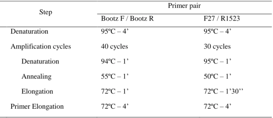
each primer, 0.2 mM of each dNTP (Fermentas), 1U Taq DNA polymerase recombinant (Fermentas) and 80 ng of template DNA. Fecal DNA samples from 2 animals housed in a conventional room, and derived from a colony known to be infected with Pasteurellaceae, were used as positive controls. Fecal DNA samples from 2 animals purchased from a commercial Pasteurellaceae-free colony (Charles River Laboratories, Spain) were used as negative controls. PCR without template DNA was performed as negative PCR control. All samples were tested with Bootz F / Bootz R and F27 / F 1523 primer pairs. PCR amplifications were carried out in a TProfessional Basic Gradient 96 thermocycler (Biometra), in triplicates, under the conditions given in Table 3.
Table 3: Reaction conditions used for PCR amplifications.
Step Primer pair
Bootz F / Bootz R F27 / R1523
Denaturation 95ºC – 4’ 95ºC – 4’
Amplification cycles 40 cycles 30 cycles
Denaturation 94ºC – 1’ 95ºC – 1’
Annealing 55ºC – 1’ 50ºC – 1’
Elongation 72ºC – 1’ 72ºC – 1’30’’
Primer Elongation 72ºC – 4’ 72ºC – 4’
2.5.1. Detection of PCR products
PCR products were detected by electrophoresis on a 1.5 % (w/v) agarose (Bioron) gel in 1x TAE buffer [40mM Tris-base pH8.0 (MP Biomedicals Europe), 20 mM acetic acid glacial (Merck), 1 mM EDTA pH8.0 (Prolabo)] containing 0.5 µg/mL ethidium bromide (Sigma-Aldrich). PCR products were visualized by UV transillumination using a GelDoc XR+ System (Bio-Rad). GeneRuler 1 kb DNA Ladder 250 – 10000 bp molecular weight marker (Fermentas) was included on each gel alongside the samples.
2.6. Cloning and sequencing of PCR products
PCR products from different animals were excised from the agarose gel and subsequently purified using illustra GFX PCR DNA and Gel Band Purification Kit (GE Healthcare) according to the manufacturer’s protocol. The purified PCR fragments were ligated in a pGEM-T Easy Vector (Promega) according to the manufacturer’s instructions and the resultant constructs were used to transform E. coli DH5α competent cells prepared according to the protocol previously described by (Hanahan, 1985). To confirm successful cloning, white colonies were checked by colony PCR using Bootz primer pair as previously described, and 5 μL of a 50 μL colony suspension as template DNA. For each ligation, 3 colonies containing the PCR derived insert were selected and plasmid DNA was isolated using GenElute Plasmid Miniprep Kit (Sima-Aldrich) according to the manufacturer’s instructions. To confirm the presence of the insert in the plasmid, a restriction pattern analysis with EcoRI (Fermentas) was performed. All assays were performed in duplicates. Sequencing of the inserts was performed by StabVida (Lisbon, Portugal) using the specific pGEM-T Easy Vector primers T7Fwd and SP6 (Table 1). Each nucleotide was sequenced a minimum of 3 times in each strand.
2.6.1. Sequencing analysis and multiple alignment
The sequences were assembled with Vector NTI software (Invitrogen). The homology search of each sequence was performed with the BLASTn tool (Altschul et al., 1990) provided by the NCBI website (http://blast.ncbi.nlm.nih.gov/Blast.cgi). Further, the assembled sequences were aligned with 16S rRNA gene sequences of the reference strains P. pneumotropica Jawetz NCTC 8141 (AY362924) and P. pneumotropica Heyl ATCC 12555 (FJ685623) using the EMBL-EBI multiple sequence alignment program ClustalW2 (Chenna et al., 2003). Multiple alignment was edited with GeneDoc software (Nicholas et al., 1997).
2.7. Comparison of the fecal PCR with standard culture methods
The animals used to develop the fecal PCR for Pasteurellaceae detection were also analysed by culture techniques in two external certified laboratories. Oral swabs from 10
animals were sent to the Charles River Laboratories (Lyon, France) for Pasteurella pneumotropica screening and 10 live animals were sent to QM Diagnostics (Nijmegen, The Netherlands) for Pasteurellaceae screening.
2.8. Prevalence of Pasteurellaceae in SOPF mice
Prevalence of Pasteurellaceae among the SOPF mice of the IBMC’s Animal House was assessed using the newly developed fecal PCR assay. A cross sectional study was performed with approximately 10% of the SOPF mice population of the IBMC’s Animal House. A total of 360 SOPF animals were analyzed during a one-year period (January to December 2010). All assays were performed as described in 2.5. Each PCR run included one positive control sample, one negative control sample and a no-DNA template control. Statistical analysis was performed with GraphPad Prism 5 Software. The Fisher’s exact test was applied to correlate the positive and negative cases with sex, age, breeding status, cage type and immune status of the animals. Statistically significant difference was considered when p < 0.05.
3. Results and Discussion
Diagnosis of Pasteurellaceae infections is usually based on bacterial culture and subsequent phenotypic characterization. As these procedures are time-consuming and sometimes yield indeterminate results due to the phenotypical diversity of the Pasteurellaceae family, molecular based techniques have been reported as alternative diagnostic tools. Several PCR assays have been described for the identification of cultured Pasteurellaceae isolated from rodents and also for their detection in clinical samples. However, the sampling methods used for DNA isolation purposes are frequently invasive and require the sacrifice of animals. PCR assays using fecal pellets have been described for several rodent pathogens, including Helicobacter spp. (Beckwith et al., 1997), Clostridium piliforme (Furukawa et al., 2002) and Citrobacter rodentium (McKeel et al., 2002). In this study we developed a simple, specific and non-invasive fecal PCR assay to detect Pasteurellaceae in laboratory mice using previously described specific primers. In that report, PCR was performed with DNA isolated either from cultured Pasteurellaceae from nasopharyngeal swabs or DNA directly isolated from pharynx, trachea and lung specimens, requiring the animals sacrifice (Bootz et al., 1998). We also compared our fecal PCR assay with standard diagnostic procedures such as bacterial isolation and identification. Furthermore we discussed the impact of this non-invasive technique in assessing the prevalence of Pasteurellaceae in laboratory rodents.
3.1. The feces as a source of bacterial DNA
Fresh fecal pellets were reliably obtained from individual mice and DNA was isolated with a QIamp DNA Stool Mini Kit (Qiagen). This commercial kit includes InhibitEX tablets for the adsorption of impurities typically present in stool samples that can degrade DNA and inhibit downstream enzymatic reactions. Such impurities can include bilirubin, bile salts (Beckwith et al., 1997) or complex polysaccharides possibly originating from vegetable materials in the diet (Monteiro et al., 1997).
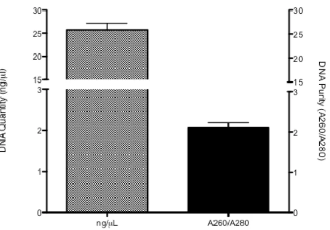
DNA isolation from 362 fecal pellets belonging to different mice yielded an average of 25.74±1.34 ng/μL per fecal pellet (range 0.32 – 184.8 ng/μL). Purity of the DNA preparation, as indicated by the OD260/OD280, had a mean value of 2.074±0.11 (range 0.46 – 37.65)
Figure 4: Mean values and standard error of fecal DNA concentration and purity from total
mice.
Regarding DNA purity, for 24 samples the OD260/OD280 had values below 1.7 and above 2.1. However, for the remaining 338 samples the OD260/OD280 showed a mean value of 1.84. As such, the obtained results indicate that fecal pellets can be used as a good source of DNA in mice.
3.2. Specificity of Bootz primer pair - in silico analysis
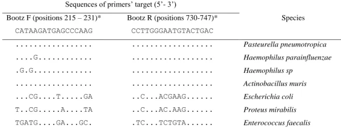
The primer pair used for the specific detection of Pasteurellaceae was described in 1998 and was selected on the basis of 16S rRNA gene sequences of various rodent isolates representing different phenotypic groups of Pasteurellaceae together with all sequences from NCBI GenBank and EMBL-EBI databases available at that time (Bootz et al., 1998). However, the number of entries in the Genbank database increased since then. As such, the primers specificity to target Pasteurellaceae was assessed in silico using the 16S rRNA gene sequences of Pasteurellaceae submitted since 1998 to date (see Table 2). In silico analysis was performed by aligning 16S rRNA gene sequences from P. pneumotropica, H. parainfluenzae, Haemophilus sp, A. muris, E. coli, P. mirabilis and E. faecalis, and subsequently searching for the sequences of primers’ targets throughout the aligned sequences. H. parainfluenzae, Haemophilus sp. and A. muris are members of the Pasteurellaceae family and have been previously isolated from laboratory rodents (Csukas, 1975; Bisgaard, 1986; Boot et al., 2005). E. coli, P. mirabilis and E. faecalis were included for their potential to interfere with the assay as they are commonly found in normal rodent’s feces (Beckwith et al., 1997). Observed nucleotide mismatches are summarized in Table 4.
Table 4: Bootz primers mismatches with target sequences of different species. Dots indicate
base identity with the primers sequence.
Sequences of primers’ target (5’- 3’)
Bootz F (positions 215 – 231)* Bootz R (positions 730-747)* Species
CATAAGATGAGCCCAAG CCTTGGGAATGTACTGAC
... ... Pasteurella pneumotropica
....G... ... Haemophilus parainfluenzae
.G.G... ... Haemophilus sp
... ... Actinobacillus muris
...CG....T...GA ..C...ACGAAG... Escherichia coli
T..CG...A....TA ..C...AC.AAG... Proteus mirabilis
TGATG....GA...GC. .TC...TCTGTA... Enterococcus faecalis
*Base positions are given according to P.pneumotropica NCTC 8141 numbering (Genbank accession no. M75083).
DNA polymerases catalyze the addition of nucleotides to the primer 3'-OH, as specified by complementarity to the template DNA. Mismatches between primers and targeted DNA can affect duplex stability, which might then hamper the ability of a system to amplify the template DNA. The effects of mismatches depend on numerous factors, such as oligonucleotide length and the nature and position of the mismatches. Several studies have investigated the effects of primer-template mismatches at the3' end of the primer sequence, and it has been demonstratedthat PCR was prevented by a single mismatched base at the 3' end. In contrast, mismatches at the 5’ end and internal mismatchescan be tolerated (Kwok et al., 1990). As shown in Table 4, the sequences of Bootz primers’ target are 100% identical to the 16S rRNA gene sequences of P. pneumotropica and A. muris at positions 215-231 for the forward primer and 730-747 for the reverse primer. Furthermore, the forward primer differs only by 1 base from the sequence of H. parainfluenzae and by 2 bases from that of Haemophilus sp near the 5’ end. The reverse primer has no mismatches with the sequences both species. However, the number of nucleotide mismatches with organisms other than Pasteurellaceae is significantly higher. Particularly in the forward primer, the mismatches are located at the 3’ end of the sequences. Therefore, the PCR reaction is not likely to occur.
The in silico analysis confirms that this primer pair specifically targets the 16S rRNA region of members of the Pasteurellaceae family, as previously described (Bootz et al., 1998). Due to its specificity, this primer pair meets the specifications to be used in PCR assays for the detection of Pasteurellaceae. Therefore it was selected to develop our fecal PCR assay.
3.3. Evaluation of the fecal PCR for Pasteurellaceae detection
To develop our fecal PCR assay, a total of 20 mice were examined (mice numbered from 1 to 20): 2 mice (numbers 1 and 2) kept in a non-barrier conventional room and 18 mice (numbers 3 to 20) with SOPF health status, kept in barrier-rooms. Samples from animals 1 and 2 were used as positive controls because they derived from colonies known to be infected with P. pneumotropica and presented clinical signs of conjunctivitis. From the 18 SOPF mice, 2 (numbers 4 and 5) were purchased from a commercial Pasteurellaceae-free colony (Charles River Laboratories, Spain) and fecal samples were collected after one week of quarantine at the IBMC’s Animal House. These samples were used as negative controls.
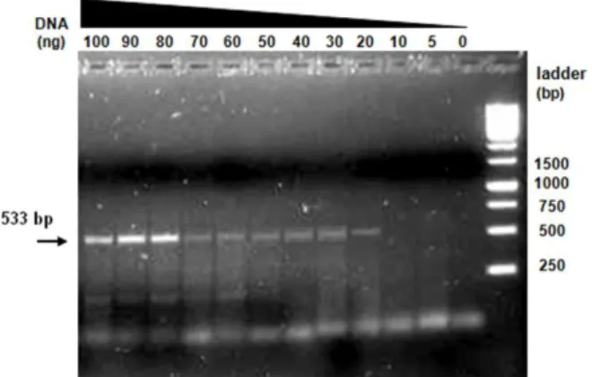
3.3.1. Assay sensitivity
To estimate the sensitivity of the fecal PCR, different quantities of template DNA ranging from 5 to 100 ng were used in a series of reactions with Bootz primer pair. In this assay we used one of the positive control samples (mouse number 2) as template DNA. Results are shown in Figure 5.
Figure 5: PCR amplification using different amounts of template DNA from mouse no.2.
Bootz primers amplified a 533 bp fragment.
According to this assay, the lower limit of detection of our test was 20 ng of template DNA. Based on the obtained results we estimate that the PCR assay should be performed with 80 to 100 ng of template DNA in each reaction, where a stronger signal is observed.
3.3.2. Detection of Pasteurellaceae in fecal samples
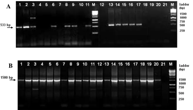
After establishing the optimal conditions for the PCR assay, all 20 samples were analyzed with Bootz primer pair using 80 ng of template DNA. The results obtained are shown in Figure 6A. The expected 533 bp DNA fragment was amplified from 12 fecal DNA samples: the positive control samples (mice 1 and 2) and samples from 10 other animals (mice 3, 6, 8, 9, 11, 13, 14, 15, 16 and 17). On the contrary, this PCR assay was not able to amplify any fragment from the negative control samples (mice 4 and 5), as expected, and from the remaining 6 samples (mice 7, 10, 12, 18, 19 and 20).
Figure 6: PCR amplification of fecal DNA samples from 20 different mice. Lanes: 1 – 20,
animals 1 to 20 respectively; 21, no template DNA; M, 1kb DNA ladder. A: PCR amplification with Bootz primer pair yielding a 533 bp fragment. B: PCR amplification with prokaryotic broad-range primer pair yielding a 1500 bp fragment.
Fecal PCR analytic methods can be used as a non-invasive means of rapidly screening large numbers of mice. However, one of the main concerns of all fecal tests is the potential of obtaining false negative results due to the presence of PCR inhibitors in feces. To exclude putative false negative results, PCR reactions with prokaryotic broad-range F27 / R1525 primer pair were performed for all samples. In Figure 6B it is shown that the expected 1500 bp fragment was amplified from all the tested samples, including those which yielded no
A
amplicon when PCR was performed with Bootz primer pair. The obtained results show that fecal DNA samples belonging to mice 4, 5, 7, 10, 12, 18, 19 and 20 are in fact Pasteurellaceae free samples, and confirm that the isolated fecal DNA is suitable to monitor bacteria in mice stool samples.
According to Bootz et al. (1998), Haemophilus influenzaemurium strains yielded an additional band approximately 1200 bp long. A band with this approximate size was amplified from the sample of animal number 3 (Figure 6A).
3.4. Sequencing analysis and multiple alignment
PCR products from randomly chosen individuals yielding the expected 533 bp DNA fragment (animals 1, 2, 3, 6, 8 and 9) were sequenced to confirm the specificity of the PCR assay and to disclose the occurrence of false positive results. In addition to the expected 533 bp amplicon, the amplicon of approximately 1200 bp obtained from animal number 3 IFigure 6) was also sequenced. Sequences from 18 clones of the 533 bp fragment (3 clones per animal) and from 3 clones of the 1200 bp fragment were assembled with Vector NTI software (Invitrogen) and compared with NCBI GenBank entries by using the BLASTn algorithm. Results for the first hit of each sequence are shown in Table 5. Further, the assembled sequences were aligned with 16S rRNA gene sequences of P. pneumotropica Jawetz and Heyl reference strains using the EMBL-EBI ClustalW2 software (Figure 7).
Table 5: NCBI GenBank sequences producing significant alignments with the sequenced 533
bp and 1200 bp fragments.
Mouse Fragment
length (bp)
Accession Organism Query
coverage
E value Maximum
identity
1 533 GU809177 P. pneumotropica Heyl strain CR5 100% 0.0 99%
2 533 GU809188 P. pneumotropica Jawetz strain CR3 100% 0.0 99%
3 533 GU809188 P. pneumotropica Jawetz strain CR3 100% 0.0 99%
1369 CP000612 Desulfotomaculum reducens MI-1 10% 6e-13 73%
6 533 GU809188 P. pneumotropica Jawetz strain CR3 100% 0.0 99%
8 533 GU809188 P. pneumotropica Jawetz strain CR3 100% 0.0 99%
533 GU809177 P. pneumotropica Heyl strain CR5 100% 0.0 99%
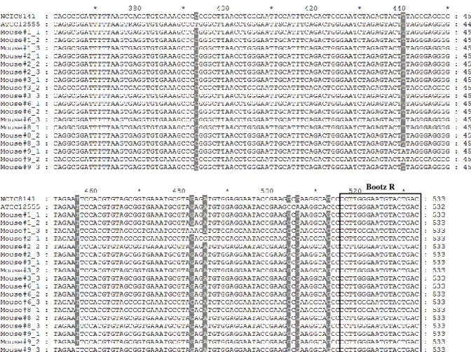
Figure 7: Multiple alignment of the 533 bp sequenced fragments and reference strains P.
pneumotropica Jawetz NCTC 8141 (AY362924) and P. pneumotropica Heyl ATCC 12555 (FJ685623). Wobble bases: R=A+G, S=C+T, N=A+G+C+T. The sequences of primers Bootz F and Bootz R are boxed.
Results from the BLASTn analysis (Table 5) show a 99% identity in the 533 bp overlap between the sequences of the fragments amplified from mice feces and GenBank sequences of the 16S rRNA gene of P. pneumotropica. Furthermore, the Expected value (E) is 0.0 for all sequences. The Expect value is a parameter that describes the number of hits one can expect to see by chance when searching a database of a particular size. The lower the E-value, or the closer it is to zero, the more significant the match is. Thus, BLASTn analysis of the sequenced 533 bp fragments confirms the specificity of the PCR assay. However, the other fragment obtained from animal number 3 was in fact 1369 bp long and revealed no identity with members of the Pasteurellaceae family.
Multiple alignment of the sequenced 533 bp fragments with 16S rRNA gene sequences of reference strains P. pneumotropica Jawetz NCTC 8141 (AY362924) and P. pneumotropica Heyl ATCC 12555 (FJ685623) (Figure 7) show 14 single nucleotide polymorphisms (SNPs)