UNIVERSIDADE DE TRÁS-OS-MONTES E ALTO DOURO Master in Biochemistry
PROTEIN OXIDATION AND CIRCADIAN RHYTHMICITY: TOWARDS THE
IDENTIFICATION OF SPECIFIC PROTEIN TARGETS
Master Dissertation presented by: Liliana Patrícia Costa Rodrigues
Under Supervision
Professor Doctor Bertrand Friguet, PhD student Audrey Desvergne and Professor Doctor Francisco Peixoto
Supervised by:
______________________________________ Professor Doctor Francisco Peixoto Chemical Department, UTAD
This work was prepared as an original dissertation application to the degree of Master of Biochemistry in the “Universidade de Trás-os-Montes e Alto Douro” in collaboration with the Laboratoire de Biologie Cellulaire du Vieillissement (UR4-Vieillissement, stress et inflamation in Université Pierre et Marie Curie- Paris VI)
ACKNOWLEDGEMENTS
Life can only be understood if we look back, but it can only be lived if we look forward – S. Kierkegaard. However, many times I would not have been able to look forward if I did not have the support of some people. Therefore I want to thank:
Universidade de Trás-os-Montes e Alto Douro, whose doors were always open to me right from the beginning and which made me feel enriched as a person.
Prof. Dr Bertrand Friguet for having accepted me in his laboratory and for all his support throughout the year.
Prof. Dr. Francisco Peixoto for having supervised this work and for all his support and help. Prof. Dra. Maria Manuel Oliveira and Prof. Dra. Carla Amaral for having given me the possibility of going on an ERASMUS programme and for all their support and dedication.
Audrey Desvergne for having been such a friendly «boss», for having taught me all the techniques which were used throughout this work, but essentially for having given me a friendly word and courage to go on.
All the UR4 team, namely Sabrina, Marine, Sofia and Fernando for all the entertainment, pieces of advice, and above all, friendship.
All my friends, especially João Morais and Diana Silva for having always backed me up with friendly words and making me believe I was able to achieve my dream.
My Parents for all their support, help, motivation and caress throughout all these years.
My Grandparents for always being by my side, believing in me and encouraging me to get here.
Davide Rodrigues for having been understanding and always been by my side trying to give me courage and determination to go ahead despite the different obstacles which appeared in my long walk here.
ABSTRACT
The circadian rhythmicity exists in all organisms and generates rhythms with a periodicity of 24 h of a significant number of biological and metabolic processes. A disruption of the circadian clock (BMAL1 and PER2) in mice, are directly associated to an increase of reactive oxygen species (ROS) which induce a premature aging phenotype (Kondratov, 2007; Ramsey, et al., 2007). On the other hand, it was described that the accumulation of oxidized (carbonylated) proteins is a hallmark of cellular aging. Moreover, protein carbonylation, an irreversible modification, involves a decrease quality of the cellular proteome which could directly affect the normal cell function.
Using HEK-293 cells synchronized with serum shock and the 1D electrophoresis technique, our working group showed that the level of carbonylated proteins has a circadian rhythmicity. This quantification of the total proteins allowed to choose two peaks of the circadian rhythm corresponding to maximum and minimum levels of protein carbonylation. In this work 2D electrophoresis were also used, to characterise some particular proteins, which seem to suffer a circadian oxidation.
After quantification of carbonylated proteins, it was observed that the same spots are common all the times, even if their level of carbonylation is noticeably different. Concluding, it was proved, although without final characterization, that some specific proteins have a circadian oxidation. For the future, it will be interesting to identify these specific proteins and compare them to biological markers of oxidative stress in order to better understand the involvement of the circadian rhythm.
RESUMO
O ritmo circadiano existe em todos os organismos e gera ritmos com uma periodicidade de 24 h em um significante número de processos biológicos e metabólicos. Uma disrupção do relógio circadiano (BMAL1 e PER) em ratos, está diretamente associada com o aumento das espécies reativas de oxigénio (ERO) que induzem um fenótipo de envelhecimento prematuro (Kondratov, 2007; Ramsey, et al., 2007). Por outro lado, foi descrito que a acumulação de proteínas oxidadas (carboniladas) é um marcador do envelhecimento celular. Para além disso, as proteinas carboniladas, com modificação irreversível, envolve uma diminuição da qualidade do proteoma celular que pode afectar diretamente a função da célula normal.
Através da utilização de células HEK293 sincronizadas com serum shock e utilização das técnicas de electroforese de ima dimensão, o nosso grupo de trabalho demonstrou que o nível das proteínas carboniladas tinham uma ritmicidade circadiana. Essa quantificação do total de proteínas levou-nos a escolher dois picos do ritmo circadiano, correspondentes ao máximo e mínimo de nível de carbonilação. Neste trabalho, utilizamos também a electroforese de duas dimensões para caracterizar algumas proteínas em particular que pareciam sofrer oxidação circadiana.
Após a quantificação das proteínas carboniladas, observou-se que os mesmos spots eram comuns em todos os tempos, mesmo se o nível de carbonilação é notavelmente diferente. Concluindo, nós provamos, embora sem proceder a uma caracterização final, que proteínas específicas possuem oxidação circadiana. No futuro, pode ser interessante identificar as proteínas alvo específicas e compará-las com os marcadores biológicos do stress oxidativo de forma a perceber melhor o envolvimento destas na ritmicidade circadiana.
Palavras Chave: Ciclo Circadiano; Serum Shock ; Dexametazona; Proteínas Carboniladas; Envelhecimento
INDEX
Acknowledgements ... iv Abstract ... v Resumo ... vi Index ... vii Index Figures ... ix Index Table ... x Abbreviations ... xi 1. Introduction ... 1 1.1. Circadian Rhythms ... 11.2. Organisation of the circadian cycle in mammals ... 2
1.3. Molecular mechanism of the mammalian circadian clock: ... 4
1.4. Metabolic processes with circadian cycles... 6
1.5. Interaction between the transcriptional clock and metabolic clock ... 7
1.6. Circadian Cycle, Aging and Diseases ... 8
1.7. Aging and oxidized proteins ...10
1.8. Circadian Synchronization in Cell Culture ...13
1.8.1. Circadian Synchronization with Serum Shock ...13
1.8.2. Circadian Synchronization with Dexamethasone ...14
2. Objectives ...16
3. Material and Methods ...17
3.1. Culture of HEK-293 cells ...17
3.2. Synchronization of the HEK-293 cells ...17
3.2.1. Synchronization with dexamethasone ...18
3.2.2. Synchronization with serum shock ...18
3.3. Analysis of the transcripts ...19
3.3.1. Extraction and quantification of RNA ...19
3.3.2. Reverse Transcription ...19
3.3.3. Quantitative PCR ...20
3.4. Analysis of Carbonylated Proteins ...21
3.4.1. Extraction and quantification of Protein ...21
3.4.2. One Dimension Electrophoresis (Oxyblot 1D) ...21
3.4.3. Two Dimension Electrophoresis (Oxyblot 2D) ...23
4. Results...26
4.1. Determination of the best type of synchronization for HEK-293 cells ...26
4.2. Quantitative PCR ...28
4.3. Determination of Carbonylated Proteins ...30
4.3.1. Quantification carbonylated proteins in 1D electrophoresis ...30
4.3.2. Quantification of Carbonylated Protein in 2D electrophoresis ...32
5. Discussion and Conclusion ...41
6. Bibliography ...43
7. Attachments ...47
7.1. Solutions for cell culture ...47
7.1.1. Culture medium (DMEM-PSG) ...47
7.1.2. Medium without serum (DMEM-PG) ...47
7.2. Solutions for quantitative PCR ...47
7.2.1. Lysis Buffer ...47 7.2.2. TAE solution ...47 7.2.3. DNA Dye 6X ...48 7.2.4. DNAmix ...48 7.2.5. Standard curve ...48 7.2.6. Solutions primers...49
7.3. Solutions for 1D and 2D electrophoresis ...49
7.3.1. Lysis Buffer ...49
7.3.2. Gel from Oxyblot 1D ...50
7.3.3. Migration buffer ...50
7.3.4. Transfer buffer ...50
7.3.5. PBS 10X ...51
7.3.6. Washing Buffer ...51
7.3.7. Fast Green Solution ...51
7.3.8. Blocking Buffer ...51
7.3.9. Re-Hydration Buffer ...52
7.3.10. Derivatization Solution ...52
7.3.11. Neutralization Solution ...52
7.3.12. Equilibration Solution ...52
7.3.13. Gels for Oxyblot 2D...53
7.3.14. Fixation Buffer ...53
7.3.15. Pre Coloration Buffer ...53
INDEX FIGURES
Figure 1- Schematic representation of the circadian cycle.. ... 3
Figure 2- Representation of the molecular mechanism of the circadian clock in mammals.. ... 6
Figure 3- Propose of interconnectivity circadian redox processes and Transcription-translation feedback loop (TTFL).. ... 8
Figure 4 –Fragmentation of the carbon skeleton.. ... 11
Figure 5- Relation between circadian rhythm aging and formation of carbonylated protein... 12
Figure 6- Possible explanation of synchronization with Serum Shock. ... 14
Figure 7- Chemical representation of dexamethasone ... 14
Figure 8- Scheme of derivatization with 2,4-Dinitrophenylhydrozine (DNPH) and reaction of the protein with the antibody anti-DNP. ... 22
Figure 9- Representation of the method of 1D electrophoresis. ... 23
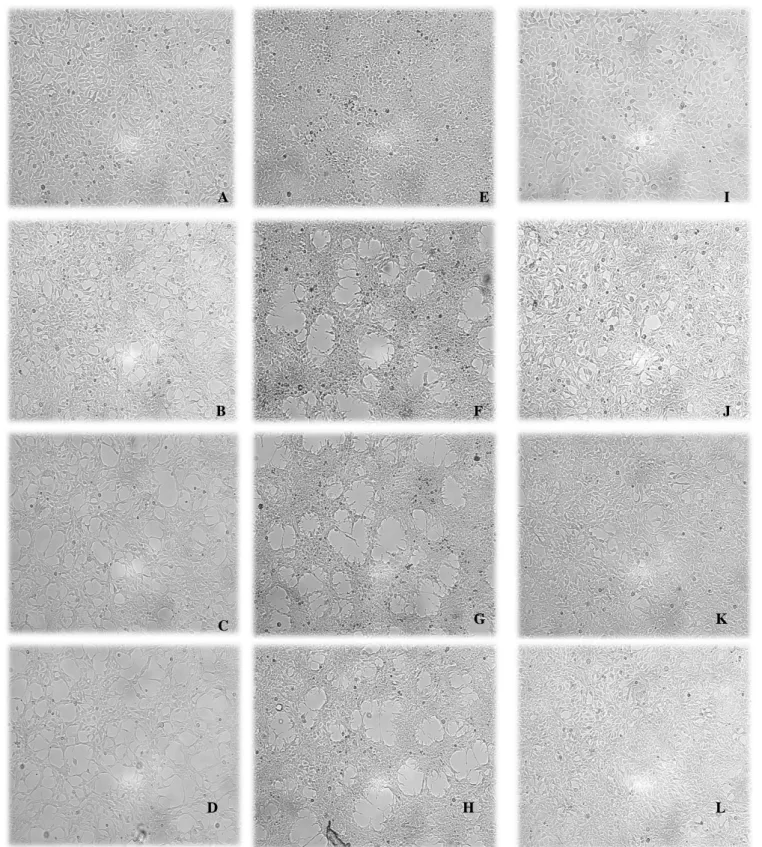
Figure 10- Microscopic analysis of HEK-293 cells in three types of treatment... 27
Figure 11- Transcription level of Clock and Per2 normalized in the three treatments tested.. ... 29
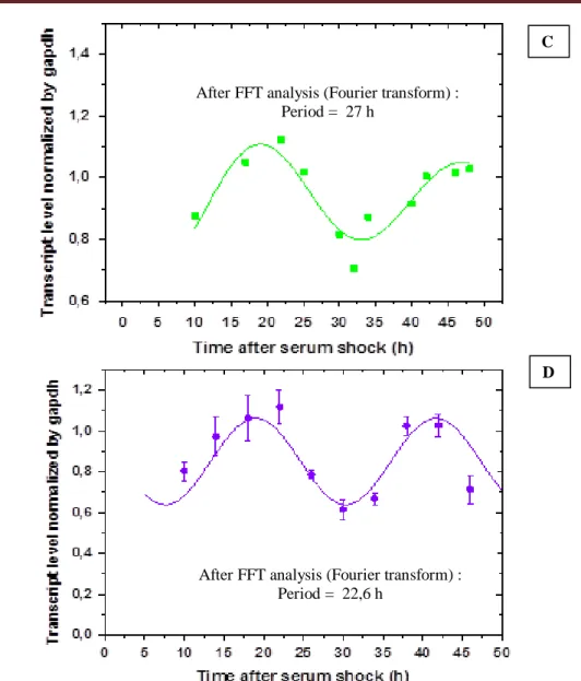
Figure 12- Transcription level of Cyclin B1 in cells submitted to serum shock treatment. ... 30
Figure 13- Levels of Carbonylated proteins in HEK-293 cells during two treatments ... 31
Figure 14- Different quantities of protein tested by Enhanced Chemiluminescence (ECL) method of a sample of normal HEK-293cells with strips of 13 cm... 33
Figure 15- Comparison between Coomassie gel and a nitrocellulose membrane revealed with the ECL method showing the carbonylated proteins of a sample of normal HEK-293 cells using strips of 13 cm.. ... 33
Figure 16- Comparison of two types of strips (11 cm and 13 cm) and two types of Lysis Buffer (Lysis Buffer I and Lysis Buffer II) in 2D electrophoresis.. ... 35
Figure 17- 2D electrophoresis of total of protein and carbonylated proteins in a sample of HEK-293 2 h after the start of the treatment (without synchronization and synchronized by serum shock) .... 36
Figure 18- Quantification of carbonylated proteins in percentage in samples 2 h after the beginning of treatment without synchronization (WS) and synchronization with serum shock (SSS) and the number of spots identified as carbonylated proteins in each sample. ... 37
Figure 19-2D electrophoresis of total of protein and carbonylated proteins in a sample of HEK-293 14 h and 26 h after the start of the treatment (synchronization by serum shock). ... 38
Figure 20- Quantification of carbonylated proteins in percentage in samples 14 h and 26 h after the beginning of treatment synchronization with serum shock (SSS) and the number of spots of the carbonylated proteins. ... 38
Figure 21-2D electrophoresis of total of protein and carbonylated proteins in a sample of HEK-293 34 h and 46 h after the start of the treatment (synchronization by serum shock). ... 39 Figure 22- Quantification of carbonylated proteins in percentage in samples 34 h and 46 h after the beginning of treatment synchronization with serum shock (SSS) and the number of spots of carbonylated proteins ... 40
INDEX TABLE
Table 1- Primers of clock controlled transcripts for qPCR ... 20 Table 2- Summary of the preparation of the different samples, with different concentrations to
ABBREVIATIONS
(NH4)2SO4 Ammonium sulfate
1D One dimension
2D Two dimension
APS Ammonium peroxodisulfate
ATP Adenosine triphosphate
bHLH basic helix-loop-helix
BMAL1 Brain and muscle ARNT-like 1
BSA Bovine serum albumin
CKIɛ Casein Kinase Iɛ
CLOCK Circadian locomotor output cycles kaput
CO2 Carbon Dioxide
CRY Cryptochrome
Cy5 Cyanine 5
DMEM Dulbecco’s Modified Eagle Medium
DMSO Dimethyl sulfoxide
DNA Deoxyribonucleic acid
DNP 2,4-dinitrophenil
DNPH 2,4-dinitrophenylhydrozine
DTT Dithiothreitol
E.coli Escherichia coli
ECL Enhance chemiluminescence method
EDTA Ethylenediaminetetraacetic acid
EGF Epidermal growth factor
ET,1 Endothelin-1
EtBr Ethidium Bromide
FGF Fibroblast growth factor
G L-Glutamine
GSK3 Glycogen Synthase Kinase3
h hours
H3PO4 Phosphoric acid
HCl Hydrochloric acid
KH2PO4 Potassium di-hydrogen phosphate
MgCl2 Magnesium chloride
Na2HPO4(H2O)7 Disodium hydrogen phosphate
NaCl Sodium chloride
NADPH Nicotinamide adenine dinucleotide phosphate
ºC degrees Celsius
P Penicilin-streptomycin
PAS PER-ARNT-SIM
PBS Phosphate buffered saline
PER Period
PGE2 Prostaglandin 2
qPCR quantitative Polymerase chain reaction REV-ERBα Reverse erythoblastosis virusα
RHT Retinohipotalamic tract
RNA Ribonucleic acid
RNS Reactive nitrogen species
ROR Related orphan receptor
RORE Related orphan receptor element
ROS Reactive oxygen species
RT Reverse transcription
S or FBS Serum or Fetal Bovine Serum
SAD Seasonal affective disorders
SCN Suprachiasmatic nucleus
SDS Sodium dodecyl sulphate
1. INTRODUCTION
1.1. Circadian Rhythms
All the organisms, from unicellular, like Neurospora crassa and cyanobacteria, to humans, show circadian rhythmicity in their biological, physiological and behavioural processes (Kondratov, 2007; Xydous, et al., 2012). The concept of circadian cycle was introduced by Halberg in 1959 and it comes from Latin “Circa Diem” which means “about a day” (Kondratov, 2007; Ramsey, et al., 2007). This concept is used to describe all the biological processes that exist in the organisms with a rhythmicity of 24 h (Kondratov, 2007; Ramsey, et al., 2007; Xydous, et al., 2012). There are other types of cycles with rhythmicity, however, those do not occur in intervals of 24 h. When a cycle has a rhythmicity bigger than 30 h, like the menstrual cycle in women, it is called infradian cycle and when something occurs in intervals lesser than 18 h, such as the case of cardiac frequency, it is called ultradian cycle (Sukumaran, et al., 2010; Yagita & Okamura, 2000). Lots of external (Zeitgebers) or internal (circadian clock) controllers are capable of controlling the circadian rhythmicity of a series of biological processes (Balsalobre, et al., 1998; Kondratov, 2007). Light is classified as the principal Zeitgeber which stimulates a series of molecular events in cascade which are capable of inducing rhythmicity in the expression of several genes (Bunney & Bunney, 2000; Fuller & Fuller, 2002). The chemical reagents and the social behaviour are some of the examples of the other controllers (Balsalobre, et al., 1998).
Important biological processes have a circadian rhythmicity as is the case of glucose, lipids and drug metabolism, proprieties of the membranes, division and cell cycle (promoted by Cyclin B1), blood pressure and many others (Balsalobre, et al., 1998; Gallego & Virshup, 2007; Kondratov, 2007; Weinert & Waterhouse, 2007). So, when a deregulation of the circadian cycle happens, lots of reactions and diseases can be induced (Froy, 2011; Gallego & Virshup, 2007; Kondratov, 2007; Reddy, et al., 2005). Many diseases and health problems exist due to the flaw of this cycle, like cardiovascular problems, cancer, hypertension or diabetes (Lévi, et al., 2010; Yu & Weaver, 2011).
Studies which have been made in shift workers, individuals subject to jet lag or people who live in northern latitudes show that these people have a deregulation of their circadian cycle which induces the appearance of many biological reactions (Albrecht & Eichele, 2003; Bunney & Bunney, 2000; Froy, 2011; Kondratov, 2007; Reddy, et al., 2005). In the first two cases the people show some sleep problems, fatigue, gastrointestinal problems and hypertension (Froy, 2011; Kondratov, 2007; Reddy, et al., 2005). However, in the last case the people have seasonal affective disorders
(SAD), also known as winter depression (Albrecht & Eichele, 2003; Bunney & Bunney, 2000). This syndrome was characterised as always appearing in the same period of the year, usually in winter, because the days are shorter and the solar intensity decreases (Bunney & Bunney, 2000). It must also be noted that persistent exposure to this type of work or life style can bring diseases like cancer (Albrecht & Eichele, 2003; Froy, 2011; Gallego & Virshup, 2007; Reddy, et al., 2005).
The circadian cycle in mammals is controlled by three different components: input pathway, the circadian master (Suprachiasmatic nucleous- SCN) and output pathway (Balsalobre, et al., 1998; Kondratov, 2007; Sukumaran, et al., 2010; Xydous, et al., 2012; Yagita & Okamura, 2000). This cycle has lots of specific proteins that control the physiological homeostasis which can influence all the tissues and cause some diseases related with aging (Kondratov, 2007; Weinert & Waterhouse, 2007).
1.2. Organisation of the circadian cycle in mammals
In mammals, the circadian system is organised in a hierarchical way (Kondratov, 2007). Anatomically and functionally, the circadian clock is characterised by having three main elements in its constitution: input pathway; pacemaker master and output pathway (Balsalobre, et al., 1998; Bunney & Bunney, 2000; Fuller & Fuller, 2002; King & Takahashi, 2000; Kondratov, 2007; Sukumaran, et al., 2010; Xydous, et al., 2012; Yagita & Okamura, 2000). The input pathway is classified as the one which is responsible for the transmission of the ambient conditions to a pacemaker master. On the other hand, the central master produces stimuli or signals by the output pathway for the whole organism and promotes the synchronization and the rhythmicity of the genes expression and the metabolic activities (Fu & Lee, 2003; King & Takahashi, 2000; Kondratov, 2007).
In 1972, Moore and Stephan, classified the suprachiasmatic nucleus (SCN) as the circadian master clock of the mammals (Froy, 2011; Kondratov, 2007; Kondratov, et al., 2003; Okamua, et al., 2010; Ramsey, et al., 2007). This central clock is located in the anterior hypotalamus, specifically in the 3rd ventricle, and it is composed of around 20000-100000 neurons (Fu & Lee, 2003; Fuller & Fuller, 2002; Kondratov, 2007; Reppert & Weaver, 2001). Although there are other controllers to command the SCN, light is considered the principal one (Balsalobre, et al., 2000; Balsalobre, et al., 1998; Kondratov, 2007; Lévi, et al., 2010). The photonic information is absorbed by the retina and it is sent by input pathway through the retinohipothalamic tract (RHT) to the SCN (Balsalobre, et al., 2000; Balsalobre, et al., 1998; Ramsey, et al., 2007; Sukumaran, et al., 2010).
This will then produce a response or signal (humoral or neuronal) that by output pathway goes to the peripheral clocks that exist in various tissues or organs of the organism promoting their synchronization (Albrecht & Eichele, 2003; Kondratov, 2007; Kondratov, et al., 2003; Okamua, et al., 2010; Ramsey, et al., 2007).
Taking into account the procedure of endogenous synchronization of the organism, the SCN is classified as being at the top of the hierarchical organisation (Figure 1), since this is what promotes the synchronization in all circadian oscillators located in the clocks of the peripheral tissues (Albrecht & Eichele, 2003; Kondratov, 2007; Kononenko, et al., 2008). Due to the importance of the SCN to synchronize the body, a deregulation of this pacemaker may promote multiple and serious health problems (Fuller & Fuller, 2002; Kondratov, 2007).
The circadian clocks that exist in the peripheral tissues in mammals answer, not only to the signalization promoted by SCN, but also to the multiple daily activities such as feeding, locomotion and others (Balsalobre, et al., 2000; Fu & Lee, 2003; Kondratov, 2007). In some situations they may act independently from the pacemaker, for example in case of damage of DNA. When this happens, they will control the local cell cycle checkpoints or they will turn on the apoptose (Fu & Lee, 2003). The similar circadian oscillators that were found in SCN exist not only in the peripheral tissues but also in the cellular culture (Froy, 2011; Gallego & Virshup, 2007). However, to promote a circadian rhythmicity of the clock genes expression in cellular culture, a first synchronization stimulus is necessary (Okamua, et al., 2010).
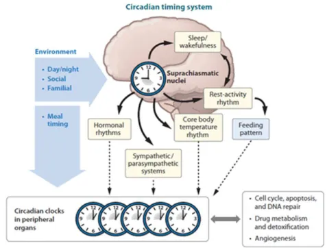
Figure 1- Schematic representation of the circadian cycle. The environmental active conditions, by input pathway, in the suprachiasmatic nucleus (SCN), which are the central pacemaker in mammals. This master promotes stimulis or signals that go, by output pathway, to the circadian clocks in peripheral organs and synchronizes lots of important biological processes (cell cycle, apoptosis, angiogenesis and others). However, the circadian clocks in peripheral organs can also be synchronized by daily activities like feeding. Adopted from (Lévi, et al., 2010).
1.3. Molecular mechanism of the mammalian circadian clock:
Molecular Biology is a science which has allowed the understanding of the behaviour and characteristics of the organisms through chemical mechanisms of activation and transcription genes. Lots of studies in molecular biology have been made to know the generation and control of the circadian rhythms, because they arise from the activation of expression and genes products (King & Takahashi, 2000; Okamua, et al., 2010). The circadian clock is characterised as being constituted by a set of specific genes, called by clock genes (Balsalobre, et al., 2000; King & Takahashi, 2000). These genes are characterised for working through the interaction between negative and positive transcriptional-translational feedback loops (TTFL) that promote the regulation of many biological activities (Albrecht & Eichele, 2003; Froy, 2011; Gallego & Virshup, 2007; Kondratov, 2007; Reppert & Weaver, 2001; Sukumaran, et al., 2010; Xydous, et al., 2012). A clock gene was defined as a necessary product to the regulation of the circadian clock, which may be expressed by circadian form (24 in 24 hours) or constantly during every day (Balsalobre, et al., 2000). In 1971 the first clock gene, period (Per), was identified in a study carried out in Drosophilas melanogaster (Balsalobre, et al., 2000; Fuller & Fuller, 2002; Ramsey, et al., 2007). In the 90s Joseph Takahashi identified the first clock gene in mammals, the transcription factor, circadian locomotor output cycles kaput (Clock). Nowadays 8 genes in the mammal’s circadian clock which are homologous to the Drosophila melanogaster have already been identified (Albrecht & Eichele, 2003; King & Takahashi, 2000; Ramsey, et al., 2007).
The molecular mechanism of the circadian cycle in mammals is constituted by two feedback loops: a positive and a negative (Albrecht & Eichele, 2003; Froy, 2011; Gallego & Virshup, 2007; Kondratov, 2007; Okamua, et al., 2010; Reddy, et al., 2005; Reppert & Weaver, 2001; Sukumaran, et al., 2010). There are two principal components of the molecular mechanism in the circadian clock in mammals, an activator heterodimer which promotes the positive feedback loop and a repressor heterodimer, which is the responsible for the negative feedback loop (Gallego & Virshup, 2007; King & Takahashi, 2000; Kondratov, 2007; Okamua, et al., 2010). The activator heterodimer is constituted by two proteins: CLOCK and brain and muscle ARNT-like protein1 (BMAL1), also called MOP3. These proteins are transcription factors basic helix-loop-helix (bHLH) PER-ARNT-SIM (PAS) that regulate the transcription of many clock genes (Albrecht & Eichele, 2003; Froy, 2011; Fuller & Fuller, 2002; King & Takahashi, 2000; Kondratov, 2007; Ramsey, et al., 2007; Reppert & Weaver, 2001; Sukumaran, et al., 2010).
When the CLOCK/BMAL1 complex binds to the E-box elements, especially in 5’-CACGTG-3’ nucleotides sequence, it promotes the transcription of three kinds of genes Period (Per1, Per2
and Per3) and two types of genes Cryptochrome (Cry1 and Cry2) (Figure 2). This mechanism has the name of positive feedback loop. And, due to this derivatization, proteins with the same name of genes are formed (Albrecht & Eichele, 2003; Froy, 2011; Fuller & Fuller, 2002; Gallego & Virshup, 2007; Ramsey, et al., 2007; Reppert & Weaver, 2001). PER and CRY proteins are formed in the cytoplasm by hyperphosphorylation promoted by Casein Kinase Iɛ (CKIɛ) and Glycogen Synthase Kinase-3 (GSK3) and when they are in the nucleus they form a PER/CRY complex (Albrecht & Eichele, 2003; Gallego & Virshup, 2007). This complex is called repressor complex and it is the responsible for the negative feedback loop, because it blocks the activity of the complex CLOCK/BAML1, so the transcription of clock genes is stopped (Albrecht & Eichele, 2003; Balsalobre, et al., 2000; Froy, 2011; Gallego & Virshup, 2007; Okamua, et al., 2010; Reppert & Weaver, 2001). The fact that the levels of Per and Cry genes decrease takes to the start of the cycle with the induction of positive feedback loop (Albrecht & Eichele, 2003; Gallego & Virshup, 2007; Reppert & Weaver, 2001). On the other hand, the increase of negative feedback limb promotes the positive feedback limb in processes that happen with a rhythmicity of 24 h (Reppert & Weaver, 2001). In diurnal animals, the positive feedback loop happens at the beginning of the day, with light period and the negative feedback loop happens more at the end of the day, with dark (Sukumaran, et al., 2010).
The positive feedback loop is also involved in a regulation of transcription of Reverse Erythoblastosis Virusα (Rev-Erbα) and Related Orphan Receptor (ROR) genes that control the expression of Bmal1 gene (Albrecht & Eichele, 2003; Froy, 2011; Gallego & Virshup, 2007; Kondratov, 2007; Ramsey, et al., 2007; Sukumaran, et al., 2010). The Rev-Erbα gene can be regulated by other processes, like adipogenesis and carbohydrate metabolism. It is characterised as being the negative regulator of the expression of Bmal1, because it stops the transcription of this gene (Albrecht & Eichele, 2003; Froy, 2011; Gallego & Virshup, 2007; Ramsey, et al., 2007). On the other hand, ROR is defined to be the positive regulator, because via retinoic acid Related Orphan Receptor Response Element (RORE) it promotes the transcription of Bmal1 (Albrecht & Eichele, 2003; Froy, 2011; Gallego & Virshup, 2007; Sukumaran, et al., 2010).
Studies which have been made over the years show that 10% of the total genes are influenced by the circadian cycle. However, some of them can be specific from some organs which underscore the importance of the regulation of the circadian cycle in life (Okamua, et al., 2010). Besides this, it was also observed that, between different species, the components of the circadian clock expression can be changed but the basic mechanisms are conserved (Gallego & Virshup, 2007).
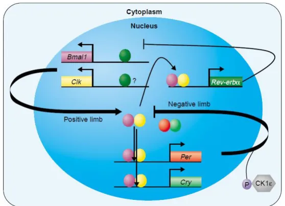
Figure 2- Representation of the molecular mechanism of the circadian clock in mammals. The positive limb happens when CLOCK and BMAL1 proteins make a heterodimer binding to the E-box elements promoting the transcription of many genes like Period (Per1, Per2 and Per3), Crypthocrhome (Cry1 and Cry2) and Rev-Erbα. These genes suffer hyperphosphorylation by Casein Kinase Iɛ (CKIɛ) and Glycogen Shyntase Kinase3 (GSK3) and form the correspondent proteins. The PER and CRY proteins bind and form a heterodimer called repressor heterodimer when they are inside the nucleus. This complex promotes the negative feedback loop because it stops the activity of the CLOCK/BMAL1 complex, so does the transcription of genes. The REV-ERBα, on the other hand it stops the expression of Bmal1 gene at the transcription level. This feedback loop happens with a rhythmicity of 24 h and a disruption can bring lots of problems to the health of the organism. Adapted from (Albrecht & Eichele, 2003).
1.4. Metabolic processes with circadian cycles
Many studies have demonstrated that the expression and activity of all the circadian proteins of the circadian cycle are important, not only to genetic processes, but also to the homeostasis of many metabolic and cytosolic processes (Froy, 2011; Kondratov, 2007; Rey & Reddy, 2013; Sukumaran, et al., 2010; Tevy, et al. 2013). A disruption of transcription-translation feedback loop (TTFL) can promote lots of diseases at the metabolic level, degenerative or carcinogenesis which can influence aging (Rey & Reddy, 2013; Tevy, et al. 2013). However, recently it was observed in the cyanobacterium Syncchococcus elongates that, even in the absence of transcription-translation feedback loop, a biochemical oscillation catalysed by several clock proteins occurs (O'Neill, et al., 2011). This fact has crested the doubt on the TTFL being the only controller of the circadian cycle. Other studies which were made before in Eukaryotic systems indicated that the cytosolic metabolism is also involved in the controlling of the circadian rhythmicity (O'Neill, et al., 2011). In
another study made in 2011 by O’Neill and his collaborators it was observed that in the human blood cells there are cells without nucleus, so without TTFL, shows the circadian rhythmicity in the peroxiredoxin oxidation cycles. Peroxiredoxins are antioxidant proteins which allow to control the levels of intracellular peroxide because they have essential catalytic cysteine residues that use thioredoxin to remove this reactive species of oxygen (Eter, et al., 2013; O'Neill, et al., 2011). It can be concluded that the circadian rhythmicity could have an involvement in the regulation of ROS. Indeed, the ROS can serve as signals coupling metabolism to other cell functions (Gyongyosi, et al., 2013).
Moreover, it was demonstrated that although the nucleus was essential to sustain the circadian rhythmicity in mammal cells it was important, however, to understand the relationship between these two types of circadian controllers (O'Neill, et al., 2011).
1.5. Interaction between the transcriptional clock and the metabolic clock
The redox cycle (NADH/NADPH) was studied in human red blood cells and it was observed that it has a period of around 24 h. The oscillation of NADH/NADPH can be associated to the modulation of DNA-binding activity of the core circadian transcription factors of two clock genes, Clock and Bmal1. Besides this when the level of ATP are studied for 48 h it shows two cycles of circadian oscillations, which confirm the idea that the cycles observed in these cells have a metabolic origin (O'Neill, et al., 2011). The Ca2+ and cAMP seem, also, to be interrelated with the clock mechanism (O'Neill, et al., 2011; Rey, et al., 2013). In another work done by Eter and other scientists, they studied the relationship between the oxidation cycles of peroxiredoxin and diabetic type II with the peripheral atherosclerotic disease (PAD). The increase of peroxiredoxin is usually associated to this dysfunction and to the resistance to insulin and both are related to aging, but it was also related to a disruption of circadian cycle (Eter, et al., 2013). Nowadays it is very important to make a relation between the TTFL and cytosolic processes to study many diseases related to them (O'Neill, et al., 2011; Rey, et al., 2013; Tevy, et al., 2013). With all these observations nowadays the nuclear rhythms are associated to the cytosolic mechanisms (Figure 3), working like two motors of rhythmicity controlling several processes that are important to our health. Considering normal cells (cells with nucleus) it was proposed that the non-transcription oscillator and TTFL drive the peroxiredoxin rhythms and in cells without nucleus the peroxiredoxin rhythms are just driven by non-transcription oscillators (O'Neill, et al., 2011). A disruption of circadian
clock is associated with an increase of ROS and promoting of aging, but this finding can also be related with the oxidation cycle of peroxiredoxin or non-transcriptional oscillations.
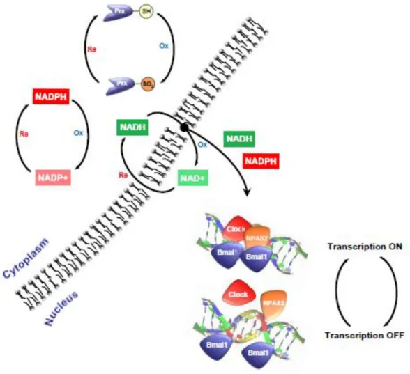
Figure 3- Propose of interconnectivity circadian redox processes and transcription-translation feedback loop (TTFL). Nowadays it is known that these two types of feedback work together to promote and control all organisms and like this control the homeostasis. The TTFL consists in a cycle and conjugation of positive and negative feedback loop. On the other hand it was discovered that the peroxiredoxin and the NADH/NADPH redox status has circadian rhythmicity and there is an interconnection between these two clocks. It was shown that NADH/NADPH redox status can directly modulate the DNA-binding activity of CLOCK/BMAL1 and control the TTFL. Adopted from (O'Neill & Reddy, 2011).
1.6. Circadian Cycle, Aging and Diseases
The circadian cycle is an important property for the good function of cellular and organism, so an anomaly in this cycle could cause serious health problems (Kononenko 2008; Lévi 2010). There are many diseases and health problems associated to dysfunctions of this cycle for example:
obesity, diabetes, degenerative diseases, aging, hypertension, cancer among others (Kondratov 2007; Lévi 2010; Yu 2011). The understanding of the molecular mechanisms of the circadian clock was the key to establish a relationship between the disruption of the circadian cycle and the development of some diseases or health problems (Kondratov, 2007; Sukumaran, et al., 2010). Many inflammatory diseases have symptoms that may be related to changes of this cycle, such as in the case of patients with a variation of rheumatoid arthritis who have more pains at night or in the early morning, or in the case of the patients with asthma, allergy, fever or rhinitis who have symptoms like nasal congestion, bronchial constriction and sneezing at night or when they wake up (Bechtold, et al., 2010). Besides this, it is possible to establish a framework of greater occurrence of cardiovascular events with periods of the day, as there are a greater number of episodes of stroke, myocardial infarction and sudden death during the morning period. This may be related to variation in blood pressure, heart rate and fibrinolysis activity that is directly dependent on the circadian cycle (Kondratov 2007).
The desynchronization can occur due to modifications of behaviour stimulus or because of changes in the expression and activity of the circadian proteins (Kondratov, 2007). Shift workers or people who are usually subject to jet lag are associated to the development of some health problems such as: insomnia, fatigue, gastrointestinal disorders, cardiovascular diseases, hypertension and diabetes, because the physiological processes and the normal hormonal state have changed (Albrecht & Eichele, 2003; Froy, 2011; Gallego & Virshup, 2007; Kondratov, 2007; Reddy, et al., 2005; Sukumaran, et al., 2010). Jet lag is the concept that is used to explain the trips made several times and for short periods of time, as the case of business people travelling from one continent to another. In these cases due to the time zone it is common for the people involved to have an "exchange of sleep", thus deregulating the circadian cycle (Bechtold, et al., 2010). The residents in countries of nordic latitudes are, also, subject to circadian dyshynchronization because in the winter the days are shorter and the 12 h cycles of light/darkness do not happen. Usually, these people develop a winter depression, also called by seasonal affective disorders (SAD) (Albrecht & Eichele, 2003; Bunney & Bunney, 2000).
The BMAL1 protein was considered as the principal element key to control the rhythmicity of the expression of the circadian genes (Kondratov, 2007; Sukumaran, et al., 2010). In 2006, Kondrakov and his collaborators verified that mice with a disruption of Bmal1 had a predisposition to the development of diseases related with premature aging. When they compared these mice with wild type mice at the same age they saw that the mutant mice had signals of sarcopenia (decreased number of muscular fibres and its diameter), osteoporosis (reduced bone mass and its composition), cataracts, chronic inflammation of the cornea and alteration of the cell composition in the blood
(Kondratov, et al., 2006; Kondratov, 2007). Since the PER proteins (PER1 and PER2) are involved in the regulation of the transcription of Bmal1, the activity and expression of PERs and BMAL1 may be related (Kondratov, 2007; Okamua, et al., 2010). In one of the studies of Lee and his collaborators it was demonstrated that in mice with disruption of PER1 and PER2 a decrease of fertility was prematurely observed, when compared with wild type mice the same age (Kondratov, 2007). It was demonstrated that a disruption in one of these two proteins could affect important metabolic processes that exist in our body, including the development of cancer (Kunieda, et al., 2006; Okamua, et al., 2010; Sukumaran, et al., 2010). A disruption of BMAL1 promotes an increase of the level of reactive oxygen species (ROS) in the organism (Kondratov, 2007; Kunieda, et al., 2006; Myers & Badia, 1995). The BMAL1 protein is involved in the control of glucose metabolism, fat metabolism and homeostasis. These metabolic processes are the principal factors to control the levels of ROS and consequently the oxidative stress that is directly related with some age associated diseases (Kondratov, 2007; Ramsey, et al., 2007).
Aging can also promote some alterations in body composition and in the circadian cycle resulting in deregulation of homeostasis (Tevy, et al. 2013). This happens because some morphological, physiological and chemical changes in the circadian clocks occur with age (Fuller & Fuller, 2002; Myers & Badia, 1995). The principal variations in the cycle are: the decrease of amplitude, increase of phase cycle, alterations of body temperature, alteration of hormonal secretion and modification of sleep-wake cycle (Fuller & Fuller, 2002; Myers & Badia, 1995). In mammals it is known that the principal hormones which show circadian rhythmicity are leptin, insulin, and glucagon that can affect the blood glycemia and are related with premature aging. Indeed, some studies in humans showed that the increase of sensibility of our body to insulin can increase longevity. However, with aging the resistance to insulin increases together with abdominal obesity and sarcopenia (Tevy, et al. 2013). In conclusion, a deregulation in circadian clocks brings lots of problems to the health at the metabolic level and has been associated, also, with premature aging.
1.7. Aging and oxidized proteins
It was well described that the accumulation of oxidized proteins is a hallmark of cellular aging. The protein oxidation is caused by reactive oxygen species (ROS) mainly generated by mitochondria. However, when they are in excessive quantities they can promote oxidative stress which is directly associated with the development of many diseases related to aging such as amyotrophic dystrophy sclerosis, Alzheimer’s disease, respiratory distress syndrome, muscular
dystrophy, cataractogenesis, Werner’s syndrome among others (Berlett & Stadtman, 1997; Butterfield & Kansi, 2001; Chondrogianni, et al., 2012; Dalle-Donne, et al., 2003). The ROS are inducing oxidative damage in many cellular components: DNA, carbohydrates, unsaturated lipids and proteins (Dalle-Donne, et al., 2003; Levine, 2002; Moller, et al., 2011). Proteins are among the most common target of oxidation (Dalle-Donne, et al., 2003; Moller, et al., 2011).
Proteins can suffer many types of damage, including the formation of carbonyl group (Chondrogianni, et al., 2012). Upon senescence of fibroblasts the oxidation of the proteins occurs essentially in four principal componentes: 44% in mitochondria, 28% in cytosol, 11% in endoplasmic reticulum and 8% in cytoskeletal. Carbonylation is one of the most common damage and is irreversible in proteins (Berlett & Stadtman, 1997; Cecarini, et al., 2007; Moller, et al., 2011). Consequently, the quantification of the level of carbonylated protein is one of the most used method to monitor the level of oxidized proteins (Chondrogianni, et al., 2012; Dalle-Donne, et al., 2003). Proteins can suffer two types of modifications when they are oxidized: first the alteration of side chains and second the cleavage of the peptide bond (Figure 4). The amino acids most susceptible to oxidative attack are: lysine, arginine, proline and theonine, leading to the formation of the carbonyl derivative proteins (Ahmed, et al., 2010; Chondrogianni, et al., 2012; Dalle-Donne, et al., 2003). The cellular homeostasis of this kind of protein damage is very important because the proteins have vital functions in many cellular processes (cellular synalisation, regulation of the cellular structure and enzymatic processes). Excess of the levels of the oxidized proteins has been associated with the development of many diseases (Berlett & Stadtman, 1997; Cecarini, et al., 2007). The mammal organism has developed mechanisms that control the accumulation of damaged proteins in order to maintain the cellular homeostasis, so when there is an increase in the level of oxidized proteins, they are eliminated by degradation or repair (Cecarini, et al., 2007; Chondrogianni, et al., 2012; Moller, et al., 2011). The oxidized proteins with reversible damage, like cysteine and methionine oxidation can be repaired by thioredoxin/thioredoxin reductase or glutaredoxin gluthation/gluthation reductase and methionine sulfoxide reductase, respectively (Ahmed, et al., 2010). On the other hand, the lysosome and proteosome are the two principal proteolytic systems responsible for the removal or degradation of the irreversibly damaged proteins (Ahmed, et al., 2010; Cecarini, et al., 2007; Chondrogianni, et al., 2012).
Figure 4 –Fragmentation of the carbon skeleton. The addition of the carbonyl group can lead to the modification of the carbon skeleton and/or to the modifications of the side chains (threonine, arginine, proline and lysine).
Usually, when the level of ROS increases, carbonylated proteins are formed, and there is a response to increase the proteasome’s activity and the cellular homeostasis is kept (Butterfield & Kansi, 2001). Moreover, with aging there is a decrease of proteasome’s activity and a decrease of regeneration of ATP, and both keep the increase of the level of ROS resulting in the accumulation of oxidized proteins (Ahmed, et al., 2010; Chondrogianni, et al., 2012). A decrease of the proteasome’s activity related with aging is mainly due to the decrease in its cellular content, accumulation of its endogenous inibitors and oxidative and glycoxidative modifications in its subunits (Ahmed, et al., 2010). Previous studies show that there is a relation between the circadian cycle and an increase of the level of ROS, essentially promoted by a disruption of circadian proteins, BMAL1 and PERs (Levine, 2002; Kondratov, 2007; Kunieda, et al., 2006; Myers & Badia, 1995). Hence it is important to understand the relation between circadian changes and age-associated diseases related with oxidative stress (Figure 5) (Kondratov, 2007).
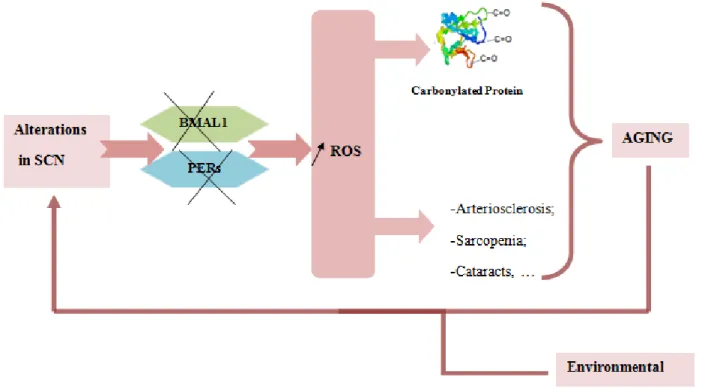
Figure 5- Relation between circadian rhythm aging and formation of carbonylated protein. When a change occurs in the SNC it may lead to a disruption of the protein BMAL1 and PERs and these are associated with an increase of the level of ROS and consequently an increase of the level of carbonylated proteins. Once the proteasome is unable to degrade all proteins which are damaged when there is a disruption accumulated as this will lead to aging. Furthermore, it is known that the increase of ROS is associated with the development of various diseases including cataracts, sarcopenia and arteriosclerosis that are associated with aging.
1.8. Circadian Synchronization in Cell Culture
Many studies associate aging or the development of some diseases related with age with a possible disruption in the circadian cycle. Thus it is becoming more and more interesting to understand how and why this relationship occurs (Fuller & Fuller, 2002; Kondratov, 2007; Kunieda, et al., 2006; Myers & Badia, 1995; Ramsey, et al., 2007). Nowadays it is known that the circadian clocks exist not only in the master pacemaker of the mammals, SCN, but also in the many peripheral tissues (Balsalobre, et al., 1998; Balsalobre, et al., 2000; Fu & Lee, 2003; Kondratov, 2007). The immortalized mammalian culture cells such as rat-1 fibroblasts and NIH3T3 are also capable of promoting circadian rhythmicity in genes expression in vitro, which was considered an asset for the studies in the circadian cycle (Balsalobre, et al., 1998; Balsalobre, et al., 2000; Izumo, et al., 2006). In mammals, the study of the circadian cycle is much more advantageous in culture cells than in cells of SCN. The culture cells are easier to sustain; can produce a large amount of material for biochemical assays and are easy access to molecular genetic tools. However, it is not possible to directly compare the studies performed in different cell types, because they can have different responses to the same treatment (Izumo, et al., 2006).
The circadian synchronization in cultured cells can be elaborated in many ways: Serum Shock, Dexamethasone, Forskolin (FSK), Epidermal growth factor (EGF) or Fibroblast growth factor (FGK), Calcium ionospheres, Glucose, Prostaglandin E2 (PGE2) and endothelin-1 (ET, 1). The first three ways are considered the most effective, depending, however, on the cell type in question (Izumo, et al., 2006). In this study HEK-293 cell was used and the best method to synchronize these cells, had to be chosen, between Serum Shock and Dexamethasone.
1.8.1. Circadian Synchronization with Serum Shock
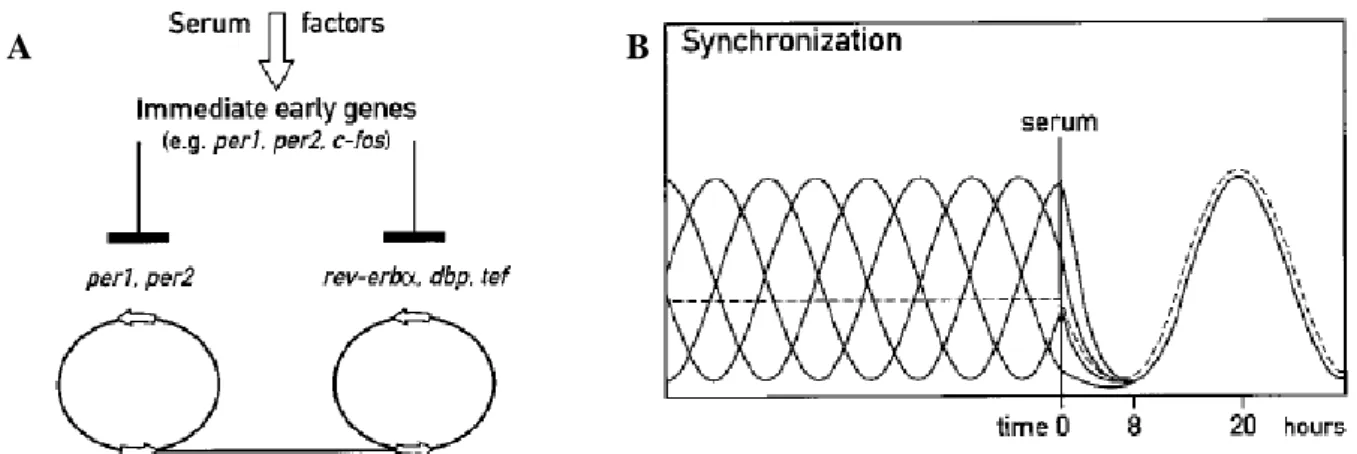
In 1998 Balsalobre and his collaborators performed a study in rat-1 fibroblasts. Initially they submitted the culture cells to higher levels of serum (50% of Serum in a usually cultured medium) for two hours and then put them in a medium without serum (Balsalobre, et al., 1998). After the treatment they used a medium without serum to avoid the cellular proliferation and placed all cells in the different phase of the cell cycle (Balsalobre, et al., 1998; Davis, et al., 2001). Through this experiment they verified that higher levels of serum induced the expression of c-fos and Per (Per1, Per2) genes, what is similar to what happened with light, which induced the expression of immediate early genes in SNC and has the same mechanisms (Figure 6). Besides this they observed
that the cultured cell kept the synchronization of the circadian genes in a medium without serum for 3 days (Balsalobre, et al., 1998). The use of this method to synchronize culture cells was carried out in many studies, which contributed to a better understanding of the circadian cycle and to what this can be associated with (Izumo, et al., 2006; Sukumaran, et al., 2010).
Figure 6- Possible explanation of synchronization with Serum Shock. A- When submitted by higher levels of serum the cells suffer an induction of immediate early genes (c-fos, Per1 and Per2) due to some serum factors and with this, synchronized the transcription of the other circadian genes. B- When in the culture cell each one of the cells is in a different phase of circadian or cell cycle but when they are submitted by a serum shock treatment shows that they can be synchronized. However, it takes around 8 hours to have all the cells synchronized, so during this time it is possible to see a decrease of levels of the genes per1 and per2. Adopted from (Balsalobre, et al., 1998).
1.8.2. Circadian Synchronization with Dexamethasone
The glucocorticoids are steroid hormones that the human body secretes in cycles and because of this they have been particularly attractive candidates for the circadian synchronization (Balsalobre, et al., 2000; Dickmeis & Foulkes, 2011). The dexamethasone (Figure 7) is a synthetically glucocorticoid that was used to promote the circadian synchronization in culture cell for the first time by Balsalobre and his collaborators in 2000 (Balsalobre, et al., 2000; Reddy, et al., 2005; Sukumaran, et al., 2010). These A B
Figure 7- Chemical representation of dexamethasone (http://commons.wikimedia. org/wiki/File:Dexamethasone _structure.png)
investigators submitted rat-1 fibroblasts for 1 hour of treatment with dexamethasone and observed that the expression of Per1 was increased very fast but the expression of Per2, contrarily to what happened with serum shock, didn’t increase (Balsalobre, et al., 2000). The fact that dexamethasone is involved in many processes of the human body could be a reason for it to be a target form in therapy (Dickmeis & Foulkes, 2011). However, in some studies it is shown that this glucocorticoid can promote oxidative stress when it is used in a concentration higher than 10 mM. This can happen because dexamethasone increases the levels of one class of redox protein, Thioredoxin 2, and the increase of these is directly related to the increase of ROS leading the cell to suffer apoptose (Dickmeis & Foulkes, 2011; Psarra, et al., 2009). So, dexamethasone is considered one of the best methods of synchronization in some types of cells, however, it needs to be used in lower concentration so as not to promote oxidative stress and affect the circadian genes expression analysis (Psarra, et al., 2009).
2. OBJECTIVES
HEK-293 synchronized with serum shock as a cellular model studies of the laboratory showed that the level of carbonylated proteins have a circadian rhythm with a period of 24 h. It is known that these damages are directly related with the increase of ROS and that during aging there exist some specific proteins which are carbonylated and defined as biological markers of this process. In this view, to understand how the circadian cycle is implicated in the cellular aging, this work has the aims to:
Determine the variation of the level of carbonylated protein during the circadian cycle and identify the time when the maximum and minimum of carbonylated protein levels occur.
Optimize the 2D electrophoresis conditions in order to characterise some proteins that are preferentially targeted by the circadian oxidation.
3. MATERIAL AND METHODS
3.1. Culture of HEK-293 cells
To cultivate HEK-293 cells a classical medium, DMEM-PSG (Dulbecco’s Modified Eagle Medium1 (DMEM) supplemented with 1% of Penicillin-streptomycin (P), 10% of Fetal Bovine Serum (S or FBS) and 1% of L-Glutamine (G))*2 were used. This medium contained all the nutrients which were needed and after the culture flasks were placed in an incubator chamber (CO2
Incubator, MCO-17A1, SANYO) at 37°C, 5% of CO2 and 95% of humidity. When the cells culture
were made a laminar flow chamber (MSC Class II, TECHGEN), an aspirator Vacusafe comfort (IBS Integra BIOSCIENCES) and a microscope (CKx41, OLYMPUS) were used. Around 30 minutes before the procedure the necessary solutions were put in the water bath (JB Aqua 12 Plus, GRANT) at 37ºC and with this a temperature shock was avoided.
The total number of cells in suspension was counted by a NucleoCounter system (CHEMOMETEC) that counts the nucleus that exist in the suspension of mammalian cells previously treated with a Lysis Buffer and a Stabilizing Buffer. The propidium iodide which came inside the NucleoCassette operates as a dye, binds to the nucleus to facilitate its counting.
The cells were kept in pellet form for later analysis and for this it was necessary to make a centrifugation 5 minutes at 500 xg and 4°C (AllegraTM X-12R centrifuge, BECKMAN COULTER).
3.2. Synchronization of the HEK-293 cells
When the cells in one of the flask T150 cm3 were in confluence (around 80-90%), 2 million cells from other culture flasks were passed and were completed with the necessary quantity of classical medium. This type of flask was used to assure that enough material would be collected for all posterior analysis (qPCR, 1D and 2D electrophoresis). The cells were first cultivated in normal conditions which were described before (3.1.) and when they were in 80% confluence the synchronization was started.
At time zero (T=0 h) an addition of DMEM-PSG with the appropriate concentration of the synchronization reagent (serum and dexamethasone) was made. 2 h after, the medium was changed
1D6546- With 4500 mg/L glucose, sodium pyruvate, and sodium bicarbonate, without L-glutamine, liquid,
sterile-filtered, suitable for cell culture (SIGMA®-ALDRICH).
2
to another one without serum (DMEM-PG)*3 and the cells could start to be collected (usually in intervals of 4 h between each one). To know until which time the cells survived in a medium without serum, a control was made and in this way it was also proved that the non-synchronized cells do not have circadian cycles. For this the cells were cultivated as usual and it was considered time 2 h when the classical medium was changed by another one without serum, like in the other types of synchronization.
The HEK-293 cells were harvested without using trypsin because with only one wash with PBS 1X (SIGMA®-ALDRICH) they would peel off from the plastic. To put the appropriate quantity of cells for analysis they were counted and after they were centrifuged (from extraction of ribonucleic acid (RNA) around 2 million cells; for 1D electrophoresis around 4 million of cells and for 2D electrophoresis around 9 million of cells).
3.2.1. Synchronization with dexamethasone
To make synchronization with dexamethasone (SIGMA®-ALDRICH) a final concentration of 10 nM of this reagent was used. A stock solution of dexamethasone of 10 mM was prepared, diluted in absolute ethanol and conserved at -80ºC. Then, two dilutions were made, the first one of 1:100 with phosphate buffered saline (PBS) 1X (SIGMA®-ALDRICH) and the second one with a reason of 1:1000 with complete medium. After this a common procedure of synchronization was made, as was described before (3.2.) until 50 h were completed.
3.2.2. Synchronization with serum shock
A synchronization with serum shock in HEK-293 cells consists in preparing a culture medium with a higher concentration of FBS (DMEM-PSG and FBS in a dilution 1:2 for each flask) and the usual procedure for synchronization was made, as was described before (3.2.) until 50 h were completed.
3
3.3. Analysis of the transcripts
A series of procedures were necessary to carry on with the analysis of the transcripts. The extraction of RNA samples and their quantification was performed in order to understand whether there was sufficient RNA to perform the analysis. Subsequently a reverse transcription (RT) was prepared so as to obtain deoxyribonucleic acid (DNA) primer sequences to be analysed by quantitative polymerase chain reaction (qPCR).
3.3.1. Extraction and quantification of RNA
The extraction of RNA of the cells harvested at different times of synchronization was executed with a NucleoSpin® RNA II kit (MACHEREY-NAGEL). The centrifugation was made in a 1-15K, SIGMA®-ALDRICH and the quantity of RNA was determined with a NanoVue (GE HEALTHCARE).
To confirm the integrity of RNA an electrophoresis was performed. First, a gel with 1% of agarose (SIGMA®-ALDRICH) in 100 mL of solution TAE 1X*4 was dissolved and after 2 µL of Ethidium Bromide (EtBr, SIGMA®-ALDRICH) was added. When the gel was ready and fixed, the mix of 5 µL of sample and 2 µL of solution DNA Dye 6X*5 was put in each and the migration with a Power Suply-Model 250/2,5 (BIORAD) was started. For the RNA to be considered good, 2 bands in gel were needed, one that corresponded to RNAr 28S (4718 pb) and the other one to RNAr 18S (1900 pb).
3.3.2. Reverse Transcription
The reverse transcription was done with a kit called SuperScript® III First – Strand Synthesis System for RT-PCR (Invitrogen, LIFE TECHNOLOGIES) which was designed to convert 1 µg of RNA to 5µg of total cDNA. To do this procedure 1µg of RNA was used in a final volume of 8 µL. In each tube 1 µL of Random Hexamers (50 mg/µL) and 1 µL of dNTP mix (10 mM) was added and after an incubation in a (Master Cycles Personal, EPPENDORF) for 5 minutes at 65ºC and 1 minute in ice was made. During this time, the solution DNAmix*6 was prepared and in each sample
4 (*) the composition is described in attachment 7.2.2. 5 (*) the composition is described in attachment 7.2.3. 6
10 µL of this solution was added and they were incubated in 3 steps. The first one corresponded to 10 minutes at 25ºC; the second one to 50 minutes at 50ºC and the last one to 5 minutes at 85ºC. When the incubation was finished it was necessary to add 1 µL of RNase H (E.coli) and the last incubation for 20 minutes at 37ºC was made so that after the samples were frozen for further studies.
3.3.3. Quantitative PCR
Before the qPCR was made, there was a need to prepare a dilution 1:10 of the samples previously subject to extraction of RNA and reverse transcription (2 µL of sample subject to reverse transcription in 18 µL of RNA-free water) and the preparation of the standard curve*7 and primers solution*8. In one specific plate for qPCR (Frame Star480, 4ATITUDE) 2 µL of each sample and 8 µL of the primers solution (Table 1) that was to be examined was added. A standard curve with dilution 1:5, 1:10, 1:20, 1:50 and 1:100 was prepared before. The dilution 1:5 was made with 10 µL of 3 different cDNA in a final volume of 150 µL.
The primer that was used for the reference was GAPDH because it did not have circadian cycles and the program of PCR consisted of four steps: denaturation, amplification, melting and cooling and it was made in Light Cycler480® Real Time PCR Systems (ROCHE). The denaturation was constituted by 1 cycle of 5 minutes at 95ºC. The amplification was founded by 43 cycles divided in 3 parts during 15 seconds each part at 95º, 60º and 72ºC, respectively. The melting step was created with just one cycle with 30 seconds at 95ºC, 30 seconds at 60ºC and after continuously until 95ºC. The program was finished with a last cycle of 30 seconds at 40ºC.
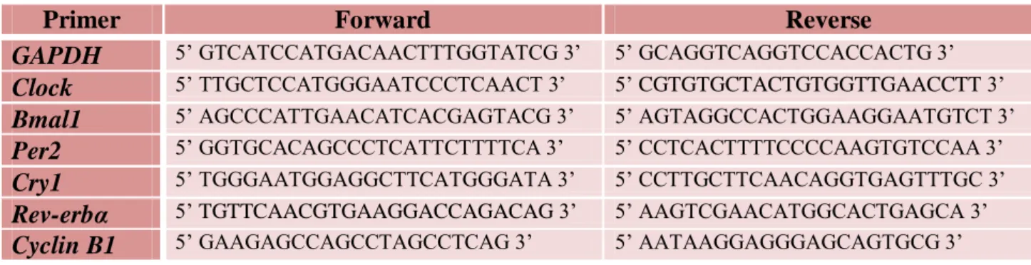
Table 1 – Primers of clock controlled transcripts for qPCR. All the primers were designed by Nicolas Ugarte with a programme AmplifX with the exception of Cyclin B1 that was designed by Audrey Desvergne.
Primer Forward Reverse
GAPDH 5’ GTCATCCATGACAACTTTGGTATCG 3’ 5’ GCAGGTCAGGTCCACCACTG 3’
Clock 5’ TTGCTCCATGGGAATCCCTCAACT 3’ 5’ CGTGTGCTACTGTGGTTGAACCTT 3’
Bmal1 5’ AGCCCATTGAACATCACGAGTACG 3’ 5’ AGTAGGCCACTGGAAGGAATGTCT 3’
Per2 5’ GGTGCACAGCCCTCATTCTTTTCA 3’ 5’ CCTCACTTTTCCCCAAGTGTCCAA 3’
Cry1 5’ TGGGAATGGAGGCTTCATGGGATA 3’ 5’ CCTTGCTTCAACAGGTGAGTTTGC 3’
Rev-erbα 5’ TGTTCAACGTGAAGGACCAGACAG 3’ 5’ AAGTCGAACATGGCACTGAGCA 3’
Cyclin B1 5’ GAAGAGCCAGCCTAGCCTCAG 3’ 5’ AATAAGGAGGGAGCAGTGCG 3’
7 (*) the composition is described in attachment 7.2.5. 8
3.4. Analysis of Carbonylated Proteins
To make an analysis of the proteins, an extraction and quantification was necessary. Initially a 1D electrophoresis was prepared to see the levels of carbonylated proteins along the circadian rhythmicity and after two time points, a maximum and minimum, were chosen to make a 2D electrophoresis. We analysed the level of carbonylated protein (Oxy ratio) when we made a ratio between the % Volume of Carbonylated Protein protein and the % Volume of total protein that exists in the sample.
3.4.1. Extraction and quantification of Protein
The extraction of protein started with adding 350 µL of Lysid Buffer I*9 for, around 107 cells. The samples were kept in ice for 15 minutes but were taken to vortex every 5 minutes or 15 minutes or more in Thermomix®Comfort for eppendorf of 1,5 mL(EPPENDORF) at 1400 rpm and 4ºC until they did not present viscosity. After, a centrifugation was made during 15 minutes at 10000xg at 4ºC. We quantified the protein that was in the supernatant with the Bradford Method (Bradford, 1976) using the Protein Kit Assay (BIORAD) and the Power Wave XS (BIOTEK). The rest of the samples were frozen at -80ºC. For the 2D electrophoresis we tested two types of Lysis Buffer I and II*10, but for 1D electrophoresis we just used the Lysis Buffer I*11.
For the quantification of protein by the Bradford Method a plate with 200 µL of final volume (5 µL sample or standart curve with 5 µL of MilliQ water or Lysis Buffer, respectively, and 190 µL of Bradford reagent) was prepared. The standard curve was made with different concentrations of bovine serum albumin (BSA) (0; 0,2; 0,4; 0,6; 0,8; 1).
3.4.2. One Dimension Electrophoresis (Oxyblot 1D)
For each sample 10 µg of proteins were prepared with MilliQ water in a final volume of 4 µL. After, 6 µL of (SDS 10% from BIORAD) was added to denature the proteins. The OxyBlot Protein
9 (*) the composition is described in attachment 7.3.1.1. 10
(*) the composition is described in attachment 7.3.1.1. and 7.3.1.2.
11 (*) the composition is described in attachment 7.3.1.1.
% Volume of Carbonylated Protein = Oxy ratio % Volume of Total Protein
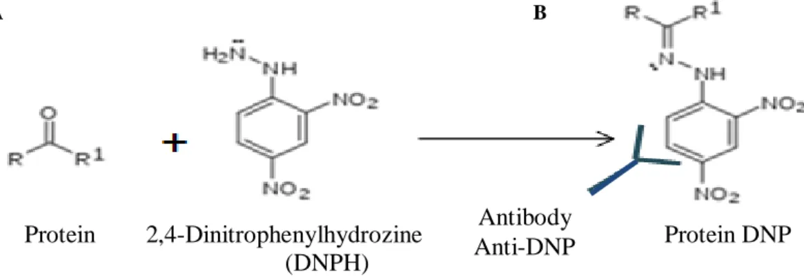
Oxidation Detection Kit (MILLIPORE) was used to start the derivatization (Figure 8, A). For this we used the 2,4-dinitrophenylhydrazine (DNPH). The protein was separated by SDS-PAGE (Figure 9, A) electrophoresis with a 12% polyacrylamide gel*12 in a Migration Buffer*13. In each well 14 µL of the samples were added. In one of them we put 7,5 µL of the marker of molecular weight (Precision Plus ProteinTM Standard, BIORAD). The electrophoresis was programmed to make 2 steps (100 V for 30 minutes; 200 V for 50 minutes) with the Power PAC HCTM (BIORAD).
The carbonylated proteins were detected by immunoblotting after being transferred to a nitrocellulose membrane. The support “sandwich” (BIORAD) was prepared inside the Tranfer Buffer*14 by placing their components in the following order: sponge, Wattman paper (AmershamTM HybondTM blotting paper, GE HEALTHCARE), gel, nitrocellulose membrane (HybondTMC Extra Amersham, BIOSCIENCES), Wattman paper and sponge (Figure 9, B). The transfer was made by Power PAC HCTM (BIORAD) which was programmed for 1 step of 100 V for 1 h. To see the total protein, the membrane was removed and placed in Fast Green solution*15 (60 mL of ethanol absolute, 20 mL of acid acetic, 120 mL of MilliQ water and 0,02 g of Fast Green) for 5 minutes and after was revealed in Odyssey programme (Odyssey3.0V, LI COR). Odyssey Buffer*16 was added to the membrane to make a blockage all night at 4ºC. Then, the membrane was incubated with 2 antibodies. The first one from the OxyBlot Protein Oxidation Detection Kit (MILLIPORE) which was an antibody against 2,4-dinitrophenyl, with a dilution 1:5000 in Odyssey Buffer (Figure 8, B). The second one was a Goat anti-rabbit IRDye®800CM from LI COR with a dilution 1:15000 in Odyssey Buffer. This incubation was made during 45 minutes but with 3 washes with Washing Buffer*17 during 10 minutes each one between each antibody. Finally a new revelation was made in Odyssey programme.
Figure 8-Scheme of derivatization with 2,4-Dinitrophenylhydrozine (DNPH) and reaction of the protein with the antibody anti-DNP. The DNPH binds to the protein and when the anti-DNP is added they form a protein DNP adduct.
12
(*) the composition is described in attachment 7.3.2.
13 (*) the composition is described in attachment 7.3.3. 14
(*) the composition is described in attachment 7.3.4.
15 (*) the composition is described in attachment 7.3.7. 16 (*) the composition is described in attachment 7.3.8.1. 17
(*) the composition is described in attachment 7.3.6.
Antibody anti-DNP
A B
Protein 2,4-Dinitrophenylhydrozine Protein DNP (DNPH)
Antibody Anti-DNP
Figure 9- Representation of the method of 1D electrophoresis (Adapted from http://www.bio-rad.com).
3.4.3. Two Dimension Electrophoresis (Oxyblot 2D)
It was necessary to adjust the Oxyblot 2D procedure. In a first step a definition of quantity of protein was tested with Enhanced chemiluminescence (ECL) method. The Lysis Buffer I and II*18 was also compared to two different strips (11 and 13 cm). For Odyssey revelation the second antibody, the anti-rabbit (LI COR) in a dilution of 1:5000 with Odyssey buffer*19 was used, for ECL the anti-rabbit GtxRb by OxyblotTM Protein Oxidation kit (MILILLIPORE) in a dilution 1:500 with BSA Buffer*20 was chosen. To have a first idea and make a quantification of carbonylated proteins, we prepared an electrophoresis 2D in a small gel, CriterionTM TGX Any kD™ Precast Gel. We used this type of gel, because it provides the separation from 10-250 kD with the best resolution of the proteins in the 20-100 kD molecular weight range.
The Re-hydratation Buffer*21 was prepared and the necessary quantity to make a solution with 300 µg of protein in a final volume of 250 µL, for 2D electrophoresis in a big gel or 100 µg of protein in a final volume of 200 µL for a 2D electrophoresis in a small gel, was calculated. This solution was added in each strip (ImmobilineTM DryStrip pH 3-10NL, GE HEALTHCARE) and after 500 µL of mineral oil (GE HEALTHCARE) was also placed and this stayed in incubation all night at room temperature without light. When this stage was finished the EttanIPGphor3 with 150 mL of mineral oil was prepared and in each well the strip was put with a Paper Wicks on each side, previously humidified with 100 µL of MilliQ water with Destreak (12 µL of Destreak for 1 mL) and the electrodes were added after. All the material used in this step of focalisation came from GE HEALTHCARE. The focalisation was programmed with 4 phases that had a different voltage and
18
(*) the composition is described in attachment 7.3.1.
19 (*) the composition is described in attachment 7.3.8. 20 (*) the composition is described in attachment 7.3.8.2. 21
(*) the composition is described in attachment 7.3.9.
A B Revelation: - Fast Green - Oxyblot 1D Quantity of carbonylated Protein