Amandine Isolina
Anastácio
Cultura de Tecido Ovárico Humano Congelado:
efeito da Activina A e GDF9
Culture of Human Cryopreserved OvarianTissue:
effect of Activin A and GDF9
Amandine Isolina
Anastácio
Cultura de Tecido Ovárico Humano Congelado:
efeito da Activina A e GDF9
Culture of Human Cryopreserved Ovarian Tissue:
effect of Activin A and GDF9
dissertação apresentada à Universidade de Aveiro para cumprimento dos requisitos necessários à obtenção do grau de Mestre em Biologia Molecular e Celular, realizada sob a orientação científica du Docteur Catherine Poirot, Professeur de la Faculté de Médecine, Paris VI, Pierre et Marie Curie e da Doutora Maria de Lourdes Gomes Pereira, Professora Associada com agregação do Departamento de Biologia da Universidade de Aveiro
o júri
presidente Prof.ª. Doutora Maria Helena Abreu Silva
Professora Auxiliar do Departamento de Biologia da Universidade de Aveiro
Prof.ª. Doutora Ana Teresa de Almeida Santos
Professora Auxiliar da Faculdade de Medicina de Coimbra
Prof.ª. Doutora Catherine Poirot
Professeur de la Faculté de Médecine Paris VI, Pierre et Marie Curie
Prof.ª. Doutora Maria de Lourdes Gomes Pereira
agradecimentos Au Professeur Catherine Poirot, pour m’avoir fais confiance encore une fois et de continuer à m’orienter dans mon travail, à sa disponibilité et son enthousiasme pour mon travail. Merci encore de me permettre de travailler avec vous et votre équipe.
Aux Docteurs Marie Prades et Benoit Schubert, pour leur amitié et leur soutien. A Aurélie, Leslie, Monia et Nicolas pour tous ces moments de détente partagés.
A toute l'équipe de l'UF de Biologie de la Reproduction de la Pitié-Salpêtrière pour m’avoir aussi bien accueille au sein de leur petite «famille».
A la Fondation Martine Midy qui a permis la réalisation de ce projet en me finançant.
A ma sœur de coeur et d’esprit, Sandrine, pour me tolérer encore chez elle malgré ma mauvaise humeur, de comprendre même mes silences, pour toutes les heures passées à m´écouter et à me conseiller.
Aos meus pais, por tudo o que fizeram para que eu pudesse seguir os meus sonhos e por me terem encorajado mesmo estando tão longe.
Aos meus irmãos, obrigada por estarem sempre presentes e pelos abraços que me dão força para voltar.
Ao Renato por nunca se negar a dar resposta às minhas perguntas, mesmo as menos pertinentes.
Ao Jacinto um obrigada muito especial pela Amizade, tolerância e força que me tem dado para a concretização deste projecto.
palavras-chave Crioconservação/ Cultura tecido Ovárico/ Activina A/GDF9
resumo A crioconservação de tecido ovárico é um dos métodos utilizados para preservar a fertilidade de mulheres em risco de menopausa precoce, devido a tratamentos gonadotóxicos.
O tecido ovárico crioconservado contém essencialmente oócitos imaturos, não fecundáveis, sendo por isso necessário induzir a sua maturação. A maturação dos oócitos contidos no tecido ovárico crioconservado pode ser realizada transplantando o tecido (in vivo) ou procedendo à sua cultura (in vitro), tema deste trabalho.
Este trabalho foi desenvolvido para avaliar a morfologia e a evolução dos folículos ováricos contidos no tecido, recorrendo a três marcadores de qualidade celular [dois marcadores da apoptose (p53 e Bcl2) e um marcador de proliferação celular (Ki67)], sem matriz de suporte, em três meios de cultura distintos durante 14 dias.
Este estudo comparativo foi realizado utilizando um meio de cultura base, também usado como grupo de controlo, ao qual se adicionaram, individualmente, duas proteínas pertencentes à Tranforming Growth Factors superfamily, GDF-9 e Activina A.
Neste trabalho, verificou-se uma diminuição da população folicular ao longo da cultura para os três meios utilizados.
No entanto, a maior proporção de folículos degenerados foram observados no grupo em cultura com Activina A contrapondo com o grupo em cultura com GDF-9 onde se observou a menor percentagem de folículos degenerados. Nos três meios de cultura utilizados apenas se observaram três tipos de folículos (primordial, early primário e primário).
Verificou-se uma marcação positiva para os marcadores p53 e Ki67 em todos os meios e ao longo da cultura. Para o marcador Bcl2 não foi observada nenhuma célula da granulosa marcada positivamente.
Com este trabalho verificamos uma melhor taxa de sobrevivência no grupo GDF-9, assim como um maior número de folículos e células da granulosa nos primeiros 10 dias de cultura.
keywords Cryopreservation/Ovarian tissue culture/Activin A/GDF9
abstract Cryopreservation of ovarian tissue is one approach to preserve fertility of young women who are at risk of premature ovarian failure, due principally to a gonadotoxic treatment.
The cryopreserved ovarian tissue has essentially immature oocytes, which need to be maturating for ulterior fertilization.
Oocyte maturation can be achieved by transplantation or by maturation in vitro, which was the subject of this work.
The present work was developed to evaluate the morphology and evolution of follicles using three markers of cellular quality [two apoptotic markers (p53 and Bcl2) and one proliferating marker (Ki67)] during 14 days of culture for three different culture media, without matrix support.
For this comparative study we used a serum free medium (control group) to which we added two proteins from the Transforming Growth Factors superfamily Activin A and GDF-9.
A diminution of the follicular population was observed throughout the culture and for all the culture media. However, the higher proportion of degenerating follicles (increasing with the time of culture) were observed at the Activin A group (A) and GDF-9 group (G) representing the lower proportion.
For the three culture media only three type of follicles were seen (primordial, early primary and primary).
All GCs in the three culture media were negative for Bcl2 immunostaining. . Positive marking were observed for the apoptotic marker (p53) and for the proliferating marker (Ki67) in all culture.
This study indicate a better survival in the group cultured with GDF-9, a higher number of follicles and cells of the granulosa in the first 10 days of culture
T
ABLE OF
C
ONTENTS
Table of Contents
I
Abbreviations
V
List of Figures
VII
List of Tables
IX
Introduction
1
Review of the Literature
3
The Ovary
3
Ovarian Reserve
4
Oogenesis and oocyte maturation
5
Cytoplasmic maturation 6
Nuclear maturation 7
Folliculogenesis and stages of follicular development
7
Apoptosis
9
Transforming Growth Factor βeta superfamily
10
Growth differentiation factor-9
11
Activin A
12
Fertility preservation
13
Embryo Cryopreservation
13
Oocyte Cryopreservation
14
Mature oocyte 14 Immature oocyte 15Fertility Restoration
17
Ovarian Tissue Transplantation
17
Heterotopic transplantation 17
Orthotopic transplantation 17
Xenotransplantation 18
Ovarian Tissue Culture
19
Aims of our study
21
Materials and Methods
23
Ovarian Tissue
23
Ovarian Tissue Thawing
23
Preparation of cortical tissue for culture
23
Different culture media composition
24
Cortical strip culture
24
Histological analysis
25
Morphological analysis
25
Follicle Counting and Classification 25
Others evaluation criteria 26
Immunohistochemical evaluation
26
Statistical analysis
27
Results
29
Morphological analysis
29
Follicle Counting and Classification
29
Others evaluation criteria
32
Degenerated/atretic follicles
33
Immunohistochemical analysis
35
Apoptotic markers (P53 and Bcl2)
35
Discussion
39
Conclusion and perspectives
43
References
45
Appendix
59
Chemicals
59
Solutions
60
Slides Reading Tables
61
ABREVIATIONS
AMH Anti-Mulerian Hormone
ART Assisted Reproductive Technology
BFGF Beta Fibroblast Growth Factor
BMP Bone Morphogenetic Proteins
BSA Bovine Serum Albumin
DBA Diaminobenzidine Tetrahydrochloride
DNA Deoxyribonucleic Acid
DMSO Dimethyl Sulfoxide
EG Ethylene Glycol
FCS Fetal Calf Serum
FD Follicular Development
FSH Follicular Stimulating Hormone
GC Granulosa Cell
GDF-9 Growth Differentiation Factor 9
GV Germinal Vesicle
GVBD Germinal Vesicle Breakdown HES Haematoxylin, Eosine and Safranin
HRP Horsedarish Peroxidase
ICSI Intra Cytoplamisc Sperm Injection IGFI Insulin like Growth Factor I
IGFII Insulin like Growth Factor II IVF In Vitro Fertilization
KL Kit-ligand
LH Luteinizing hormone
MI Methaphase I
MII Methaphase II
mRNA Messenger Ribonucleic Acid
PGC Primordial germ cells
POF Premature ovarian failure
PrOH PropanedioL
RNA Ribonucleic Acid
L
IST OF
F
IGURES
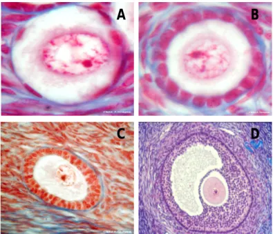
Figure 1: Schematic representation of an ovary showing its internal configuration, and the different types of follicles (Adapted from http://academic.kellogg.cc.mi.us).
Figure 2: Diagram of ovarian reserve and outcomes of the formed follicles throughout women life (adapted from Kaipa & Hsueh, Annu Rev Physiol 1997, 59:349-63).
Figure 3: Meiosis of oogonia (adapted from Pearson education Inc., publishing as Benjamin Cummings).
Figure 4: Human ovarian follicles at the primordial stage (A), the primary stage (B), the secondary stage (C) and the antral stage (D)(by Gougeon in the photothèque fertilité Ferring®).
Figure 5: Diagram of p53 apoptosis pathway (“+”= stimulation; “-“= inhibition; Mit = mithocondria) (adapted from Gillham et al. World Journal of Surgical
Oncology 2007).
Figure 6: The BMP and TGFβ/activin signaling pathway (adapted from http://www.rr-research.no).
Figure 7: Total number (n) of follicles counted for each culture media at the different time of culture.
Figure 8: Microscopy visualization of a histological section with HES staining of primordial follicle and primary follicle (A) and early primary follicle (B), in ovarian cortical strip. Indicate the cuboidal cells; Indicate the flattened cells.
Figure 9: Distribution of the different types of follicles observed during the culture, for the three culture media, and for the not cultured ovarian fragments.
Figure 10: HES histological section of a primary and a degenerating follicle of not cultured strips; original magnification X 400.
Figure 11: Proportion of degenerating follicles counted in the different culture media, at different times of culture and in not cultured strips.
Figure 12: HES sections of cultured stroma after 5 days of culture (A) and 14 days of culture.
Figure 13: Cells staining brown are positively marked, cells staining blue are negatively marked to the p53.
Figure 14: GCs marked for Ki67, cells staining brown are positive and cells staining blue are negative.
Figure 15: General view, with the inverted microscope (200X) of follicles in a cortical strip, in the GDF-9 medium, at the 5th Day (A) and of one follicle in the peripheral zone (B)(400x)
L
IST OF
T
ABLES
Table 1: Autotransplantations of cryopreserved human ovarian tissue
Table 2: Characteristics observed and follicle stage associated
Table 3: Average number of granulosa cells per follicle, calculated, by the histological counting of granulosa cells surrounding classifiable follicles, for each culture media at the different times of culture and for not cultured follicles
Table 4: Proportion of the different aspects of the cytoplasm in the oocytes observed, for each culture media and at different times of culture and for not cultured follicles
Table 5: Proportion of the follicles with a diffuse or a dense chromatin configuration, for the not cultured follicles and those cultured in the different culture media, at different time of the culture
Table 6: Follicle counting and proportions of positive granulosa cells marked for p53 antigene in a total of GCs in the three culture media, during the culture and in the not cultured strips
Table 7: Follicle counting and proportions of positive granulosa cells marked for Ki67 antigen in a total of GCs in the three culture media, during the culture and in the not cultured strips
(adapted from Poirot et al., 2008) Table 2: Characteristics observed and follicle stage associated
I
NTRODUCTION
Women have a finite number of follicles that sustain ovarian function until the menopause. Any situation affecting the follicles pool can induce a premature ovarian failure. This failure can be caused by genetics disorders and more commonly as a consequence of anticancer treatments exposure, being extremely important to propose methods to preserve their fertility.
One approach to preserving the potential fertility of young women who risk losing their follicles reserve is the cryopreservation of ovarian tissue containing immature oocytes.
The cryopreserved tissue can be transplanted into the pelvic cavity (orthotopic) or in a heterotopic site such the abdominal wall or the forearm. However in some types of cancer the transplantation has a high risk of reintroduce malignant cells, subsequently is very important to study and develop alternative methods such as the maturation in vitro of the follicles and their oocyte.
Over the last two decades, culture systems have been developed with the aim of growing follicles from the earliest stage into maturation of the oocyte and fertilization.
In mice offspring it has already been obtained with maturation of primordial follicles and their fertilization in vitro but in Human this process reveals being more complex due to the long time needed for the folliculogenesis, the little knowledge of the factors involved in the initiation of the growth of small follicles and the dense structure of the ovarian tissue.
Nevertheless a number of factors produced in the ovary itself, as the Transforming Growth Factor beta family, are known to control the initiation of the growth of small follicles.
At present time, it is known that the GDF-9 plays an important role in the initiation of the growth of small follicles as well as the survival of these throughout the culture while activin A seams to promote the growth and development of secondary follicles to antral follicles.
The aim of our study was to analyze the effects of growth differentiation factor 9 and activin A during the culture of human cryopreserved ovarian tissue, since to date, the published studies show results obtained essentially with culture of fresh ovarian tissue, using morphological characteristics, apoptotic markers (p53, Bcl2) and proliferating marker (Ki 67).
R
EVIEW OF THE
L
ITERATURE
T
HE
O
VARY
The ovary is the reproductive organ in female mammals, it measures about 4 cm long, 2 cm wide, 1 cm of thick and has a dual role of gonad and endocrine gland. Usually, each woman has two ovaries located in the pelvic zone, one in the right side and the other in the left side of the uterus to which they are attached by a fibrous cord called the ovarian ligament.
The outermost layer of the ovaries is the germinal epithelium constituted by simple cuboidal cells. Underneath this, a layer of dense connective tissue constitutes the tunica albuginea and covers the ovarian cortex.
In the cortex, we found the ovarian follicles and between them the stroma, a peculiar soft tissue consisting mainly of connective tissue enriched with blood vessels. The innermost layer of the ovary, called medulla, is essentially loose connective tissue with abundant blood vessels, lymphatic vessels and nerve fibers (Fig.1).
The ovary performs several tasks that include the production, storage and nurturing of the oocytes and segregation of hormones that promote follicle/oocyte maturation and development of secondary sex characteristics.
O
VARIAN
R
ESERVE
In Humans, the follicular reserve is mainly comprised of primordial follicles, containing an oocyte I, and begins to form at the fourth month of fetal life (Baker, 1963), around the 20 weeks of gestation, about seven millions of oocytes are formed in each ovary, and it is believed those are all the oocytes that woman will ever have. At birth the reserve drops to one to two millions and at the onset of puberty only 300 000 to 400 000 remain (Baker, 1963; Faddy et al., 1992; Faddy, 2000). From there, the number of follicles gradually decreases until the age of 37-38 years old, when rapidly drops and ends in menopause several years later with only 1000 follicles in each ovary (Faddy et al., 1992). However, during reproductive life of a woman, only 400 follicles mature and ovulate (Gougeon, 1996), whereas the remainders go through atresia and degenerates.
The depletion of the ovarian stock occurs as a result of two major processes: atresia, by apoptosis, and entry in growth phase. In the Human the depletion before puberty and after 30 years of age is mainly due to atresia whereas between those ages this follicles loss is due to entrance of resting follicles in growth phase (Gougeon, 1996).
Figure 2 : Diagram of ovarian reserve and outcomes of the formed follicles throughout women life (adapted from Kaipa & Hsueh, Annu Rev Physiol 1997, 59:349-63)
O
OGENESIS AND OOCYTE MATURATION
The oogenesis starts with the formation on the oogonia and involves several processes that lead to their development and maturation.
The oogonia are originated from primordial germ cells (PGC) in a common path of gametogenesis in females and males.
At the end of the third week of development PGC’s migrate from the primary ectoderm into the yolk sac (Baker et al., 1963). Between the fifth and the sixth weeks, under the influence of cytokines such as Kit-ligand (KL) and transforming growth factor β (TGFβ), these cells continue to migrate until they reach the genital ridge, where they stay and colonize the developing gonads, by mitosis (Motta and Makabe, 1986).
After colonization, around the seventh week of gestation, the sex specific differences appear and the germ cells differentiate into oogonia or spermatogonia according to chromosomal constitution and the environment in which they develop. Henceforth oogonia continue to undergo mitosis and stimulate cells of the adjacent epithelium to form follicular cells (Faddy et al., 1992), but around the eleventh week of development they enter in meiosis and stop in the prophase of the first meiotic division, becoming oocytes I. At the same time, they became enclosed in one layer of pregranulosa cells surrounded by a basal membrane forming a complex called primordial follicle (Gosden and Bownes, 1995). The nucleus of these dormant oocytes I becomes very large and it is called a germinal vesicle (GV).
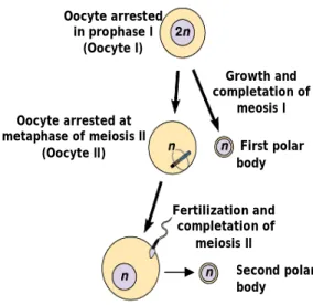
When the follicle is recruited for the growing pool the oocyte undergoes an important increase of volume from 35µm in primordial follicle to 120µm in a fully developed follicle (Gougeon, 1996; Picton et al., 1998) associated with the follicular growth. However the oocyte I persists in prophase of the first meiotic division until the time of ovulation, when meiosis is resumed and the first polar body is formed and extruded, becoming a fertilizable oocyte. This oocyte is arrested in the metaphase of meiosis II and only completes his second meiotic division after fertilization (Fig 3).
Oocyte arrested in prophase I
(Oocyte I)
Oocyte arrested at metaphase of meiosis II
(Oocyte II) First polar
body Second polar body Fertilization and completation of meiosis II Growth and completation of meosis I
Figure 3 : Meiosis of oogonia (adapted from Pearson education Inc., publishing as Benjamin Cummings)
The fully and competent maturation of the oocyte depends on two crucial events, the long and imperceptible cytoplasmic maturation and the short and clearly observable nuclear maturation.
Cytoplasmic maturation
Cytoplasmic maturation includes accumulation of mRNAs and proteins, cytoplasmic reorganization and epigenetic modifications, necessary to prepare the oocyte for fertilization and normal embryo development, and it is divided in two major events, the acquisition of competence to development (before resumption of meiosis) and the oocyte activation prior to ovulation.
During the oocyte growth, an extensive production and reorganization of the cytoplasm takes place. At this period, the oocyte stores glycogen granules, lipid droplets and proteins likely to provide energy and substrate to the formation of new membranes after fertilization (Picton et al., 1998), the number of mitochondria increases drastically and become more vacuolated, sign of loss activity (Wassarman and Josefewicz, 1978) as well as the number of ribosome, important to the accumulation of mRNA and proteins.
As the oocytes get larger organelles migrate to the periphery of the oocyte, the Golgi apparatus enlarges and becomes active in secreting proteins to the production of the zona pellucida (Mehlmann et al., 1995) and the endoplasmic reticulum forms more cortical distribution where it facilitates the exocytosis of cortical granules by releasing calcium.
During oocyte maturation a large quantity of RNA is stored in the cytoplasm. In a human immature oocyte there is 2 ng of RNA (Neilson et al., 2000) of which 8% is mRNA, a bigger quantity than in a somatic cell (Gosden, 2002). Throughout maturation mRNA is degraded and in a mature human oocyte there is 40% less mRNA than in an immature oocyte (Dobson et al., 2004). The synthesis and expression of the mRNA are controlled by a complex system and depends on when during the maturation process they are needed (Bachvarova, 2002).
The oocyte activation designs the ultrastructural modifications occurred in the oocyte just before the ovulation, which are essential for his capacity to be fertilized and to develop embryo (Hyttel et al., 1997). Thus, in this oocyte, a diminution of the size of the Golgi apparatus, an increase of the number of lipid vesicles and a regrouping of cortical granules is observed (Cran, 1985).
Nuclear maturation
During meiotic division, the genetic material is halved completing two nuclear divisions, but only a single replication of the nuclear DNA.
The immature oocyte is blocked at the diplotene stage of the first meiotic division, and his nucleus is called GV.
The nuclear maturation is characterized by the germinal vesicle break down (GVBD) and the cells enters metaphase I (MI) and progresses with meiosis until it is arrested once again, in the metaphase II (MII) and the first polar body is extruded. At this stage, the oocyte can be fertilized. When the oocyte is fertilized it restarts meiotic division and completes its maturation with the expulsion of a second polar body.
F
OLLICULOGENESIS AND STAGES OF FOLLICULAR DEVELOPMENT
Follicles, the basic functional units of the ovary, are comprised in two functional pools, resting and growing follicles. It’s from the resting pool that follicles will be recruited for maturation throughout life.
The recruitment is regulated by paracrine and autocrine signals produced in the ovary itself, while proliferation and differentiation are controlled by internal signaling and endocrine signals from outside the ovary (Thomas et al., 2003).
By unknown reasons, follicles are gradually and continuously recruited and start to growth, unfortunately, the factors and hormones which stimulate or inhibit this process remain to be identified. Even if gonadotrophins receptors were identified at this stage (Bao and Garverick, 1998), this initial growth appears to be independent of pituitary gonadotrophins since the primordial follicles can still develop to the early antral stage in their absence (Awotwi et al., 1984; Gong et al., 1996). Others studies indicate that maybe several members of the growth factor beta superfamily are implicated (Erickson GF and Shimasaki S
, 2001; Reddy et al., 2008;
Trombly et al.,
2009
).The quiescent follicle consists of an immature oocyte surrounded by a single layer of flattened granulosa cells and it is called primordial follicle (Gougeon, 1996) and measure approximately 30µm.
When these follicles are recruited into the growing pool, the granulosa cells grow and become cuboidal but continuous to form only one layer – primary follicle, this appearance is the first sign of activation and initial recruitment. The granulosa cells of
Figure 4: Human ovarian follicles at the primordial stage (A), the primary stage (B),the secondary stage (C) and the antral stage (D)
(by Gougeon in the photothèque fertilité Ferring®)
this follicle, that measure now approximately 60 µm, start to secrete mucopolysacharides around the oocyte to form the zona pellucida.
The granulosa cells proliferation forming multiple layers around the oocyte, and oocyte size increasing, are the secondary or pre-antral follicle characteristics. When there are three or more layers of granulosa cells, the surrounding stromal cells differentiate to form theca interna and theca externa, with vessels blood between it (Reynolds et al., 1992). At this stage the diameter of the follicle is 100-200 µm and granulosa cells demonstrate very high mitotic activity. Once the follicle reaches a specific size, approximately 500 µm, it stars to develop a space fluid filled within granulosa cells called antrum (Telfer and Gosden, 1987) and become acutely dependent on gonadotrophins for further growth and development (Nayudu and Osborn, 1992) – antral follicle. At this stage the granulosa cells differentiate to mural granulosa cells, forming a thin layer along the periphery of the follicle, and to cumulus granulosa cells that surround the oocyte, forming the pre-ovulatory follicle.
As soon as the follicle enters the growing pool the oocyte starts to grow and communicate with the surrounding follicular cells, which communicate each other, through gap junctions (Eppig, 1982). The gap junctions allow the bidirectional transfer of nutrients, metabolic precursors and signal molecules (Eppig, 1991; 1992).
This interaction is the essential factor for the oocyte and follicular development.
A
D
C
A
POPTOSIS
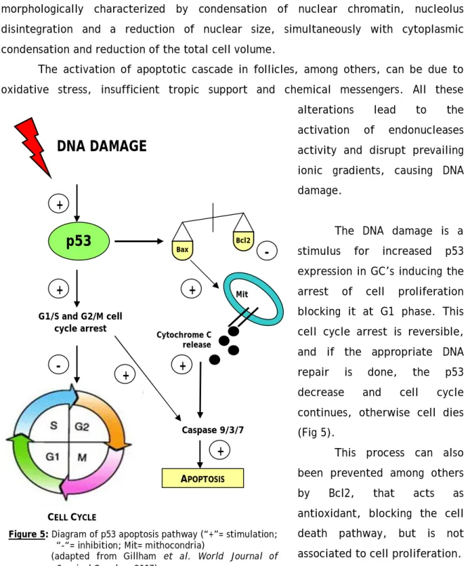
Contraction followed by clumping of the chromatin, wrinkling of the oocyte nuclear membrane and dislocation of the granulosa cells (GC’s) is considered signs of atresia (Baker, 1963). This atresia condition is the result of an apoptotic process that is morphologically characterized by condensation of nuclear chromatin, nucleolus disintegration and a reduction of nuclear size, simultaneously with cytoplasmic condensation and reduction of the total cell volume.
The activation of apoptotic cascade in follicles, among others, can be due to oxidative stress, insufficient tropic support and chemical messengers. All these alterations lead to the activation of endonucleases activity and disrupt prevailing ionic gradients, causing DNA damage.
The DNA damage is a stimulus for increased p53 expression in GC’s inducing the arrest of cell proliferation blocking it at G1 phase. This cell cycle arrest is reversible, and if the appropriate DNA repair is done, the p53 decrease and cell cycle continues, otherwise cell dies (Fig 5).
This process can also been prevented among others by Bcl2, that acts as antioxidant, blocking the cell death pathway, but is not associated to cell proliferation.
Figure 5: Diagram of p53 apoptosis pathway (“+”= stimulation; “-“= inhibition; Mit= mithocondria)
(adapted from Gillham et al. World Journal of Surgical Oncology 2007) Bax Bcl2
DNA DAMAGE
p53
G1/S and G2/M cell cycle arrest+
-
+
CELL CYCLE-
+
Mit Cytochrome C release Caspase 9/3/7 APOPTOSIS+
+
+
Briefly, the fate of GC’s during development may be decided by the balance of death repressors and death inducers.
The proliferation of GC’s is essential to follicle and oocyte development, being an important way to evaluate their viability. The proliferating status can be proved by the expression of Ki67 that is expressed in all phases of cell cycle excepting G0 phase.
T
RANSFORMING
G
ROWTH
F
ACTOR ΒETA SUPERFAMILY
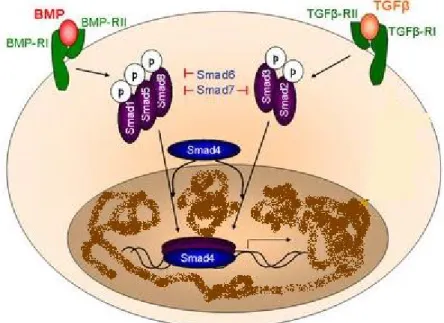
Among the many growth factors that have been identified as being important in early follicular growth several are members of the Transforming Growth Factor-β superfamily (Erickson and Shimasaki, 2000).
The TGF-β superfamily is a large group of 25kDa proteins, which share commons structural motifs (Chang et al., 2002). They are all synthesizes as prepropeptides with one signalpeptide, a large proregion and a smaller biologically active mature region, and exists as homo or heterodimers (Massagué, 1990; Chang et al., 2002). They are categorized into 23 distinct gene types that fall into four major groups, i.e., bone morphogenetic proteins and growth differentiation factors; activins and inhibins; TGF-β itself and a group encompassing various divergent members as anti Mulerian hormone (AMH) (Burt, 1992; Herpin et al., 2004).
Two types of transmembrane serine/threonine kinase receptors (Type I and Type II) have been identified for these polypeptides (Massagué and Wotton, 2000; Chang et al., 2002; Miyazawa et al., 2002). When a ligand binds, the kinase of the type II receptor phosphorylate and thereby activates the kinase of the type I receptor, which in turn will activate the intracellular signaling molecules called Smad proteins (Shi and Massagué, 2003; ten Dijke and Hill, 2003; Caestecker, 2004). There are eight distinct Smad proteins known, divided in three functional classes, which are, receptor regulated Smads (Smad 1,2,3,5,8), collaborating Smads (Smad 4) and inhibitory Smads (Smad 6,7) (Shi and Massagué, 2003).
There are basically two main pathways, the TGF-β/activin pathway where the ligand has a higher affinity for the type II receptors and the BMP pathway with ligands that have a higher affinity for the type I receptor (Shi and Massagué, 2003; Caestecker, 2004; ten Dijke and Hill, 2004). The TGF-β/activin pathway induce the activation of Smads 2/3 while the BMP pathway activate the Smads 1/5/8, however both pathways lead to the formation of the complex regulated Smad actived/collaborating Smad (Smad
4) (Zhang and Derynck , 1999). This complex then translocates into the nucleus and activates transcription of specific genes (Zhang and Derynck, 1999). The Smads 6/7 can inhibit the Smads of the BMP pathway while the Smads of the TGFβ/activin pathway only are inhibiting by the Smad 7 (Massagué, 1998; Miyazawa et al., 2002) (Fig 6).
G
ROWTH DIFFERENTIATION FACTOR
-9
This growth factor was identified in 1993 (McPherron and Lee, 1993) and is expressed and secreted by the oocytes in mostly species, including humans (McGrath et al., 1995).
Mutations in the GDF-9 gene are associated with various reproductive abnormalities, among the most serious is that although the oocytes grow in their follicles, the growth of the follicular cells is retarded which results in an arrest in the follicle growth and infertility (Carabatsos et al., 1998), proving its essential role during follicular development (Dong et al., 1996).
The expression of this factor differs among the species with GDF-9 expression observed in primordial follicles of sheep, cattle, possum and hamster (Bodensteiner et
Figure 6 : The BMP and TGFβ/activin signaling pathway (adapted from http://www.rr-research.no)
al., 1999; Eckery et al., 2002; Wang and Roy, 2004) and in primary follicles in rats, mice and humans (Laitinen et al., 1998; Jaatinen et al., 1999).
This protein function as a paracrine factor in the regulation of granulosa cell proliferation and differentiation (Elvin et al., 1999; Eppig, 2001; Gilchrist et al., 2006), and is correlated with recruitment of follicles for growth being essential for fertility (Dong et al., 1996). The higher proportion of viable human follicles in organ culture, suggest that this factor enhances the survival of the follicles (Hreinsson et al., 2002).
The role of GDF-9 in each stage of follicular development remains unclear but several studies pointed to its extreme importance for the proper development of follicle/oocyte (Dong et al., 1996; Elvin et al., 1999; Hanrahan et al, 2004; Kovanci et al., 2007), becoming crucial to understand his action mechanism .
A
CTIVIN
A
Activins are hetero or homo dimmers comprising two different beta subunits (A or B) with activin A being the predominant activin isoform. In the ovary, activins are produced by the GC’s and are important in folliculogenesis (Findlay et al., 2002), but the exact stages of development that activin A regulated are unclear. However, it has been shown to promote follicle growth by increasing GC proliferation and enhancing antral formation (Mizunuma et al., 1999; Zhao et al., 2001).
Activin stimulated follicle in vitro has been demonstrated in preantral ovine follicles (Thomas et al., 2003), caprine follicles (Silva et al., 2006), rodent follicles (McGee et al., 2001) and in human follicles (Telfer et al., 2008). Furthermore, it has been observed in vitro that activin A stimulates the meiotic maturation of the oocytes in human, rat and rhesus monkey (Itoh et al., 1990; Sadatsuki et al., 1993; Alak et al., 1998).
There is no evidence for the role of activin A in primordial follicle activation, but his expression on this early stage is a reason to think that it can have a relevant role at this stage (Mizunuma et al., 1999),thus, further investigations will be necessary.
F
ERTILITY PRESERVATION
The survival rate of children and young women after treatment against malignant diseases has increased in the last decades, due to improvements in the treatment therapies (Landis et al., 1998). Nevertheless one of the side effects of these treatments (chemotherapy/radiotherapy) is the frequent loss of fertility and the onset of a premature ovarian failure (POF) ((Donnez et Bassil, 1998; Yeung et al. 1998; Smitz et Cortvrindt, 1999).
The ovaries are very sensitive to cytotoxic treatment, especially to alkylating agents that increase the risk of POF by a factor of 9 (Byrne et al., 1992).
For the abdominal radiation it has been stated that a dose of 5-20 Gy is sufficient to completely impair gonadal function (Wallace et al., 2005), moreover, the association of abdominal radiation and chemotherapy with alkylating agents could rendering patients infertile in almost 100% of the cases (Donnez et al., 2006).
Beyond cancer and autoimmune diseases treatments such as radiotherapy or chemotherapy POF can be induced by chromosomal abnormalities such Turner’s syndrome (characterized by partial or total loss of one of the X chromosomes) (Sylven et al., 1993) and gene defects such as FSH receptor mutations (Aittomaki et al., 1995). Thus it is important to develop options for fertility preservation for these women.
Currently, there are three options proposed for fertility preservation: embryo cryopreservation, oocyte cryopreservation and ovarian tissue cryopreservation (Gosden, 2005; Meirrow et al., 2005). Nevertheless the choice of the most convenient strategy for preserving fertility depends on the type of the disease, the patient’s age and in some countries their partner status.
E
MBRYO
C
RYOPRESERVATION
This technique is the most widely available because she has become a routine in all Assisted Reproductive Technology (ART) centers and has proven its efficacy in terms of pregnancy and rates of delivery babies. However there are several drawbacks to the application of this technique.
First it is only applicable to post pubertal patients, who have a partner or, status law depending, are willing to use donor sperm.
Secondly, the In Vitro Fertilization (IVF) needs a cycle of hormonal stimulation for about a month that can represent an unacceptable delay to the beginning of the treatment, and at last, if the patient has a hormonal-sensitive tumour the traditional ovarian stimulation may be harmful. Recently, tamoxifen and letrozole were proposed as a safe alternative to the traditional ovarian stimulation (Oktay et al., 2005).
O
OCYTE
C
RYOPRESERVATION
Oocyte cryopreservation is an alternative for patients with the same characteristics as those described to the embryos but who are not with a partner and/or do not wish, or can not use sperm donation. The oocyte cryopreservation can be doing at the mature or immature state but there are different advantages and inconveniences associated with each state.
Mature oocyte
The metaphase II (MII) oocyte is a large and highly specialized cell that is extremely fragile due, among other things, to the spindle apparatus that is easily damaged by intracellular ice formation (Mandelbaum et al., 2004) inducing a risk of loss of chromosomes.
Several years after the first report of a live birth after cryopreservation of matures oocytes and thawing followed by IVF (Chen et al., 1986) the success rate remained low, but the combination with intra cytoplamisc sperm injection (ICSI) slightly improved the success rate (Porcu et al., 1997; Fabbri et al., 2001) since it avoid the difficulty of the spermatozoas to penetrate the zona pellucida hardened during the freezing process.
Improvements of the freezing protocols such the concentration of cryoprotectants (Fabbri et al., 2001), the time of exposure to pre-equilibration and thawing (Yang et al., 2002) also improved the survival rate of the mature oocytes after thawing (Sonmezer and Oktay, 2004), however, the mean pregnancy rate per thawed oocyte does not exceed 1.8% (Borini et al., 2006. Levi Setti et al., 2006).
Recently the ultrarapid freezing with vitrification offer advantages over conventional slow cooling protocols by improving post-thawing survival rates avoiding the formation of intracellular ice. A study demonstrates a rate of 5.1% live birth per warmed oocyte after vitrification (Kim et al., 2009).
Immature oocyte
This oocytes at the GV stage, survive better to the cryopreservation procedure (Boiso et al., 2002) but there maturation in vitro is still suboptimal with only one live birth reported (Tucker et al., 1998).
O
VARIAN TISSUE CRYOPRESERVATION
This is the only technique that can be proposed to children and patients who need immediate chemotherapy (Poirot et al., 2007).
Cryopreservation of ovarian tissue is considered a promising method for preservation fertility due to the large number of immature oocytes existing in the ovarian cortex.
After the first studies with animals in the 1950s (Parkes et al., 1953) and offspring obtaining in mice after transplantation (Parrott, 1960) the revival of the interest in cryopreservation of follicles in ovarian cortical tissue was in the 1990s.
The first human ovarian tissue cryopreservation was in 1996 (Hovatta et al., 1996; Newton et al., 1996) using different cryoprotectants to prevent the ice crystals formation in the cells. The cryoprotective agents commonly used are dimethyl sulfoxide (DMSO) and propanediol (PrOH) or ethylene glycol (EG) at 1.5M with sucrose 0.1M (Hovatta et al., 1996; Newton et al., 1996; Gook et al., 1999). No significant differences have been observed between DMSO, PrOH in terms of follicle survival (Newton et al., 1996).
The follicles are cryopreserved embedded in the ovarian tissue using a controlled-rate cooling followed by storage in liquid nitrogen at -196°C. The tissue is previously divided into small fragments to facilitate the permeation of the cryoprotectant.
Studies concerning follicular viability report that at least 70% of the follicles can be expected to survive the process of cryopreservation and thawing (Hovatta et al., 1996; Oktay et al., 1997; Gook et al., 1999; Cortvrindt and Smitz, 2001; Nisolle et al., 2000).
The quiescent primordial follicles are the most able to survive due to their size and low number of granulosa cells being easily equilibrate with the cryoprotectant. Those follicles have a low metabolic rate, the zona pellucida is not yet formed and their oocyte is arrested in the prophase of the first meiotic division (Shaw et al., 2000).
Recently, vitrification was proposed as an alternative procedure to slow cryopreservation. This technique uses a high concentration of cryoprotective agents and an immediate freezing, transforming the aqueous parts of the cells in a solid amorphous vitreous stage and avoids the formation of ice crystals.
The handicap of those options is that the follicles surviving the cryopreservation process contain immature oocytes. These oocytes can not be immediately fertilized, they need further maturation. This maturation can be achieved in vivo by transplantation or in vitro by culture of the ovarian tissue.
F
ERTILITY
R
ESTORATION
O
VARIAN
T
ISSUE
T
RANSPLANTATION
This is to date the most viable utilization of the ovarian cryopreserved tissue allowing reestablishing the hormonal balance and thereby delay menopause and also restore fertility.
The transplantation can be done in its original site (orthotopic), in a well vascularized site in the woman body (heterotopic) and to another species (xenotransplantation).
The viability of this technique was demonstrated with offspring obtained in animals and in humans.
Heterotopic transplantation
This technique involves the transplantation of the tissue to another part of the body as the forearm (Oktay et al., 2001) or the abdomen wall (Oktay et al., 2004). With these options the women resumed normal menstrual cycle avoiding the hormone replacement therapy and oocytes have been obtained. Nevertheless, this technique didn’t result in evolutive pregnancy but it was shown for the first time that oocytes and embryos could be obtained from cryopreserved/thawed ovarian tissue, in human (Oktay et al., 2004).
Orthotopic transplantation
In this case the tissue is implanted at its original site. The first human transplantation was reported by Oktay and Karlikaya (2000), who obtained ovarian function after sixteen weeks and even an ovulation after stimulation. This study proved that the cryopreservation protocol was not totally harmful for human ovary.
The first live human birth was obtained in 2004 (Donnez et al., 2004) in a patient without previous treatment before the cryopreservation. This, open the discussion of the really nature of the pregnancy; “oocyte from the remaining ovary or from the
orthotopic transplantation of slices of ovary cryopreserved after a cancer treatment (Meirow et al., 2005).
Since twenty four transplantations were reported and eight pregnancies obtain with six babies born.
Xenotransplantation
This technique is commonly done for viability studies and not as a clinical option. The studies, using immunodeficient mice, show good recovery of follicles and follicular growth to late and antral stages (Newton et al., 1996; Gook et al., 2001; Kim et al., Table 1 : Autotransplantations of cryopreserved human ovarian tissue
(adapted from Poirot et al., 2008) REFERENCE
AGE BEFORE FREEZING
GRAFT TYPE OUTCOME
Oktay K (2000) 29 Orthotopic FD after stimulation
Radford J (2001) 36 Orthotopic Ovarian function
Callejo J (2001) 47 Heterotopic Endocrine ovarian
function
Oktay K (2004) 30 Heterotopic Embryo
Tryde Schmidt KL (2004) 32 Orthotopic MII oocyte
Donnez J (2004) 25 Orthotopic Live birth
Kim S (2004) 37 Orthotopic Ovarian function
Meirow D (2005) 28 Orthotopic Live birth
Schmidt KL (2005) 28 25 32
Orthotopic & Heterotopic Orthotopic & Heterotopic
Orthotopic
Ovarian function Embryo
Embryo
Wolner-Hanssen P (2005) 30 Heterotopic FD after stimulation
Donnez J (2006) 21 Orthotopic Ovarian function
Rosendhal M (2006) 28 Orthotopic & Heterotopic Pregnancy Demeestere I (2006) 24 Orthotopic & Heterotopic Pregnancy Demeestere I (2007) 24 Orthotopic & Heterotopic Live birth Donnez J (2008) 22 28 22 Orthotopic Oocytes Cycles Cycles Andersen CY (2008) 26 27 36 25 Orthotopic Orthotopic Orthotopic
Orthotopic & Heterotopic
Live birth Live birth Ovarian function
Silber SJ (2008) Orthotopic Live birth
2002). Ovulation and the formation of corpus luteum has also been observed with this technique (Gook et al., 2003).
O
VARIAN
T
ISSUE
C
ULTURE
For some diseases, the transplantation of the thawed fragments of ovarian tissue represents a high risk of reintroducing malignant cells (Shaw et al., 1996; Gosden et al., 1997; Aubard et al., 1999). The maturation of the follicles in vitro could avoid this risk.
The development of primordial follicles to an antral stage and consequent oocyte maturation is a complex procedure and until now, completely successful only in mice with live offspring born (Eppig and O’Brien, 1996; O’Brien et al., 2003).
In Humans the in vitro development, with or without cryopreservation of ovarian tissue was reported but with fewer results than those observed in rodents, but still hopeful for further investigation. The complete follicular development was not yet report, but the various stage of development obtained separately shows that human folliculogenesis in vitro is possible. The long time required for follicle development (Gougeon et al., 1987) and the dense ovarian stroma make this culture more challenging (Hovatta et al., 1997; Lass et al., 1997).
Under the last ten years multiple studies regarding culture of fresh and cryopreserved ovarian tissue tried to discover and establish the perfect conditions to lead to complete and successful in vitro follicle development.
In Humans several works described ovarian follicles growth from a state to the following state. Some develop techniques using preantral follicles isolated mechanically (Abir et al., 1997) or by enzymatic digestion (Roy and Tracy, 1993) that after culture observed antral cavity formation. However those achievements were hardly reproduce with primordial and primary follicles (Abir et al., 1999; Hovatta et al., 1999), showing that for this type of follicles the culture within ovarian tissue is recommended (Hovatta et al., 1997, 1999; Hreinsson et al., 2002; Scott et al:, 2004).
Recently a team reported a two-step culture (Telfer et al., 2008), trying to reproduce the good achievements obtained with this technique in mice (Eppig and O’Brien, 1996; O’Brien et al., 2003). In this protocol the small follicles, were in a first time cultured within a thin slice of ovarian tissue until they reach the secondary state. From there, the secondary follicles obtained were mechanically isolated and cultured until antral cavity formation was observed (Telfer et al., 2008). The particularity of this study is the short time need to all this process (10 days), and the largest diameter
obtained for follicles cultured in a medium with activin A than in follicles cultured without this additional factor.
The addition of different growth factors and/or hormones in the culture medium as been study trying to improve the development and survival of the follicles in vitro (Wright et al., 1999; Louhio et al., 2000; Hreinsson et al., 2002; Telfer et al., 2008).
Among the factors that are pointed to regulate the initiation and progression of primordial development, Kit Lingand (KL) and b-fibroblast growth factor (bFGF) seem to be essential for their progression to primary follicles (Parrot and Skinner, 1999; Nilsson and al., 2004) while GDF-9 are fundamental to their development into the secondary stage (Hreinsson et al., 2002). Insulin, insulin like growth factor I (IGFI) and IGFII act as trophic factors for follicles and simultaneously stimulate follicular growth (Fabbri et al., 2006; Louhio et al., 2000) and gonadotropins, FSH and LH are essential for progression from the preantral to the antral follicle stage (Wrigth et al., 1999).
The utilization of an extracellular matrix in the culture of isolated follicles to mimics the ovarian conditions were described successful (Roy and Treacy, 1993; Abir et al., 1999, 2001; Woodruff and Shea, 2007) but Telfer shows that it is not essential to obtained follicle growth (Telfer et al., 2008).
The presence or absence of serum in the culture medium is equally discussed with follicular growth observed in the two cases.
Although all progress made in the culture of human follicles, this reality and clinical application is still far from the optimum conditions. Further experimentation is needed to fully understand the entire continuum of follicle development, optimize timings of exposure to key factors and to evaluate the viability of oocytes obtained to be fertilized and develop into normal embryo.
A
IMS OF OUR STUDY
While most of previously works were realized on fresh ovarian tissue, the clinical reality is the future utilization of cryopreserved ovarian tissue, being important to realize and evaluate culture of cryopreserved ovarian tissue.
The aim of our study was to analyze the effect of two growth factors added into medium, evaluating 4 parameters of the follicle growth.
To accomplish it we realized:
Culture of human cryopreserved tissue simultaneously in three different culture media
Counting and morphological analyze of follicles
Observation of eventually changes in the morphological aspect of the stromal and vascular tissue into the ovarian fragments, along the culture Immunohistochemical analyze of the follicles, stromal and vascular tissue
using two markers involved in apoptosis (p53 and bcl2) and one marker of cell proliferation (Ki67)
M
ATERIALS AND
M
ETHODS
O
VARIAN
T
ISSUE
Human ovarian fragments frozen for fertility preservation of one 15 years old patient were used.
The patient and her parents, before cryopreservation, signed an agreement for postmortem utilization, for research, of the frozen tissue.
The isolated ovarian cortex has been cryopreserved as little fragments of about 0,5 cm3 following the slow freezing method described by Gosden (Gosden et al., 1994) using DMSO and sucrose as cryoprotectants.
All manipulation of the human ovarian tissue was done with sterile material and under sterile conditions.
O
VARIAN
T
ISSUE
T
HAWING
The ovarian fragments were thawed one by one as follow.
The cryovial was pull out of the liquid nitrogen and placed for a few seconds at room temperature followed by five minutes in the incubator at 37ºC, till complete defrost of the cryopreservation medium.
After this process the fragment was washed, at room temperature, successively in 4 different solutions containing a decreasing concentration of DMSO (2M; 1,5M; 1M; 0M). The thawing solutions were composed by Leibovitz medium (Eurobio®, Courtaboeuf, France), DMSO (Sigma Aldrich®, St Quentin Fallavier, France), fetal calf serum (FCS) (Eurobio®) and 0.2M sucrose (Sigma Aldrich®).
P
REPARATION OF CORTICAL TISSUE FOR CULTURE
After thawing the fragments were placed in a prewarmed Leibovitz medium (Eurobio®) supplemented with sodium pyruvate (2mM,), bovine serum albumin, BSA, (Fraction V, 3mg/mL), ascorbic acid (50 µg/mL,) and stabilized solution of L-Glutamin,
penicillin G and streptomycin (2mM, 75µg/mL, 50 µg/mL, respectively) all purchased from Sigma-Aldrich®.
In this medium the fragments were cut into smaller pieces (~ 0,5 mm3) with a scalpel under a stereo microscope and pulled mechanically with 25 gauges needles (Sherwood médical®, Evry, France) to flatten out the tissue and to minimize the underlying stromal tissue.
D
IFFERENT CULTURE MEDIA COMPOSITION
For this work three different culture media were used.
The medium used as control group was composed of McCoy’s 5a medium (Sigma-Aldrich®) supplemented with HEPES (20mM, Eurobio®), BSA (0,1%), stabilized solution of L-Glutamin, penicillin G and streptomycin (3mM; 0,1mg/mL; 0,1g/mL, respectively), transferrin (2,5µg/mL), selenium (4ng/mL), insulin (10ng/mL) and ascorbic acid (50µg/mL), all purchased in Sigma Aldrich®.
The two other media were composed by the control medium which has been supplemented with 200ng/mL of GDF-9(CliniSciences®, Montrouge, France) or with 100ng/mL of activin A (R&D Systems®, Lille, France).
The different culture medium gave name for the different groups, so there were formed 3 groups:
C group – control culture medium G group – culture medium with GDF9 A group – culture medium with activin A
C
ORTICAL STRIP CULTURE
This work was accomplished in four different time cultures.
The cortical strips were cultured individually in 24-well culture plates (Costar®,), containing 300 µL of the respectively culture medium, using a culture plate for each medium.
The three groups are incubated (at the same time) at 37°C in humidified air with 5% CO2. Half of the medium, of each well, was changed every 2 days. This pulled
Along the culture the strips were observed at the inverted light microscope, to verify the presence of developing follicles.
A total of 247 cortical strips were cultured (82 in the control group; 82 in the GDF-9 group and 83 in the activin A group).
H
ISTOLOGICAL ANALYSIS
Before placing the cortical strips in the different culture media, 4-8 strips (from each fragment used), were fixed in a 10% formaldehyde solution overnight.
At day 5, 10 and 14, of culture, eight strips of each group were fixed in a 10% formaldehyde solution overnight.
At each time of fixation the strips of the same medium were fixed together. After fixation with 10% formaldehyde the samples were dehydrated in ethanol, using increase purity baths (70%, 90% and 100%). The absolute alcohol was then replaced by xylene and followed by paraffin inclusion of the samples.
The blocks were sectioned manually at the microtome (3 or 4µm), mounted on charged slides and left to dry for at least one night at 37ºC. Each slide contained 2 or 3 serial sections.
M
ORPHOLOGICAL ANALYSIS
For this analysis the serial sections obtained were staining with haematoxylin, eosine and safranin (HES).
Every section in the slide was examined, by screening, to make the counting, morphological classification and evaluation of the follicles and intuitive evaluation of the stromal and vascular tissue.
Follicle Counting and Classification
The global counting of follicles includes all follicles observed, but for the classification only follicles with visible nucleus were counted, for evaluation of all parameters of the oocyte. The sections with visible nucleolus were chosen in preference.
Table 2: Characteristics observed and follicle stage associated
F
OLLICULAR STAGEC
HARACTERISTICSPrimordial Oocyte surrounded by one layer of flattened GCs
Early primary Oocyte surrounded by one layer of flattened and cuboidal GCs Primary Oocyte surrounded by one layer of cuboidal GCs
Secondary Oocyte surrounded by two or more layers of cuboidal GCs
Others evaluation criteria
The morphological evaluation of follicles was made only in the classified follicles by counting the GC’s, observing chromatin distribution (diffuse/dense) and oocyte cytoplasm configuration (normal/vacuolar/retracted).
I
MMUNOHISTOCHEMICAL EVALUATION
The immunohistochemical staining was performed by an automated immunohistochemical processor (Benchmark XT, Ventana®, Illkirch, France) using HRP (horsedarish peroxidase) as reactive and DAB (diaminobenzidine tetrahydrochloride). The counterstaining was realized with haematoxylin II, all reagents were purchased in Ventana®.
The primary antibodies used were monoclonal mouse anti-Human Ki67 (clone MIB-1) 1/50 diluted, p53 (clone DO-7) 1/25 diluted and Bcl2 (clone 124) 1/50 diluted, all obtained in Dako®, Trappes, France.
The immunostaining was considered positive when the cells were observed with brown coloration, for all antibodies, and negative when cells observed are blue.
Immunohistochemical evaluation of follicle was realized counting the number of GC’s positively marked versus GC’s negatively marked.
S
TATISTICAL ANALYSIS
Quantitative values were treated with ANOVA with effect medium/day of culture.
Binary variable were treated with logistic model with effect medium/day and interceptions within it.
Ordinal variables were treated with logistic model ordinal with 3 or 2 modalities. For all tests the significatif value considered was p<0.05.
R
ESULTS
M
ORPHOLOGICAL ANALYSIS
Follicle Counting and Classification
For this analyze 46 slides with each 2 or 3 serial sections of 8 grouped strips were observed.
The counting of all follicles show a significant (p< 0.05) decrease in the number of follicles observed since the day 10 of culture for all culture media (Fig 7).
Even if the different culture media did not present a significant difference it was observed a higher number of follicles in the G group. At the end of the culture this was the group with a higher number of follicles and a lower proportion of degenerate follicles.
Throughout the culture three types of follicles, primordial, early primary and primary were observed (Fig 8).
The distribution of the different types of follicles varies in the three culture media and throughout the time but without significant difference and no evidence of follicle growth (Fig 9).
Histological sections of the cortical strips (n=28) fixed after thawing (not cultured) showed 57 follicles, distributed in 12 strips, where follicles were visualized. According to our counting method, from these follicles, only 31 were classifiable in the different stages (19 primordial, 3 early primary and 9 primary).
At the 5th day of culture, in the obtained sections, an increase of the proportion of early primary and primary stage were registered, especially in the GDF-9 group (G) where 65% of the classifiable follicles were at the early primary stage. The activin A group (A) present the higher proportion of primary follicles (42% vs 12% for G and 33% for the control group (C) and 30% for not cultured). The primordial stage of development appears, at this time, in low number in the three culture media.
A
B
Figure 8:Microscopy visualization of a histological section with HES staining of primordial follicle and primary follicle (A) and early primary follicles (B),in ovarian cortical strip.
At the 10th day of culture only 13 of the observed follicles were classifiable, and were equally distributed in the three groups. The observation of the histological sections showed an increased of the primary follicles for de G group and a decrease of the proportion of early primary follicles in the same day, compared with the 5th day of culture. For the others two culture media, the number of the three different types of follicles (primordial, early primary and primary) observed decreased comparing to the 5th Day but a similar proportional expression was registered.
After 14 days of culture only a few number of follicles were classified (7), with the presence of one follicle for the C and the A groups (primary and early respectively). The five remainder follicles were observed in the G group and were distributed among the 3 types of follicles with proportions that are close to those found at the 5th Day.
In all sections analyzed no secondary follicles were seen.
Figure 9: Distribution of the different types of follicles observed during the culture, for the three culture media, and for the not cultured ovarian fragments
Others evaluation criteria
The average of granulosa cells that surrounded the oocyte forming the follicle presents small variations in each medium during the culture (Table 3). However in the 10th day of culture the G group shows a mean of 11 GCs for each follicle instead of the 7 observed at the 5th day of culture. In the opposite the activin A group at the end of the culture only presented 3 GCs.
Table 3: Average number of granulosa cells per follicle, calculated, by the histological counting of granulosa cells surrounding classifiable follicles, for each culture media at the different times of culture, and for not cultured follicles
5
thDay
10
thDay
14
thDay
NotCultured C G A C G A C G A
GC/foll (n) 7 5 7 5 6 11 5 6 7 3
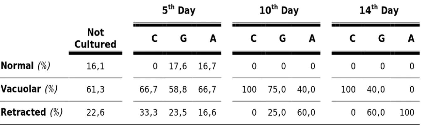
The chromatin configuration and the cytoplasm aspect are morphological markers of the oocyte quality.
The variations observed throughout the culture are shown in the table 4. Highlighting that, the normal aspect of the cytoplasm was mostly observed at the not cultured follicles, or in the follicles that were cultured only for 5 days. However, only at the end of the culture we register a higher proportion of retracted cytoplasm. In generality, we observe that in this culture, the oocyte presents a vacuolar cytoplasm.
Table 4: Proportion of the different aspects of the cytoplasm in the oocytes observed, for each culture media and at different times of culture, and for not cultured follicles
5th Day 10th Day 14th Day
Not
Cultured C G A C G A C G A
Normal (%) 16,1 0 17,6 16,7 0 0 0 0 0 0
Vacuolar (%) 61,3 66,7 58,8 66,7 100 75,0 40,0 100 40,0 0 Retracted (%) 22,6 33,3 23,5 16,6 0 25,0 60,0 0 60,0 100
The proportion of oocyte with dense or diffuse chromatin didn’t differ in the not cultured oocyte, however after 5 days of culture it was observed a higher proportion of oocyte with diffuse configuration of chromatin. This behavior was inverted at the 10th day of culture for the C and G groups that have a higher proportion of oocytes with dense chromatin. At the end of the culture only the G group shows oocytes with dense chromatin, however the observation for the C and A groups was made on a single oocyte for each group (Table 5).
D
EGENERATED
/
ATRETIC FOLLICLES
For calculate the proportion of the degenerating follicles we used as total follicle population the classifiable, no classifiable and degenerating follicles (Fig 10).
Table 5: Proportion of the follicles with a diffuse or a dense chromatin configuration, for the not cultured follicles and those cultured in the different culture media, at different time of the culture
5
thDay
10
thDay
14
thDay
NotCultured C G A C G A C G A
Diffuse (%) 48,3 100 82,3 91,2 25,0 25,0 75,0 100 40,0 100
Dense (%) 51,7 0 17,7 8,8 75,0 75,0 25,0 0 60,0 0
Figure 10: HES histological section of a primary and a degenerating follicle of not cultured strips; original magnification X 400