IN ST IT U T O D E C IÊ N C IA S B IO M ÉD IC A S A B EL S A LA Z A R
Ana
Margarida
Costa
. Establishment of a three-dimensional
in vitro
model of air-blood barrier to assess the translocation of
nanoparticles targeted to the lung
Establishment of a
t
hree-dimensional
in vitro
model of air-blood barrier to
assess the translocation of nanoparticles targeted to the lung
Ana Margarida Costa
2018
D
.ICBAS
2018
CIÊNCIAS BIOMEDICASEstablishment of a
three-dimensional in vitro model of
air-blood barrier to assess the
translocation of nanoparticles
targeted to the lung
Ana Margarida Costa
Establishment of a three-dimensional in vitro model of air-blood
barrier to assess the translocation of nanoparticles targeted to the
lung
Tese de Candidatura ao grau de Doutor em Ciências Biomédicas submetida ao Instituto de Ciências Biomédicas Abel Salazar da Universidade do Porto
Orientador
Doutor Vítor Manuel Fernandes Seabra da Silva
Categoria – Professor Associado
Afiliação – IUCS - Instituto Universitário de Ciências da Saúde
Coorientador
Doutor Bruno Filipe Carmelino Cardoso Sarmento
Categoria – Investigador Auxiliar/Professor Auxiliar
Afiliação – i3S - Instituto de Investigação e Inovação em Saúde, INEB - Instituto de Engenharia Biomédica, Universidade do Porto & IUCS - Instituto Universitário de Ciências da Saúde
Tutor
Doutor Pedro Lopes Granja
Categoria – Investigador Principal/Professor Associado
Afiliação – i3S - Instituto de Investigação e Inovação em Saúde, INEB - Instituto de Engenharia Biomédica & Instituto de
Ciências Biomédicas Abel Salazar (ICBAS), Universidade do Porto
Nanomedicines & Translational Drug Delivery i3S – Instituto de Investigação e Inovação em Saúde
INEB – Instituto de Engenharia Biomédica Universidade do Porto, Porto, Portugal
Rua Alfredo Allen, 208 4200-135 Porto, Portugal
Drug Discovery, Delivery & Toxicology
IINFACTS – Institute of Research and Advanced Training in Health Sciences and Technology
Rua Central de Gandra, 1317 4585-116 Gandra, Paredes, Portugal
DDEL – Department of Drug Delivery
HIPS – Helmholtz-Institut für Pharmazeutische Forschung Saarland Universitätscampus E8 1
“A mente que se abre a uma nova ideia nunca mais volta ao seu
tamanho original”
“The mind that opens to a new idea never returns to its original size”
ACKNOWLEDGMENTS
I could not reach the end of this stage without expressing my gratitude and acknowledgments to all the people and institutions that were involved directly and indirectly in this journey:
To my supervisors Professor Vítor Seabra and Professor Bruno Sarmento, first for the invitation and the opportunity to enroll on a doctoral program; second, for all the scientific discussion, guidance and support during the last four years. It was not an easy path and walking through it required a lot of patience from their side. I am grateful for their trust, freedom, encouragement and for the opportunities that allowed me to grow as a researcher. A special thanks to Professor Bruno Sarmento for the close supervision at Instituto de Investigação e Inovação em Saúde (i3S)/Instituto de Engenharia Biomédica (INEB), always with a good sense of humor that I appreciated;
To Professor Claus-Michael Lehr for the kindness of letting me perform part of my work at Department of Drug Delivery (DDEL) from Helmholtz-Institut für Pharmazeutische Forschung Saarland (HIPS), and for the scientific discussion that allowed me to see my work with a different point of view; also Dr. Cristiane Carvalho-Wodarz for the close supervision, for all ideas and suggestion given, and mostly, for her friendship, trust and the motivation which were important to carry on this work during my stay in Germany;
To Professor Pedro Granja for being my co-supervisor/tutor on this work and for providing me, as well as all INEB members, a constant positive energy;
To all my work colleagues from Nanomedicines & Translational Drug Delivery from i3S/INEB, Universidade of Porto, that supported my work on a daily-basis, and for the funny and good moments that I will not forget. I would like to give my deepest acknowledge to Rute Nunes for the fully support, trust, encouragement and her friendship that started from the beginning. No doubt you were like sister for me during this period of my life. Also, to all members that belonged to this research group on the past, in particular Pedro Fonte who introduced me the interest for research, and
also, to Cassilda Cunha-Reis, for all the advice, encouragement, and for teaching me how to see life in a different perspective;
To my HIPS foodies, my family when I was far from Portugal. To all I am grateful for the help and the funny moments inside and outside the lab, for the friendship and for making me feel at home. A special thanks to Stephanie Kletting (for the scientific discussions and help with performing my in vitro model), Carlos Montefusco and Adriely de Góis (my Brazilian brothers), Remi Hendrix-Jastrzebski (my crazy Zumba partner that shared the same questions and doubts about cells), Sara Menina (for the friendship, joy and the good time spent together) and Hanzey Yasar (for the time together during our coffee talks on Sundays afternoons);
To Andreia Almeida, my fellow citizen (from Gandra) and my sweetest friend from the lab, that always helped me, even on small things. Thanks for the all information regarding the permeability studies. You were always there, ready for everything, even when I was in Germany;
To Francisca Araújo for the assistance during the preparation of histology slices at i3S;
To Vírginia Gonçalves from Instituto de Investigação e Formação Avançada em Ciências e Tecnologias da Saúde (IINFACTS) for all the help with the high-performance liquid chromatography (HPLC) experiments;
To Maria Gomes Lázaro for the assistance at confocal laser scannining microscopy
(CLSM) ImageStream®X imaging flow cytometry analyses performed at Bioimaging
i3S Scientific Platform;
To Rui Fernandes and Francisco Rosário Figueiredo from the assistance at transmission electron microscopy analyses performed at Histology and Electrom Microscopy i3S Scientific Platform; It was not easy to get the cut, but we got it!!;
To Biointerfaces and Nanotechnology i3S Scientific Platform for the support with particle size and zeta potential analyses;
To the technicians Petra König and Jana Westhues for the support on cell culture and all the help during my stay on Germany;
To Chiara de Rossi for the help, friendship and for the support with scanning electron microscopy experiments;
To Marijas Jurisic for the availability, patience, for teaching me how to work with CLSM and last, for the help during the histology experiments;
To all research institutions involved in this work and their members, namely i3S and INEB from University of Porto, IINFACTS/CESPU and last to HIPS from Saarland University;
To my close friends for the friendship, motivation and encouragement throughout my life, in particularly for the last four years. Thank you for spending times listening to my failures, doubts, concerns, sadness, and always believing that I would be able to give one more step ahead;
To my family for the unconditional support, patient and love throughout my life, and for understanding my absence during the hard times. I always appreciated the trust of my family on me and the freedom in choosing my own career, even knowing I would be far from home. I am grateful for having you remembering me all the time that “knowledge does not occupy space, there is always space for more”, which were essential values for carrying on my studies for four more years.
FINANCIAL SUPPORT
Ana Costa gratefully acknowledges Fundação para a Ciência e a Tecnologia (FCT), Portugal for the financial support (Grant SFRH/BD/95227/2013).
This thesis was partially supported by the project NORTE-01-0145-FEDER-000012, supported by Norte Portugal Regional Operational Programme (NORTE 2020), under the PORTUGAL 2020 Partnership Agreement, through the European Regional Development Fund (ERDF). This work was financed by FEDER - Fundo Europeu de Desenvolvimento Regional funds through the COMPETE 2020 - Operacional Programme for Competitiveness and Internationalisation (POCI), Portugal 2020, and by Portuguese funds through FCT - Fundação para a Ciência e a Tecnologia/ Ministério da Ciência, Tecnologia e Ensino Superior in the framework of the project "Institute for Research and Innovation in Health Sciences" (POCI-01-0145-FEDER-007274
PUBLICATIONS
Ao abrigo do disposto do nº 2, alínea a) do artigo 31º do Decreto-Lei n.º 115/2013 de 7 de Agosto, fazem parte integrante desta tese de doutoramento os seguintes trabalhos já publicados ou submetidos para publicação:
Costa A, Sarmento B, Seabra V. Targeted Drug Delivery Systems for Lung Macrophages. Current Drug Targets. 2015;16(14):1565-81;
Costa A, Andrade F. Tissue-based in vitro and ex vivo models for pulmonary permeability studies In: Sarmento B, editor. Concepts and Models for Drug Permeability Studies. Amsterdam: Woodhead Publishing; 2016. p. 255-72;
Costa A, Pinheiro M, Magalhães J, Ribeiro R, Seabra V, Reis S, Sarmento B. The formulation of nanomedicines for treating tuberculosis. Advanced Drug Delivery Reviews.2016;102:102-15;
Costa A, Sarmento B, Seabra V. Mannose-functionalized solid lipid nanoparticles are effective in targeting alveolar macrophages. European Journal of Pharmaceutical Sciences. 2017; 114:103-13;
TABLE OF CONTENTS
ABSTRACT ... XVII RESUMO ... XXI ABBREVIATIONS AND LIST OF ACRONYMOUS ... XXV
CHAPTER I
STATE-OF-ART ... 1
1. Morphology and physiology of respiratory tract ... 3
1.1. Lung physiology and tissue biology ... 3
1.2. The importance of lung macrophages on host-defense ... 8
1.3. The importance of lung as a route for drug delivery ... 12
2. Nanotechnology for pulmonary drug delivery against Mycobacterium tuberculosis ... 13
2.1. Physiopathology of Mycobacterium tuberculosis infection ... 13
2.2. Tuberculosis therapy and limitations ... 14
2.3. Nanocarriers for the pulmonary delivery of antituberculosis drugs ... 15
3. Functionalized nanosystems for alveolar macrophages targeting ... 22
3.1. The relevance of surface receptors of alveolar macrophages for drug delivery ... 22
3.2. Lipid-based nanoparticles for mannose receptors targeting ... 24
3.3. Polymeric nanoparticles for alveolar macrophages targeting ... 26
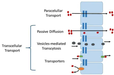
4. In vitro cell culture models as a tool to assess the lung permeability ... 33
4.1. General considerations ... 33
4.2. Mechanisms involved on drug absorption at lung tissue ... 33
4.3. In vitro cell based models to assess the pulmonary permeability ... 36
4.4. Microengineering for lung-on-a-chip ... 39
CHAPTER II OVERVIEW AND AIMS OF THE WORK ... 43
1. Overview ... 45
DEVELOPMENT AND CHARACTERIZATION OF MANNOSE-FUNCTIONALIZED
SOLID LIPID NANOPARTICLES FOR ISONIAZID DELIVERY ... 47
1. Introduction ... 49
2. Materials and Methods ... 51
2.1. Materials ... 51
2.2. Preparation and characterization of SLN ... 52
2.3. Physical-chemical characterization of Isn-SLN ... 53
2.4. Morphological characterization of SLN ... 54
2.5. In vitro release studies ... 55
2.6. Mannose functionalization ... 55
2.7. Cell culture maintenance ... 56
2.8. Metabolic activity and cytotoxicity ... 57
2.9. In vitro uptake studies ... 58
2.10. Statistical analysis ... 60
3. Results and discussion ... 60
3.1. Preparation and physical-chemical characterization of SLN ... 61
3.2. Preparation of SLN with different amounts of stearylamine ... 66
3.3. In vitro release studies ... 67
3.4. Fourier transform infrared characterization of M-SLN ... 68
3.5. Metabolic activity and cytotoxicity ... 70
3.6. In vitro uptake studies ... 71
4. Conclusions ... 73
CHAPTER IV ESTABLISHMENT AND CHARACTERIZATION OF A THREE-DIMENSIONAL IN VITRO MODEL OF AIR-BLOOD BARRIER ... 75
1. Introduction ... 77
2. Materials and Methods ... 78
2.1. Materials ... 78
2.2. Cell culture maintenance ... 79
2.3. Evaluation of growth pattern in different culture conditions ... 80
2.4. Establishment of the number of NCI-H441 and HPMEC-ST1.6R cells .... 82
2.6. Transepithelial electrical resistance of in vitro cell culture models ... 84
2.7. Morphological and ultrastructural analysis of in vitro cell culture models . 85 2.8. Influence of insulin-transferrin-selenium and time of culture on the multi-layers formation ... 88
2.9. Statistical analysis ... 91
3. Results and discussion ... 91
3.1. Evaluation of growth rate for each cell line in different culture medium ... 91
3.2. Optimization of the cell number of NCI-H441 and HPMEC-ST1.6R ... 93
3.3. Transepithelial electrical resistance and morphology of in vitro models with 8 days of culture ... 96
3.4. Surface of in vitro cell culture models with 8 days of culture ... 100
3.5. Cross section and ultrastructural characterization ... 101
3.6. Influence of insulin-transferrin-selenium and time of culture on the formation of multi-layers ... 104
4. Conclusion ... 107
CHAPTER V THREE-DIMENSIONAL IN VITRO AIR-BLOOD MODEL AS A TOOL TO EVALUATE THE LUNG TRANSLOCATION OF NANOCARRIERS ... 111
1. Introduction ... 113
2. Materials and Methods ... 114
2.1. Materials ... 114
2.2. Cell culture maintenance ... 115
2.3. Triple co-culture model of air-blood barrier ... 116
2.4. Transepithelial electrical resistance assessment of in vitro cell culture models ... 117
2.5. Morphological and ultrastructural analysis of in vitro cell culture models 118 2.6. Apparent permeability of sodium fluorescein ... 119
2.7. Translocation of fluorospheres... 120
2.8. Translocation of Isn-SLN and Isn permeability ... 122
2.9. Influence of lipopolysaccharide on the Isn permeability ... 122
2.10. Quantification of Isn ... 123
2.11. Interleukin measurement ... 123
2.12. Statistical analysis ... 124
3.2. Surface and ultrastructural characterization ... 126
3.3. Permanent permeability of sodium fluorescein ... 128
3.4. Translocation of fluorospheres... 131
3.5. Translocation of Isn-SLN and Isn permeability ... 134
3.6. Influence of lipopolysaccharide on the release of interleukin-8 ... 136
3.7. Influence of lipopolysaccharide on Isn permeability ... 138
4. Conclusion ... 140
CHAPTER VI GENERAL CONCLUSIONS AND FUTURE PERSPECTIVES ... 143
CHAPTER VII REFERENCES ... 149 APPENDICES APPENDIX I ... 189 APPENDIX II ... 193 APPENDIX III ... 198
ABSTRACT
The use of in vitro models for drug screening as an alternative to animal experiments is increasing over the last years, in particular, models to assess the permeation through biological membranes. Initially, cell culture models were constituted by one type of cells forming a confluent monolayer, but due to its oversimplicity they are being replaced by three-dimensional (3D) in vitro models, that present a higher complexity and reflect more the in vivo-like conditions. Being the pulmonary route one of the most studied approaches for drug administration, several in vitro models of alveolar epithelium have been used to assess the drug permeability and the translocation of nanocarriers. This work aims to develop a 3D in vitro model of air-blood barrier, comprising alveolar epithelial cells, capillary endothelium and macrophages, to test the translocation of nanocarriers. For that purpose, in this work was developed a solid lipid nanoparticles (SLN) and isoniazid (Isn), an antituberculosis drug, was encapsulated inside of the lipid matrix of this system. The formulation was further functionalized with mannose, in order to target SLN towards the mannose receptors expressed on the surface of alveolar macrophages (AMs), the main cells responsible for the Mycobacterium
tuberculosis survival during the latent states of infection, and at the same time, the
therapeutic target.
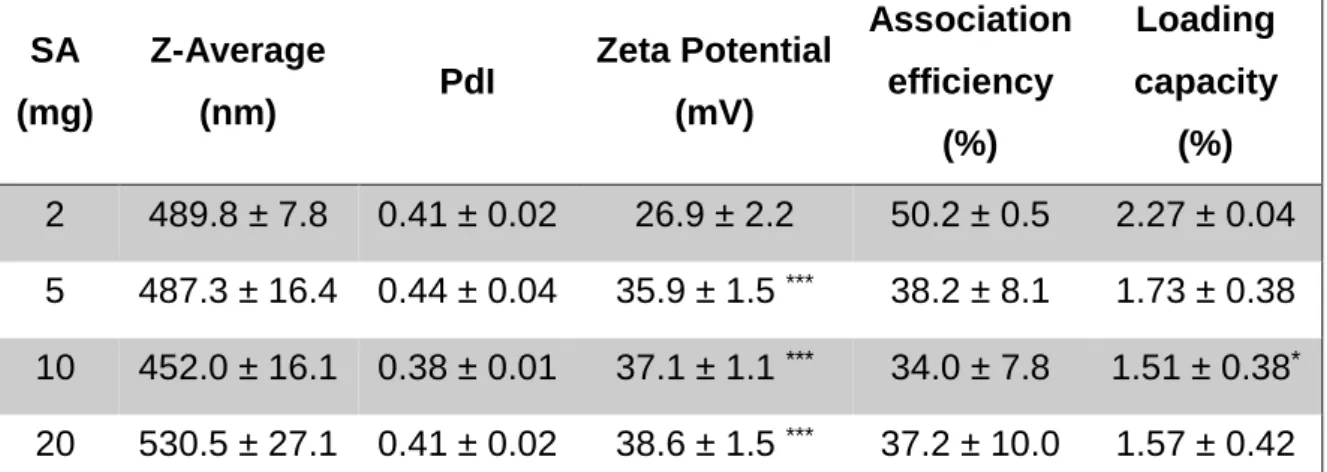
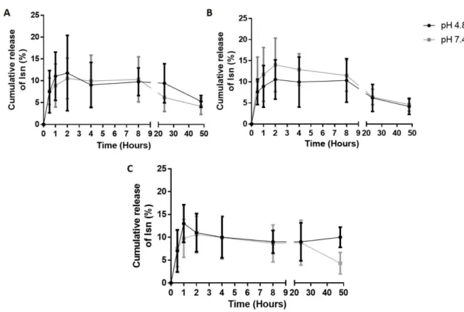
Different SLN formulations were developed, varying the type of lipid, the inner phase composition, the surfactant agents, the organic solvent and the stearylamine concentration. The selected formulation presented an average particle size around 500 nm, a polydispersity index between 0.3-0.4, a positive surface charge (around + 37 mV), an Isn association efficiency close to 35%. and a slow drug release kinetics (less than 10%) up to 48 hours. The functionalization of SLN with mannose was confirmed through fourier transform infrared spectroscopy. Uptake studies on differentiated THP-1 cells showed that mannosylated SLN were efficiently internalized through receptor-mediated endocytosis.
The 3D in vitro model of air-blood barrier was initiated by establishing the suitable conditions for the co-culture of a single monolayer of epithelial cells (NCI-H441) and a monolayer of endothelial cells (HPMEC-ST1.6R), seeded at the apical side and
pore size of 3.0 µm) aims to mimic the basement membrane. Finally, differentiated THP-1 cells were added apically, on the top of NCI-H441 cells, in order to mimic the AMs that are presented at alveolar space.
During the optimization, this of 3D in vitro model (being also designed as triple co-culture in this work) was characterized regarding transepithelial electrical resistance, morphology, topography and the analysis of its ultrastructural was also performed. A
successful model was established based on 1.0 x 10⁵ NCI-H441 cells/Transwell®, 5.0
x 104 HPMEC-ST1.6R cells/Transwell® and 1.0 x 10⁵ dTHP-1 cells/Transwell®,
cultured for 5 days, and translocation of different systems were tested across this new model.
Fluorospheres with an average size of 50 nm could translocate across the 3D in vitro model (18%), but fluorospheres with a size of 1.0 µm were retained on the apical side of the Transwell®, and less than 1% of these fluorospheres were found at basolateral compartment of this model. Based on these outcomes, it was concluded that this air-blood barrier model can be used to study the retention of 1.0 µm-sized inhaled particles, that may deposit on the alveoli, but still allows to study the translocation of small nanocarriers.
In this work SLN were produced to enable the local delivery of Isn at alveolar airways, so that this system was also used for validation the retention capacity of the 3D in vitro model. The majority SLN (with Isn loaded) did not translocate, while around 35% of free Isn could permeate this in vitro model.
Inflammatory response through the release of interleukin (IL)-8 was assessed after lipopolysaccharide (LPS) incubation. 3D in vitro model exhibited a higher IL-8 release regarding the NCI-H441 monoculture, which showed to be unresponsive on the presence of LPS. During the inflammatory process the permeability of alveolo-capillary membrane is generally enhanced. Thus, the Isn permeability across each monoculture, bi-cultures (NCI-H441+dTHP-1 and NCI-H441+HPMEC-ST1.6R) and across the 3D model was also assessed after LPS incubation. Overall, the permeability of free Isn through different in vitro cell culture model was not influenced by the LPS treatment.
In conclusion, the 3D in vitro model presented a better morphology that resembles the air-blood barrier when compared with epithelial cell monolayer, but based on different
translocation studies, no significant differences were observed comparatively to NCI-H441 monocultures and both bi-cultures. However, due to
higher release of IL-8 after incubation of a proinflammatory stimulus, this new 3D model can be a potential in vitro tool to assess the inflammatory response of drugs/nanocarriers due to its higher sensibility regarding monocultures of epithelial cells.
Keywords: Nanotechnology; macrophages targeting; pulmonary administration; 3D
RESUMO
O uso de modelos in vitro como alternativa à experimentação animal tem vindo a aumentar durante a fase de investigação de novos fármacos, em especial, os modelos para avaliar a permeação de membranas biológicas. Inicialmente, os modelos de cultura celular eram constituídos por um único tipo de células que formavam uma monocamada confluente, mas devido à sua simplicidade estes têm sido substituídos por modelos in vitro tridimensionais (3D), que apresentam uma maior complexidade e melhor refletem as condições in vivo. Sendo a administração pulmonar uma das estratégias mais estudas para a administração de fármacos, vários modelos de epitélio alveolar têm sido utilizados para avaliar a permeabilidade de fármacos e a translocação de nanotransportadores.
Este trabalho pretende desenvolver um modelo in vitro 3D de barreira alvéolo-capilar, constituído por células epiteliais alveolares, endotélio capilar e macrófagos, de forma a avaliar a translocação de nanotransportadores. Para esse efeito, neste trabalho foram desenvolvidas nanopartículas lipídicas sólidas (SLN) e a isoniazida (Isn), um fármaco antituberculoso, foi encapsulado dentro da matriz lipídica deste sistema. A formulação foi posteriormente funcionalizada com manose, com o objetivo de direcionar as SLN para os recetores de manose expressos na superfície dos macrófagos alveolares (AMs), que são principais células responsáveis pela sobrevivência da Mycobacterium tuberculosis nas fases latentes da infeção, e ao mesmo tempo, o alvo terapêutico.
Diferentes formulações de SLN foram desenvolvidas, variando o tipo de lípido, a composição da fase interna, o surfactante, o solvente orgânico e as concentrações de estearilamina. A formulação selecionada apresentou um tamanho médio aproximado de 500 nm, um índice de polidispersão compreendido entre 0.3-0.4, uma superfície com carga positiva (por volta dos + 37 mV), uma eficiência de associação de Isn próxima dos 35% e uma cinética de liberação do fármaco lenta (menos de 10%) durante 48 horas. A funcionalização das SLN com manose foi confirmado por espectroscopia de infravermelho por transformada de fourier. Os estudos de uptake realizados em células THP-1 diferenciadas demonstraram que as SLN manosiladas foram eficientemente internalizadas através de endocitose mediada por recetor.
condições de co-cultura de uma única monocamada de células epiteliais (NCI-H441) e uma monocamada de células endoteliais (HPMEC-ST1.6R), cultivadas do lado apical e basolateral do Transwell®, respetivamente. A membrana do Transwell® (com um diâmetro de poro de 3.0 µm) pretende mimetizar a membrana basal. Por último, células THP-1 diferenciadas foram adicionados apicalmente, no topo das células NCI-H441, de forma a mimetizar os AMs que estão presentes no espaço alveolar.
Durante a otimização, este modelo in vitro 3D (sendo designado também por co-cultura tripla neste trabalho), foi caracterizado relativamente à resistência elétrica transepitelial, morfologia, topografia e a análise da sua ultra-estrutura foi também
realizada. O modelo foi estabelecido com sucesso, baseado em 1.0 x 10⁵ NCI-H441
células/Transwell®, 5.0 x 10⁴ HPMEC-ST1.6R células/Transwell® e 1.0 x 10⁵ dTHP-1
células/Transwell® cultivadas por apenas 5 dias, e a translocação de diferentes sistemas foi testado através deste novo modelo.
Fluorosferas com tamanho médio de 50 nm conseguiram translocar através do modelo
in vitro 3D (18%), mas as fluorosferas com tamanho de 1.0 µm ficaram retidas no lado
apical do Transwell®, e menos de 1% destas fluorosferas foram encontradas no lado basolateral deste modelo. Com base nestes resultados, concluiu-se que este modelo de barreira alvéolo-capilar pode ser usado para estudar a retenção de partículas inaladas com de tamanho de 1,0 μm, que podem depositar nos alvéolos, mas ainda permite estudar a translocação de pequenos nanotransportadores.
Neste trabalho foram produzidas SLN para permitir a libertação local de Isn nas vias aéreas alveolares, de modo que este sistema foi também utilizado para validação da capacidade de retenção deste modelo in vitro 3D. A maioria das SLN (com Isn
encapsulada) não translocaram, enquanto que cerca de 35% de Isn na sua forma livre conseguiu permear este modelo in vitro.
A resposta inflamatória através da produção de interleucina (IL)-8 foi avaliada após incubação de lipopolissacarídeo (LPS). O modelo in vitro 3D exibiu uma maior libertação de IL-8 em relação à monocultura de NCI-H441, que não apresentou uma resposta na presença de LPS. Durante um processo inflamatório a permeabilidade da membrana alvéolo-capilar está aumentada. Deste modo, a permeabilidade de Isn através de cada monocultura, bi-culturas (H441+dTHP-1 and NCI-H441+HPMEC-ST1.6R) e através do modelo 3D foi também avaliada após a
incubação de LPS. De um modo geral, a permeabilidade da Isn através dos diferentes modelos celulares in vitro não foi influenciada com o tratamento de LPS.
Em conclusão, o modelo in vitro 3D apresentou uma morfologia que melhor se assemelha à barreira alvéolo-capilar quando comparada com uma monocamada de células epiteliais, mas baseados nos diferentes estudos de translocação, não foram observadas diferenças significativas em relação à monocultura de NCI-H441 e a ambas bi-culturas. Contudo, devido à maior libertação IL-8 após a incubação de um estímulo pró-inflamatório, este novo modelo 3D poderá ser uma potencial ferramenta
in vitro para avaliar a resposta inflamatória de fármacos/nanotransportadores, devido
à sua maior sensibilidade em relação às monoculturas de células epiteliais.
Palavras-chave: Nanotecnologia; direcionamento para os macrófagos; administração pulmonar; modelos in vitro 3D, estudos de translocação.
ABBREVIATIONS AND LIST OF ACRONYMOUS
ACN – Acetonitrile ALI – Air-liquid interface AMs – Alveolar macrophages Anti-TB – Antituberculosis ASL – Airway surface liquid ATI – Alveolar type I
ATII – Alveolar type II
ATCC – American Type Culture Collection BCS – Biopharmaceutical classification system BM – Basement membrane
Chol – Cholesterol
CLSM – Confocal laser scanning microscopy CRDs – Carbohydrate-recognition domains CRs – Complement receptors
DAPI – 4′,6-diamidino-2-phenylindole DCM – Dichloromethane
Dex – Dexamethasone DMSO – Dimethyl sulfoxide dTHP-1 – Differentiated THP-1
ECGS – Endothelial cell growth supplement EDTA – Ethylenediamine tetraacetic acid ELISA – Enzyme-linked immunosorbent assay EMA – Ethyl methacrylate
End:epi – endothelial:epithelial cells ET – Empty Transwell®
ET (+ Matrigel) – Empty Transwell® with Matrigel™ coating ET (- Matrigel) – Empty Transwell® without Matrigel™ coating FBS – Fetal bovine serum
FcRs – Fc receptors
GAM-h – Galactomannan hydrolyzed
hAELVi – Human alveolar epithelial lentivirus immortalized hAEpC – Human alveolar epithelial cells
HBSS – Hank's balanced salt solution
HPLC – High-performance liquid chromatography ICAM-1 – Intercellular adhesion molecule-1 ICC – Immunocytochemistry Ig – Immunoglobulin IL – Interleukin IFN – Interferon ITS – Insulin-transferrin-selenium Isn – Isoniazid
Isn-SLN – Isoniazid-loaded solid lipid nanoparticles KRB – Krebs-Ringer Buffer
LDH – Lactate dehydrogenase LLI – Liquid-liquid interface LPS – Lipopolysaccharide ManLAM – Lipoarabinomannan
MBSA – Maleylated bovine serum albumin MDR-TB – Multidrug-resistant tuberculosis MHC – Major histocompatibility complex MRs – Mannose receptors
M-SLN – Mannosylated SLN
MTB – Mycobacterium tuberculosis
MTT – 3-(4,5-Dimethylthiazol-2-yl)-2,5-Diphenyltetrazolium bromide MV – Microvillus
NaFlu – Sodium fluorescein
NHBE – Normal human tracheal/bronchial epithelial NPs – Nanoparticles
OATs – Organic anion transporters
OATPs – Organic anion-transporting polypeptides OCTs – Organic cation transporters
OPP – O-palmitoylated pullulan O-SAP – O-steroyl amylopectin
Papp – Apparent permeability
PAM – Aminophenylmannopyranoside Pen/Strep - Penicillin-Streptomycin
PAMPs – Pathogen-associated molecular patterns PBS – Phosphate buffered saline
PdI – Polydispersity Index PFA – Paraformaldehyde
PLGA – Poly(lactide-co-glycolide) acid PM – Physical mixture
PMA – Phorbol 12-myristate 13-acetate
PMA-dTHP-1 – Phorbol 12-myristate 13-acetate-differentiated THP-1 PVA – Poly(vinyl alcohol)
Pza – Pyrazinamide Rif – Rifampicin
RLDPI –Rifapentine-loaded proliposomal dry powder for inhalation SA – Stearylamine
SD – Standard deviation
SEM – Scanning electron microscopy SLC – Solute carrier
SLN – Solid lipid nanoparticle
4-SO4GalNAc – 4-sulfated-N–acetylgalactosamine SP – Surfactant protein
SV – Secretory vesicles TB – Tuberculosis
TEER – Transepithelial electrical resistance TEM – Transmission electron microscopy TFA – Trifluoroacetic acid
TJ/AJ – Tight junctions/adherent junctions TLRs – Toll-like receptors
TGF – Transforming growth factor TNF – Tumor necrosis factor
WHO – World Health Organization W/O – Water/oil
W/O/W – Water/oil /water
XDR-TB – Extensively drug-resistant tuberculosis ZO – Zonula occluden
CHAPTER I
This chapter was partially published in the following publications:
- Costa A, Sarmento B, Seabra V. Targeted Drug Delivery Systems for Lung Macrophages. Current Drug Targets. 2015;16(14):1565-81;
- Costa A, Andrade F. Tissue-based in vitro and ex vivo models for pulmonary permeability studies In: Sarmento B, editor. Concepts and Models for Drug Permeability Studies. Amsterdam: Woodhead Publishing; 2016. p. 255-72; - Costa A, Pinheiro M, Magalhães J, Ribeiro R, Seabra V, Reis S, Sarmento B.
The formulation of nanomedicines for treating tuberculosis. Advanced Drug Delivery Reviews.2016;102:102-15;
STATE-OF-ART
1. Morphology and physiology of respiratory tract
1.1. Lung physiology and tissue biology
The respiratory system is the main entrance of exterior air, enabling the body oxygenation through gas exchange between environment and organs. It is a complex system that possesses several airways parts (nose, pharynx, larynx, trachea, bronchi, bronchiole and alveoli) with different characteristics that conduct the inhaled air until it reaches the alveoli. Airways at distal portions become more branched, smaller and thinner, with an increase of the surface area that allows the gas exchange between alveoli and blood capillaries (1).
Inhaled air may contain microorganism and particles that can be dangerous to the organism, but the respiratory system contains defense mechanisms that are able to remove and/or neutralize possible threats. The same defense mechanisms that prevent the development of diseases are also responsible for the clearance of particles or even pharmaceutical systems when administered by pulmonary route, influencing this way, the bioavailability of the inhaled drugs. Cough, mucociliary escalator, macrophages and epithelium are the main defense mechanisms of lungs that maintains its health, and their inefficiency or failure could result in several lung diseases (2, 3). The epithelium throughout airways forms a physical barrier; the presence of ciliated cells and the production of mucus and antimicrobial compounds in the bronchial region constitute important defense mechanisms, that in conjugation with macrophages and surfactant at alveolar region, clear the potential noxious substances and pathogens (3).
Respiratory system is anatomically divided in upper airways (nose, pharynx and larynx), and lower airways, composed by trachea, bronchioles and alveoli. (1). Upper airways are responsible for conducting inhaled air towards lower alveoli where the gas exchange occur, but also presents the function of air filtration, heating and humidification (4). Each part of respiratory system present physiological, morphological
fulfill its functions (1). Special attention is given to epithelium, since it delimits all respiratory tract acting as a barrier against injury and is also responsible for pathogens and particles clearance from inhaled air. According with location, this barrier is composed by different types of epithelium with different cell proportions and morphologies (5, 6).
1.1.1. Nasal cavity
Nasal cavity is the main entrance of air into respiratory systems, and it is characterized by having different types of epithelium, with different morphologies and functionalities: at nasal vestibule there is a squamous epithelium that changes towards a pseudostratified columnar ciliated epithelium at the nasopharynx region; a transitioned epithelium with non-ciliated cuboidal cells and an olfactory epithelium can be also found (7). Basal cells, responsible for renew and differentiation of new populations (8), and goblet cells, which segregate mucus (7, 9), are also presented on the respiratory epithelium of nasal mucosa.
Nasal mucosa promotes the humidification and heating of the air; additionally, nasal cavity shape creates a turbulent flow on the inhaled air that causes the impact and deposition of particles (10). It is covered by mucus layer and through underlying cilia beating, the mucus layer moves distally towards the oropharynx being posteriorly swallowed (7).
Nasal cavity is highly vascularized and permeable, being an interesting route for drug administration. However, the excessive secretion of mucus observed in some occasions compromise the proper drug absorption (11-13).
1.1.2. Tracheobronchial tree
Inhaled air passes through nose or month and reaches the pharynx, where large particles can impact; thereafter it flows towards the trachea, a cylindrical and semiflexible tube of 12-17 mm internal diameter and 10-13 cm in length (10, 14). The pseudostratified columnar epithelium of trachea with 50 µm of thickness is constituted by goblet cells that produce mucus, ciliated and brush cells, which allows the transport
of particles and debris towards upper airways. It is also possible to find a subpopulation of basal cells with the main function of cell renewing and replacing in the events of injury. The wall of trachea is also constituted by a basement membrane (BM) and connective tissue, which is surrounded by incomplete C-shaped rings of hyaline cartilage (14-16).
Trachea has three main functions: warming and humidification of air, clearance of particles and conduct the air between larynx and bronchi (14). At distal portion, trachea divides into two main bronchi, which in turn branch themselves, so that the number of airways increases and became smaller, forming bronchioles (1, 10).
Main bronchi still have cartilage with the function of support, but with branching and decrease of size at distal portions, cartilage becomes scarce until be inexistent in the terminal bronchioles (10). Epithelium at bronchi varies upon the location: at upper bronchial region epithelium is similar with the one of trachea, with large ciliated cells; but at distal portions it changes for a simple ciliated epithelium with cuboid cells and ends with a simple non-ciliated cuboidal epithelium at terminal bronchiole (figure 1.1) (1, 17). Bronchiole’s walls present smooth muscle and connective tissue (composed by collagen I/III, elastin and fibroblast, which regulate the diameter of airway and airflow (6)), and are also irrigated by blood from bronchial circulation (1).
Figure 1.1. Morphology and thickness of epithelium in different lung regions. Reprinted with
At tracheobronchial airways there are goblet cells at epithelial surface, which are characterized by presenting secretory granules that produce mucins (19, 20), the main components of mucus, that form a barrier and maintain the homeostasis between environment and lung epithelium. In a general way, when mucins are overexpressed cause airway obstruction, such in the case of asthma, cystic fibrosis and chronic obstructive pulmonary disease (21).
At submucosal level are present the submucosa glands, which comprises a secretory tubule, a collecting duct and last, a ciliated duct which opens at airway surface. At most distal and acinar portion of secretory tubules there are serous cells, whereas mucus cells are usually located at proximal region of tubules and ducts (figure 1.2) (22, 23). Submucosa glands produce gel forming mucins by mucus cells, especially MUC5B (19), while the antimicrobial components are secreted by serous cells (24).
Submucosa glands are innervated, being regulated by autonomic nervous system (22, 24) through neural and also by humoral mediators, such histamine, tumor necrosis factor (TNF)-α, interleukin (IL)-1β, among others (24). They are mainly located at nasal cavity, trachea and bronchi, decreasing in number at terminal bronchi. Moreover, in this region there are some histological changes, since epithelium becomes simple cuboidal with less ciliated cells and with no goblet cells, being Clara Cells the
predominant secretory cells in this region (20, 25, 26).
Figure 1.2. Representative image of submucosal gland presented at submucosa. Reprinted with permission from (23).
Clara Cells are non-ciliated secretory cells presented at small airways, with several functions: synthesis and production of Clara cell secretory protein (CC16) and other immunomodulatory substances (such surfactant protein (SP), anti-microbial peptides, cytokines and chemokines, and galectin-3), metabolic activity, and mucus secretion when exposed to some injury (27, 28). In addition, a subset of Clara cells are responsible for the maintenance of epithelium due to its activity as a bronchiolar tissue-specific stem cell (27).
Tracheobronchial epithelium possesses ciliated and mucus producing cells, acting as a mechanism of mucociliary clearance (3). Serous cell and mucus cells continuously secrete mucus that trap inhalable particle (29), and through cilia movements, the mucus and large diameter inhalable particles are transported towards oropharynx where they are swallowed (1, 30, 31). Nonetheless, fine particles can overcome these barriers and reach the lower airways (1, 30).
Mucociliary clearance mechanisms depends not only the action of mucus and ciliary beating, but also on the volume of airway surface liquid (ASL) (32). ASL is constituted by a sol phase, also called periciliary liquid layer which is composed by a non-viscous serous fluid placed adjacently to the epithelial cells. On the top of periciliary fluid there is a mucus layer (designate as gel phase). This is constituted by a viscous and elastic layer with glycosylated macromolecules (mucins) that enable the entrapment of bacteria and airborne particles (33, 34). Periciliary liquid presents the function of mucus lubrication and maintain an optimum distance between mucus and underlying epithelia, allowing the mucus clearance through the cilia beating or even by cough (33, 34). A disorder on the production and transport of ASL (such imbalance of ASL composition) impairs the ciliary beating and can cause the failure of mucociliary clearance, causing lung diseases (35).
1.1.3. Air-blood barrier tissue
Bronchioles end at alveolar space containing about 480 millions of alveoli where the gas exchange occurs (17). Epithelium in this region is composed by two different types of alveolar epithelial cells (6, 17): alveolar type I cells (ATI) cells and alveolar type II cells (ATII) cells. The first ones are large squamous cells that cover the majority of
reproduce, representing only 3-5% of alveolar surface. They are also responsible for the surfactant production, a thin liquid layer that covers the epithelial cells, whose functions are the reduction of surface tension inside of alveoli and to help the clearance of inhalable particles (36, 37).
The alveolar epithelium forms part of the air-blood barrier, a physiologic structure with
about 140 m2 and a thickness between 0.2-2 µm (17, 38) that enables the gas
exchange between alveoli and blood capillaries. The air-blood barrier also comprises a continuous endothelium, that is separated from the epithelium by the BM, a layer of extracellular matrix and connective tissue. In several zones of air-blood barrier the alveolar epithelium and capillary endothelium are fused (17) (figure 1.3).
Figure 1.3. Air-blood barrier structure: epithelium at alveoli is mainly covered by ATI cells and
a few percentage of ATII cells, that produce the surfactant. Alveolar macrophages (AMs) are presented at alveolar space. The capillary endothelium is in close contact with alveolar epithelium, being only separated by the BM.
1.2. The importance of lung macrophages on host-defense
In the lower respiratory tract, it is possible to find resident lung macrophages.
main phagocytic cell at the alveolar space, constituting the first line of defense at lower airways (37), and the interstitial macrophages which are located at lung interstitial spaces of lung parenchyma (39, 40). Some species reveal the presence of constitutive pulmonary intravascular macrophages, residing on pulmonary capillaries, but it is believed that they are absent humans (41), Nonetheless, there is a lack of studies regarding these macrophages, due to the difficulty to perform in vitro isolation, as they are attached to the endothelium (42).
Besides pulmonary macrophages, there are other mediastinal macrophages which can be found at pleura space and at local lymph nodes (43). Pleura membrane covers the lung surface and constitute a barrier against pathogens agents. Its cavity contains different cells among them, mast cells, lymphocytes, and resident macrophages, also called pleural macrophages, representing 50% of total free cells in the pleural fluid (44, 45). These macrophages revealed functionality and phenotype similar to peritoneal macrophages (46), and are involved in some pathologies associated with lung diseases (43).
AMs and interstitial macrophages are both formed in the bone marrow as monocytes (37), and through competent migration processes are recruited to the lung where they maturate (47). However their source is still controversial, as recent studies demonstrated that monocytes may not be the source of resident macrophages in the steady state (48). Furthermore, it was described that the establishment of resident macrophages occurs before the birth and its maintenance and renewing is not dependent of blood monocytes (49).
Interstitial macrophages are considered the precursor of AMs (50), presenting morphology, function and phenotypic differences from AMs. They present half of diameter of AMs and are more heterogeneous in terms of morphology, but its phagocytic capacity is lower, despite of presenting higher levels of intercellular adhesion molecule-1 (ICAM), complement C3 protein and better antigen presentation properties (47, 50). Nonetheless, information regarding these macrophages are limited, whereas due to easy acquisition through bronchoalveolar lavage, AMs are extensively studied (50).
AMs are in quiescent state through the release of anti-inflammatory IL-10 (51) and transforming growth factor (TGF)-β produced by epithelial cells (52), avoiding the continuous activation of host-defense mechanisms when exposed to harmless
pathogens or particles, triggers an inflammatory response, with the release of inflammatory mediators and phagocytic mechanisms (37). At surface of AMs is possible to found several receptors responsible for phagocytosis, namely immunoglobulin (Ig) receptor, complement receptors (CRs), MRs and also scavenger receptors (53). They also have the potential of producing proinflammatory mediators responsible for local inflammatory response at lungs, such as granulocyte-macrophage colony-stimulating factor, IL-6, IL-1β, IL-8 and TNF-α (54, 55).
Activated macrophages can have two distinct polarized states, M1 and M2, which is influenced by different stimuli. AMs are activated for M1-like classically activated or M1 macrophage, after being exposed at different components, such lipopolysaccharide (LPS) or interferon (IFN)-γ. An immune response is initiated, characterized by the detachment of AMs from alveolar epithelial surface, production of inflammatory substances (TNF-α, IL-1β, IL-12, IL-23), reactive oxygen species, nitrogen radicals and an improvement of phagocytic capacity. In this M1 stage, cells present the ability to kill the harmful agent through phagocytosis and antigen presentation through major histocompatibility complex (MHC) receptors (47, 56, 57).
M2-like alternatively activated macrophages can be divided in different subsets. M2a is a wound-healing phenotype induced by IL-4 and IL-13, being responsible for cell matrix organization. In this phenotype, there is low phagocytic ability and low production of proinflammatory cytokines and reactive oxygen species. Tumor-associated macrophages expressed the M2b phenotype, being responsible for the initiation and development of tumor. M2c phenotype presents regulatory features through downregulation of IL-12 and up-regulation of IL-10, but AMs still present their main functions, such as phagocytosis and antigen presentation in this stage (47, 56, 58). Table 1.1 describes the features of each polarized macrophage.
Phagocytosis occurs after opsonization of an antigen by complement proteins or antibodies followed recognition trough CRs and Fc receptors (FcRs), respectively. Recognition of microorganism or apoptotic cells can be opsonin-independent, through recognition by other surface receptors, namely MR, scavenger receptors, integrins or asialoglycoprotein receptors (59).
Table 1.1. Characteristic and functional properties of different polarized macrophages*
M1 M2a M2c M2b
Classically activated
Wound-healing Regulatory Tumor-associated
Drivers of
phenotype INF-γ + LPS/TNF-α
4 and IL-13 TGF-β and αvβ6 integrin Dependent on tumor microenvironment Cytokine secretion Proinflammatory (IL-12, IL-1β, IL-6,
IL-23)) Reduced cytokine secretion Anti-inflammatory (IL-10) Pro-angiogenic (TNF-α, vascular endothelial growth
factor, IL-1), IL-10
Enzyme activity Inducible nitric oxide synthase, matrix metallopeptidase-9 Arginase, chitinases ______________ Matrix metallopeptidase 9
Phagocytosis Enhanced Reduced Normal
Dependent on tumor microenvironment
Physiological
role Host defense Tissue repair
Immunological homeostasis ______________ Associated pathologies Chronic inflammation; tissue damage promoting neoplasia Fibrosis; asthma; susceptibility to infection Susceptibility to infection Neoplasia
* Adapted with from (47)
For the phagocytosis process, size is a critical factor. Phagocytosis is actin-dependent mechanism, where cytoplasmic membrane of AMs engulfs the particle, forming the phagosome. It enables the uptake of particles with a diameter ranging between 0,5 and 6 µm (60), but this process is more efficient when particles have size between 2-3 µm (61, 62). There are other mechanisms that enable the clearance of small particles, microorganisms and macromolecules (63). Among the endocytic mechanisms which enable the internalization of particles with small size, are of importance clathrin-mediated endocytosis, caveolae-mediated endocytosis and clathrin- and caveolae-independent endocytosis (64).
Besides the phagocytic skills, AMs are also responsible for antigen processing and antigen presentation. When the antigen is processed through endocytic route, it will combine with MCH II, to form a MCH II complex in order to be expressed at cell surface of antigen presentation cells, followed by activation of CD4+ T cells (65, 66).
In a technical laboratorial perspective, AMs refer to the macrophages obtained from bronchoalveolar lavage of the lung, and they are not characterized to be an homogenous population, but instead they are a macrophage population with different functions and atypical features (37). Moreover, on a healthy individual, AMs represent more than 80% of total cells acquired from the bronchoalveolar lavage (67).
Therefore, AMs are the first line of defense at lower airways, since they have the capacity to phagocyte inhaled particles or pathogen agents, segregation proinflammatory cytokines and antigen presentation function, being responsible for activation of the innate immunity and adaptive immune response.
1.3. The importance of lung as a route for drug delivery
Lung is the main entrance of air, pollutants, allergens and microorganism through the inhaled air. Due to its unique features, inhalation is for long time considered one of the most important non-invasive routes for drug delivery. The high surface area and vascularization at bronchioles and alveoli, the thin epithelium, low metabolism and enzymatic activity makes the respiratory system a target of growing interest as a non-invasive route for drug administration with both local and systemic action (68).
To achieve a successful delivery of drugs through the pulmonary route, several aspects must be taken into consideration during the design of drug delivery systems. The first aspect is to overcome the defense mechanisms presented throughout the respiratory tract. Cough is a protective mechanism to clear the upper respiratory tract from particles and to help the mucus removal. The cilia beating and the presence of mucus on upper airways at bronchioles also allow the removal of entrapped particles through mucociliary escalator (69). At lower airways the epithelial surface is covered by SP, that helps to regulate the tension inside of the lungs, and also the entrapment of particles that could not be removed by the defense mechanisms on upper airways. Moreover, the presence of resident macrophages (AMs) enable the phagocytosis of particles and the pathogen clearance (37). Throughout respiratory system, the different
types of epithelium also constitute a barrier to the entrance of particles and pathogens, as well as to the drug permeability into the bloodstream (69, 70).
The physical-chemical properties of drugs (aerodynamic size, density, hygroscopicity), as well the pattern of inhalation/exhalation have a great influence on drug deposition and absorption at different parts of respiratory airways (71, 72). The aerodynamic size distribution of particles is one of the main characteristic that will influence the drug deposition. Large particles, with an aerodynamic size higher than 5 µm, can impact at oropharynx and mouth and be posteriorly swallowed, while particles with an aerodynamic size between 1-3 µm can have a deep lung deposition through inertial impaction and sedimentation mechanisms (68, 73). Particles with an aerodynamic size below 1 µm can reach the alveoli through diffusion mechanism but can be exhaled (74, 75), nevertheless, particles with a size lower than 100 nm can translocate through the alveolar epithelium and be distributed through extra-pulmonary organs, such as brain, liver, kidney or heart (76, 77). By other hand AMs can efficiently phagocyte particles with an average size of 500 nm (60, 78).
For the pulmonary administrations an inhaler must be used. Pressurized metered-dose inhalers, dry powder inhalers and nebulizers are the most well-known inhalers. The device can influence the deposition pattern after aerosolization, so that its selection must be done in order to achieve the highest efficacy for a specific formulation (68).
2. Nanotechnology for pulmonary drug delivery against Mycobacterium tuberculosis
2.1. Physiopathology of Mycobacterium tuberculosis infection
Tuberculosis (TB) is the second most prevalent human infectious disease caused by the etiologic agent Mycobacterium tuberculosis (MTB) (79). According to World Health Organization (WHO), the number of deaths is globally falling, but in 2015 were estimated 10.4 million new cases of TB (80).
The infection starts by inhalation of aerosol droplets containing the bacteria. Once the bacillus reaches the alveolar space, it can be phagocyted by the AMs (81), and resides inside the phagosome, through the preventing of phagosome-lysosome fusion (82).
Infected macrophages induce the release of inflammatory cytokines leading to the attraction of new phagocytic cells, despite their inability to remove the previous phagocyted mycobacteria, forming focal lesions called granulomas (83). Early granulomas are composed by a core of infected macrophages surrounding by other cells, such foamy macrophages, (granuloma-specific cell population with high lipid content) (84), lymphocytes and other mononuclear cells (85). With the maturation of granuloma, a fibrous capsule is formed containing infected macrophages, lymphocytes, fibroblasts, endothelial cells and foamy macrophages (85).
At latent stage of this disease, TB infection is asymptomatic and non-transmissible, granuloma can heal forming small fibrous and calcified lesions at immunocompetent patients. Granuloma is useful to limit the spreading of bacteria, but during an immunosuppressive state, granuloma can liquefy, enabling the activation and replication of bacteria, so that TB can spread to other organs (liver, kidney, genitalia, central nervous system) through lymphatic and blood circulation, giving rise to extrapulmonary TB (83).
AMs are crucial for TB infection and resolution, since are the main target of TB, their long-standing reservoir, and they can promote an immune response which might lead to the infection resolution.
2.2. Tuberculosis therapy and limitations
Current therapy for TB starts by daily oral administration of first-line antituberculosis (anti-TB) drugs (isoniazid (Isn), pyrazinamide (Pza), rifampicin (Rif) and ethambutol) for a period of 2 months, followed the administration of Isn and Rif for more 4 months (86). Second-line anti-TB drugs, which includes aminoglycosides, fluoroquinolones and oral agents (prothionamide, ethionamide, terizidone, D-cycloserine, and para-amino salicylic acid), can be used in association with first-line anti-TB drugs (87), especially in the case of drug-resistant TB.
Resistant to TB strains is one of the main drawbacks of current TB therapy. It is defined as multidrug-resistant tuberculosis (MDR-TB) when TB presents in vitro resistance to Isn and Rif, or as extensively drug-resistant tuberculosis (XDR-TB) when in vitro resistance is observed to four drugs, at least one injectable second-line anti-TB drug and fluoroquinolone, in addition to Isn and Rif (88, 89). To overcome the TB resistant,
two new drugs, delamanid and bedaquiline, have been approved by the European Medicine Agency in 2014 (90, 91), for treatment of MDR-TB and XDR-TB in adults, for a maximum period of 6 months (88). Delamanid was also approved to be used on children with more than 6 years of age (92).
TB treatment is associated with several toxic effects, which associated with long-term therapy, can cause low patient compliance. Hepatotoxicity is the main secondary effect caused by the first-line anti-TB drugs, but the second-line anti-TB drugs are recognized for being more toxic (93): aminoglycosides may cause nephrotoxicity and ototoxicity (94), fluoroquinolones can cause cardiotoxicity and D-cycloserine may originate central nervous system toxicity (95)
Anti-TB drugs are mainly administered through oral route (some drugs are administered through parenteral route), requiring high dosages to reach an effective drug accumulation at lung for the bacteria killing (71, 96). The first-pass metabolism by the liver is also responsible for the side-effects and low drug bioavailability. Therefore, several approaches are under investigation for pulmonary delivery of anti-TB drugs, with the main goal of reduce the side effects, improve the lung bioavailability, reducing the time of therapy and lastly, the anti-TB drug resistance. The use of nanotechnology to design anti-TB drugs-loaded nanocarriers can be a suitable approach for the local treatment of pulmonary TB (71, 96).
2.3. Nanocarriers for the pulmonary delivery of antituberculosis drugs
Nanotechnology is an area of science regarding the design and study of structures, called nanoparticles (NPs), in which at least one of the dimensions is measured at the nanoscale range (1 to 1000 nm). NPs can be used for medical purposes, namely as nanocarriers for therapeutic and diagnostic agents by means of encapsulation, covalent attachment, or surface adsorption of these agents (97). The use of NPs as a strategy for pulmonary drug delivery is a promising area of research for several reasons. First, the size of these particles can be fine-tuned to reach different areas of the lung, allowing for successful passive targeting strategies. Second, their surface can be modified and ligands attached to actively target bodies of interest, such as AMs (98). Third, studies have demonstrated that pulmonary delivery of nanosuspensions
favor higher lung tissue concentrations and markedly raise the lung to serum ratio of drugs, compared with other routes of administration (99, 100). This could improve bioavailability and consequently dosing frequency, which ultimately leads to the improvement of patient compliance and better efficacy of treatment (100).
Inhalable nanocarriers offers a potential value in local and passive delivery of anti-TB therapy (68, 71). The most common carriers to achieve pulmonary delivery are liposomes, lipid-based (Solid lipid nanoparticles (SLN) and nanostructured lipid carriers(NLC) and polymer-based NPs (figure 1.4).
Figure 1.4. Different types of nanocarriers for anti-TB drug delivery.
2.3.1. Liposomes
Liposomes are vesicular structures, constituted by phospholipid bilayers enclosing an aqueous medium (101, 102). They were discovered in 1965 and possess a unique and versatile structure, with a lipid and aqueous regions, which can be altered to make them better suited to carry hydrophilic, lipophilic or amphiphilic particles (103, 104). Their size can be fine-tuned to achieve different regions of the lung by passive targeting, and their structure and composition can be changed to achieve active targeting to specific cells, namely AMs (105-108). Liposomes seem particularly appropriate for pulmonary delivery, since they can be formulated with endogenous compounds, such as the components of pulmonary surfactant (109). However, many aerosolization techniques can compromise liposome structure and integrity. In most
aerosolized liposome formulations for pulmonary delivery, liposomes are formed before packaging. This usually results in rupturing of vesicle structure during administration, thereby losing the ability of promoting a sustained release. However, it had already been demonstrated that PEGylated and plain tuftsin-conjugated liposomes are stable enough to undergo nebulization in the course of an inhalational therapy (110). For all the above-mentioned reasons, it is therefore not uncommon to find a vast number of studies involving pulmonary delivery of drugs with liposomal formulations, many of them focusing on antibiotics and particularly anti-TB drugs. Recently, Chattopadhyay et al. (2012) showed that changing their composition, by incorporation of charged lipids into the bilayer, such as cholesterol (Chol) molecules, prevented the particle aggregation and preserved bilayer integrity after air-jet nebulization (111). Another approach is based on the fact that a drug–lipid mixture solubilized in chlorofluorocarbon will form liposomes upon hydration in small airways. Gaur et al. (2012) explored the hypothesis of forming liposomes in situ, since the lung has a wet surface, which could provide an aqueous phase for spontaneous formation (112). The formulation encapsulating Rif with a loading efficiency of 29-38% was formed by egg phosphatidylcholines, Chol and dicetylphosphate. They have reported that no vesicle rupture was observed with in situ formed liposomes and a prolonged drug release was achieved.
Other anti-TB drugs have already been encapsulated in liposomes. Liposomes made of dipalmitoylphosphatidylcholine for the delivery of Isn (loading efficiency of approximately 37%) were developed and evaluated in vitro by Chimote et al. (2009) (113). They observed a sustained release of Isn encapsulated into liposomes, held over 24 hours after a burst release in the first 5 hours of 50% of the drug. They have also conducted biocompatibility and stability studies, and found the formulations to be haemocompatible, cytocompatible, and stable for the duration of at least one month. Justo et al. (2003) studied the possibility of co-encapsulation of several anti-TB drugs (i.e., Isn, Pza, Rif, ethionamide, and streptomycin) in liposomes made of
distearoylphosphatidylcholine and Chol (109). However, under the tested conditions,
Rif and ethionamide were not successfully encapsulated. Low encapsulation efficiency was achieved to both Isn and Pza, with loading efficiency respectively of 3 and 2%, being the encapsulation of streptomycin the highest (42%). Gaur et al. (2010) published a feasibility study where they used Rif as the model drug (112). In this study,
sustained release profile than the preformed liposomes, but both aerosolized liposomes showed an improved delivery of Rif over plain drug aerosols, with an encapsulation efficiency around 30%. Recently, Patil et al. (2015) developed Rif-loaded freeze-dried liposomes and their results confirmed that as the concentration of Chol increased, the drug release decreased (114). The optimized formulation had 79.25% of drug entrapment efficiency and showed a slow and sustained release of drug. Aerodynamic characterization data suggested that the developed NPs were within respirable size range. In vitro results revealed an enhanced solubility of drug and higher anti-TB activity, when compared to pure drug alone (114). Patil-Gadhe et
al. (2014) successfully applied the principles of quality by design to develop
rifapentine-loaded proliposomes for inhalation by spray drying in a single step. Their results demonstrated a sustained drug release with a longer retention of drug in lungs (115). One year later, Patil-Gadhe et al. (2015) evaluated the anti-TB activity, in vitro cytotoxicity and in vivo toxicity of rifapentine-loaded proliposomal dry powder for inhalation (RLDPI) (114). The results confirmed anti-TB potential of rifapentine in spray-dried RLDPI. Moreover, the direct delivery of rifapentine in the form of RLDPI to the lungs, achieved higher drug concentration in the lungs than free drug (114). These studies clearly indicate that anti-TB-encapsulated liposomes have remarkable potential as direct drug delivery system to the lungs.
2.3.2. Lipid Nanoparticles
Lipid NPs show higher drug loading capacity, higher stability, and may not require the use of organic solvents during production in contrast to liposomes and polymeric NPs (79). On the other hand, like liposomes and most polymeric NPs, these nanocarriers are biocompatible, can be produced with appropriate size and morphology for lung targeting and deposition (116), being studied as a viable pulmonary drug delivery strategy (117). SLN and NLC are the most common lipid NPs used, and their surface can be modified to achieve active targeting of AMs.
Several studies have showed that SLN are suitable systems for pulmonary drug anti-TB delivery. Jain et al. (2008), who compared four different nanocarriers for the incorporation of ciprofloxacin, showed that SLN are capable of prolonged drug release
(118). Pandey and Kuller (2005) have prepared SLN constituted by stearic acid for pulmonary delivery through nebulization (117). They incorporated Isn, Rif and Pza with loading efficiencies of roughly 50% for each drug. All the formulations promoted a sustained drug release with a burst drug release less than 20% in the first 6 hours, and 11-15% during 6-72 hours, in the case of Isn and Pza; for Rif, 9% of release was observed in the first 6 hours, while 11% occurred during 6-72 hours. The nebulized SLN were successfully deposited in the lungs and were detected in other organs up to 7 days after administration. Administrated free drug was cleared within 24-48 hours. Chuan et al. (2013) developed Rif-loaded SLN as an AMs-target drug delivery system. Their Rif -loaded SLNs had an average size of around 800 nm and showed relatively low cytotoxicity when their concentration was higher than 20 µg/mL. Moreover, their results demonstrated that Rif-loaded SLN were internalized more selectively in AMs than in ATII cells (119).
NLC are a novel type of lipid NPs, composed by a mixture of solid and liquid lipids which creates an imperfect crystal matrix, enhancing the drug loading and minimizing the drug expulsion during storage (120). Studies already explored the encapsulation of Rif (121) and rifabutin (122) into NLC, as a strategy for pulmonary TB treatment. Both systems presented a good encapsulation efficiency (above 80%), a minimum toxicity and they could be mannosylated with the aim to target AMs. Due to their characteristics, lipid NPs offer an economical approach for pulmonary administration of anti-TB drugs.
2.3.3. Polymeric NPs
Natural and synthetic polymers are used for the purpose of producing polymeric NPs as nanocarriers for drug delivery (97). Common examples of natural polymers suitable for pulmonary delivery are alginate, chitosan and gelatin. Synthetic polymers include poly(lactide-co-glycolide) acid (PLGA), poly lactic acid, poly anhydride and poly acrylate (123).
Polymeric NPs are among the most widely researched systems for drug delivery in general, and many reports focus on pulmonary delivery in particular. These delivery systems fulfill the requirements for pulmonary delivery, such as: sufficient association