Title: Clinical Course and Outcomes of Small Supratentorial Intracerebral Hematomas
Author: Réza Behrouz, Vivek Misra, Daniel A. Godoy, Christopher H. Topel, Luca Masotti, Catharina J.M. Klijn, Craig J. Smith, Adrian R. Parry-Jones, Mark A. Slevin, Brian Silver, Joshua Z. Willey, Jaime Masjuán Vallejo, Hipólito Nzwalo, Aurel Popa-Wagner, Ali R. Malek, Shaheryar Hafeez, Mario Di Napoli, the MNEMONICH Investigators
PII: S1052-3057(17)30024-1
DOI: http://dx.doi.org/doi: 10.1016/j.jstrokecerebrovasdis.2017.01.010 Reference: YJSCD 2977
To appear in: Journal of Stroke and Cerebrovascular Diseases
Received date: 26-12-2016 Accepted date: 13-1-2017
Please cite this article as: Réza Behrouz, Vivek Misra, Daniel A. Godoy, Christopher H. Topel, Luca Masotti, Catharina J.M. Klijn, Craig J. Smith, Adrian R. Parry-Jones, Mark A. Slevin, Brian Silver, Joshua Z. Willey, Jaime Masjuán Vallejo, Hipólito Nzwalo, Aurel Popa-Wagner, Ali R. Malek, Shaheryar Hafeez, Mario Di Napoli, the MNEMONICH Investigators, Clinical Course and Outcomes of Small Supratentorial Intracerebral Hematomas, Journal of Stroke and
Cerebrovascular Diseases (2017), http://dx.doi.org/doi:
10.1016/j.jstrokecerebrovasdis.2017.01.010.
This is a PDF file of an unedited manuscript that has been accepted for publication. As a service to our customers we are providing this early version of the manuscript. The manuscript will undergo copyediting, typesetting, and review of the resulting proof before it is published in its final form. Please note that during the production process errors may be discovered which could affect the content, and all legal disclaimers that apply to the journal pertain.
CLINICAL COURSE AND OUTCOMES OF SMALL SUPRATENTORIAL INTRACEREBRAL HEMATOMAS
Réza Behrouz, DO, PhD1; Vivek Misra, MD1; Daniel A. Godoy, MD2; Christopher H. Topel, DO1; Luca Masotti, MD3; Catharina J.M. Klijn, MD, PhD4; Craig J. Smith, MD5; Adrian R. Parry-Jones, MD, PhD5,6; Mark A. Slevin, PhD7; Brian Silver, MD8; Joshua Z. Willey, MD, MS9; Jaime Masjuán Vallejo, MD10; Hipólito Nzwalo, MD11; Aurel Popa-Wagner, MD, PhD12;Ali R. Malek, MD13; Shaheryar Hafeez, MD1; Mario Di Napoli, MD14; the MNEMONICH Investigators
1. Department of Neurology, School of Medicine, University of Texas Health Science Center, San Antonio, Texas, USA
2. The Neurointensive Care Unit, Sanatorio Pasteur; and the Intensive Care Unit, Hospital Interzonal de Agudos ''San Juan Bautista,” Catamarca, Argentina
3. Department of Internal Medicine, Santa Maria Nuova Hospital, Florence, Italy
4. Department of Neurology, Donders Institute for Brain, Cognition and Behaviour, Centre for Neuroscience, Radboud University of Nijmegen Medical Centre, Nijmegen, the Netherlands 5. Comprehensive Stroke Centre, Manchester Academic Health Sciences, Salford Royal NHS
Foundation Trust, Salford, UK
6. The Stroke and Vascular Centre, Institute of Cardiovascular Sciences, University of Manchester, UK
7. School of Healthcare Science, Manchester Metropolitan University, Manchester, UK 8. Department of Neurology, Alpert Medical School, Brown University, Providence, Rhode
Island, USA
9. Department of Neurology, Columbia University College of Physician and Surgeons, New York, New York, USA
10. Department of Neurology, Ramón y Cajal University Hospital, Alcalá University, Madrid, Spain
11. Stroke Unit, Centro Hospitalar do Algarve; and Biomedical and Medicine Department, University of Algarve, Algarve, Portugal
12. University of Medicine and Pharmacy of Craiova, Craiova, Romania; Department of Psychiatry, Aging and Psychiatric Disorders Group, Rostock University Medical School, Rostock, Germany
13. The Palm Beach Neuroscience Institute; Comprehensive Stroke Center, St. Mary’s Medical Center, West Palm Beach, Florida, USA
14. Neurological Service, San Camillo de’ Lellis General Hospital, Rieti, Italy; and the Neurological Section, SMDN, Centre for Cardiovascular Medicine and Cerebrovascular Disease Prevention, Sulmona, L’Aquila, Italy
CORRESPONDENCE
Réza Behrouz, DO, PhD, FAAN, FAHA – Medical Arts & Research Center, University of Texas Health Science Center, 8300 Floyd Curl Drive, MC-7883, San Antonio, Texas 78229, USA. Telephone: 210-450-0500; Facsimile: 210-562-9366; Email: behrouz@uthscsa.edu
KEY WORDS
Intracerebral hemorrhage Prognosis
Outcomes
All cerebrovascular diseases WORD COUNT
Abstract 249
Text (including references, tables, and legends) 3,656
ABSTRACT
Background and Purpose
Intracerebral hemorrhage (ICH) volume, particularly if ≥30 mL, is a major determinant of poor outcome. We used a multinational ICH data registry to study the characteristics, course, and outcomes of supratentorial hematomas with volumes <30 mL.
Methods
Basic characteristics, clinical and radiological course, and 30-day outcomes of these patients were recorded. Outcomes were categorized as early neurological deterioration (END), hematoma expansion, Glasgow Outcome Scale (GOS), and in-hospital death. Poor outcome was defined as composite of in-hospital death and severe disability (GOS ≤3). Comparison was conducted based
on hemorrhage location. Logistic regression using dichotomized outcome scales was applied to determine predictors of poor outcome.
Results
Among 375 cases of supratentorial ICH with volumes <30 mL, expansion and END rates were 19.2% and 7.5%, respectively. Hemorrhage growth was independently associated with END (odds ratio: 28.7, 95% confidence interval [CI]: 8.51–96.5; p<0.0001). Expansion rates did not differ according to ICH location. Overall, 13.9% (exact binomial 95% CI: 10.5–17.8) died in the hospital and 29.1% (CI: 24.5–34.0) had severe disability at 30 days; a cumulative poor outcome rate of 42.9% (CI: 37.9–48.1). Age, admission Glasgow Coma Scale, intraventricular extension, and END were independently associated with poor outcome. There was no difference in poor outcome rates between lobar and deep locations (40.2% versus 43.8%, p=0.56).
Conclusion
Patients with supratentorial ICH <30 mL have high rates of poor outcome at 30 days, regardless of location. Nearly one in five hematomas <30 mL expands, leading to END or death.
INTRODUCTION
Intracerebral hemorrhage (ICH) is the most pernicious form of stroke, wherein approximately 1/3 of patients die within the first 30 days [1]. Several characteristics independently predict early death and poor functional outcome in ICH patients [2,3]. Initial ICH volume and hematoma enlargement are among the strongest determinants of mortality and disability [4,5]. As such,
attenuation of hematoma expansion has been the objective of many interventional clinical trials in ICH and remains a tantalizing therapeutic target. It is unclear, however, whether this strategy has a beneficial effect on outcomes notwithstanding ICH size. Very large hemorrhages
inherently have less favorable outcomes. The rates of mortality and poor outcome especially increase when ICH volume exceeds 30 mL [2-4]. This cut-off is used as a prognostic item in estimation of the ICH Score [2,3]. Although outcomes in patients with the initial ICH volume ≥30 mL have been adequately delineated in various studies, less is known about supratentorial ICH <30 mL. The aim of this study was to determine the characteristics, clinical course, and outcomes of patients with small (<30 mL) supratentorial hematomas.
METHODS Study Design
A cohort design was applied, wherein ICH occurrence defined the qualifying event. We used the Multi-National survey of Epidemiology, Morbidity, and Outcomes iN Intra-Cerebral
Haemorrhage (MNEMONICH) registry to compile the data for this study. MNEMONICH (NCT2567162 ClinicalTrials.gov) is an ongoing, international, multicenter, observational, collaborative database of consecutive adult (≥18 years of age) patients with spontaneous ICH from participating centers in Europe, Latin America, and the United States [6,7]. It consists of existing and ongoing datasets from international collaborators who have agreed to a mutual peer-to-peer exchange of collected data on spontaneous ICH patients aged ≥18 years. Approval by the each center’s ethics committed or review board was obtained. Data compilation and retention are based on informed consent provided by patients or their legal representatives. Information from various databases are anonymized and recorded in a unique format before inclusion in the
and neuro-radiological findings collected at the participating centers. Quality control and consistency of methodology and data, such as agreement on computed tomography (CT) scan interpretation between participating institutions are monitored and checked regularly by the coordinating center in Rieti, Italy. Any inconsistency is discussed and resolved with agreement. Data of interest included age, sex, ICH volume, ICH location (thalamus, basal ganglia/internal capsule, and lobar), intraventricular hemorrhage (IVH), Graeb Score (if IVH present), initial Glasgow Coma Scale (GCS) scores, initial International Normalisation Ratio (INR), 30-day outcomes based on Glasgow Outcome Scale (GOS) scores, and prior antiplatelet agent and anticoagulant use.
Case Ascertainment
We specifically extracted data on patients who presented with supratentorial ICH <30 mL in volume. Patients with anticoagulant-associated ICH are also included. To prevent any potential confounding effects, patients who had undergone surgical evacuation were excluded.
Neuroimaging
In MNEMONICH, spontaneous ICH is defined as acute intraparenchymal bleeding confirmed by CT scan, in the absence of secondary etiology (e.g. brain tumors/infections, vascular
malformations, aneurysms, hemorrhagic transformation of cerebral infarcts, and trauma). ICH volume was calculated using the ABC/2 method [8]. No data on CT angiography or specifically the “spot sign” were collected.
We gathered data on early neurological deterioration (END) and ICH expansion. END was defined as ≥3 points decrease in the GCS score for non-comatose patients (GCS >8) or ≥2 point decrease for comatose patients (GCS ≤8), presence of a new neurological deficit or worsening of previous deficit, or the appearance of clinical signs of brain herniation within 24 hours of
admission [9]. ICH expansion was defined as any increase in the original hematoma volume by 33% or more.
Statistical Methods
We divided ICH cases into 3 categories based on location: thalamus, basal ganglia/internal capsule (including the caudate nucleus), and lobar. Data were presented as mean ± standard deviation for normally distributed continuous variables, median and interquartile range for non-normally distributed continuous variables, and as frequencies for categorical variables. Baseline characteristics between patient groups were compared using χ2-test, ANOVA, and Kruskal-Wallis test, as appropriate. Multiple comparisons were conducted using Marascuillo's post hoc procedure for proportions, ANOVA with Bonferroni corrections for normally distributed variables, and Kruskal-Wallis test with Dunn-Bonferroni post hoc method for non-normally distributed variables. We further divided the cases into volume tertiles, <10 mL, 10-20 mL, and >20 mL, and used with Marascuillo's post hoc analysis following comparing multiple
proportions to compare hematoma expansion rates of each volume tertile according to location.
The primary outcomes was the composite endpoint of severe disability (defined as GOS ≤3) and 30-day mortality. To determine whether ICH location or other known ICH prognostic factors were associated with the primary outcome, multivariable logistic regression was performed. In
this model, we adjusted for prespecified baseline characteristics known potentially to influence survival and outcomes in ICH: gender, IVH, END, INR >1.4 on admission were included as binary variables; age, baseline ICH volume (log transformed), serum glucose at admission, and GCS score were treated as continuous variables. Goodness of the model for assessment of multivariate collinearity was tested using the variance inflation factor with exclusion in the final model of collinear variables. We reported the results as unadjusted and adjusted odds ratios (OR) with 95% confidence intervals (CI). Statistical significance for all analyses was set at
p<0.05. Statistical analyses were performed using SPSS® 22.0 (IBM® Inc.).
RESULTS
Baseline Characteristics
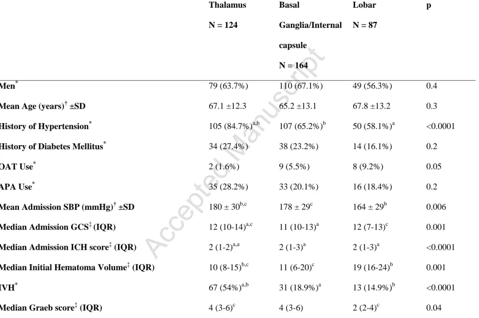
Among 716 ICH patients included in the registry, there were 375 (52.4%) cases of supratentorial ICH with volumes <30 mL. The mean age for this group was 67.1 ±12.3 years and 63.7% were men. Table 1 compares the baseline characteristics of the study cohort according to specific hematoma location (Supplementary Table I compares deep [thalamus plus basal ganglia/internal capsule] versus lobar). Overall, compared with other ICH locations, patients with thalamic ICH presented with higher mean systolic blood pressure, had significantly higher prevalence of hypertension, and a higher rate of IVH. Furthermore, thalamic ICH cases with IVH had higher median Graeb scores than lobar and basal ganglia/internal capsule hematomas with
intraventricular extension. However, median initial hematoma volume was lowest in patients with thalamic ICH and highest in the lobar group. Thalamic ICH patients also presented with higher median GCS score than the rest of the cohort (basal ganglia/internal capsule patients had
the lowest). Lobar ICH patients were more likely to be on oral anticoagulants than the remainder of the cohort, but the proportion of patients with INR >1.4 in each group was not different.
Clinical Course
Hematoma expansion and END rates for the total cohort were 19.2% (9.2% had missing data) and 7.5%, respectively. The rates and measure of expansion and END did not differ between the three locations (Table 2) or deep versus lobar (Supplementary Table II). After adjusting for age, gender, baseline ICH volume (log transformed), admission GCS, IVH, and initial INR >1.4, hematoma expansion was strongly associated with END (OR: 28.7, 95% CI: 8.51 – 96.5;
p<0.0001). Within each volume tertile, the rates of ICH growth were not different when stratified according to specific hematoma location (Table 3). However, in the lobar and basal
ganglia/internal capsule groups, ICH volume categories 1–9 mL showed significantly higher rates of expansion than volumes >9 mL (Table 3). No significant difference between volume tertiles was noted in the rates of thalamic hematoma expansion.
Outcomes
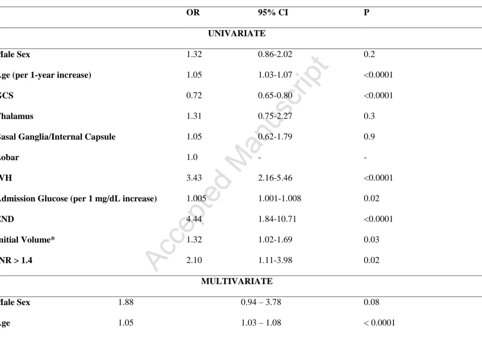
Fifty-two patients (13.9%) died in the hospital, and 109 (29.1%) had a severe disability (GOS scores, 3 and 2) at 30 days. The overall rate of poor outcome (GOS ≤3) was 42.9%. We found no difference between the three ICH location in the rates of in-hospital death, severe disability at 30 days, or poor outcome (Table 2). ICH location was also not a predictor of outcome in either univariate or multivariate analysis. Specifically, there was no difference in the rates of poor outcome between lobar versus deep locations (40.2% versus 43.8%; p = 0.56) (Supplementary Table III). In the univariate model, age, admission GCS, initial volume, IVH, END, and INR
>1.4 were associated with poor outcome (Table 4). Initial serum glucose had a modest but statistically significant predictive power. However, in multiple logistic regression analysis, only age (OR: 1.04, 95% CI: 1.03-1.08; p<0.0001), admission GCS (OR: 0.68, 95% CI: 0.59-0.79; p<0.0001), IVH (OR: 2.63, 95% CI: 1.28-4.42; p=0.008), and END (OR: 8.1, 95% CI: 2.55-25.78; p<0.0001) remained associated with the poor outcome (Table 4).
DISCUSSION
This study demonstrated that supratentorial hematomas that are <30 mL in volume comprise a large proportion of patients with ICH. Within this category, 42.9% died in the hospital or were left with severe disability at 30 days. Although this figure may be lower than the overall estimated rate of poor outcome for ICH, it highlights the fact that even smaller-volume
supratentorial hematomas can expand and have devastating outcomes. A recent study looking at 315 ICHpatients stratified by ICH volume showed that the rates of >33% expansion in volume categories of 3–10 mL and 10–20 mL were 26% and 32.9%, respectively [10]. At 90 days, in patients with ICH volumes 3–20 mL, the rate of poor outcome (modified Rankin Scale score ≥4) was 41.9%, which is very close to our number. In our study, expansion was independently associated with END and ultimately, poor outcome. Moreover, we found that known
determinants of mortality and poor outcome (age, initial GCS score, ICH volume, and IVH) appropriately apply to this subset of ICH patients [2,3].
One of the earliest studies looking at the significance of ICH volume in predicting outcomes showed that the overall 30-day mortality rates for ICH volumes <30 mL was 23% for deep and 7% for lobar hemorrhages [4]. A combination of hematoma volume and the initial GCS score
was a strong predictor of 30-day morbidity and mortality. The probabilities of death by 30 days with ICH volume <30 mL and initial GCS score ≥9 and ≤8 were 19% and 44%, respectively [4]. Our study, too, showed that GCS score on presentation was a strong predictor of outcome. While prior studies have shown initial ICH volume as an independent determinant of GCS score on presentation, we found low GCS to be a powerful predictor of poor outcome even in patients with smaller-volume hematomas [11].
Another important finding in our study was that in hematomas <30 mL, specific ICH locations were not major determinants of mortality or outcome. Lobar hemorrhages were, on average, larger in volume in our study, which is consistent with prior reports [12]. However, the prognostication capacity of deep localisation may be volume-dependent, consummating at volumes above 30 mL, where deep hematomas have worse outcomes than lobar hemorrhages but not for volumes below 30 mL [13]. In addition, the mortality rate in patients with thalamic hemorrhages was similar to that of patient with basal ganglia/internal capsule hematomas,
contrary to previous investigations, suggesting that the prognostication capacity of different deep localisation is prevalently volume-dependent [14-16].
Surgical evacuation of deeply situated hematomas with volumes <30 mL, even with minimally invasive techniques, does not seem to carry a substantial benefit, regardless of the initial GCS score [17,18]. In a study of 400 patients with spontaneous putaminal and thalamic hemorrhages who underwent conservative treatment versus surgical evacuation (including endoscopic and stereotactic aspiration), mortality rates were lower for the conservative management group, compared with surgical treatment when the GCS score was 3 to 12 and ICH volume <30 mL
[18]. Currently, minimally invasive hematoma evacuation strategies under investigation only include patients with ICH volume ≥30 mL, consequently leaving a treatment gap for hematomas with smaller volume [19]. This subgroup of ICH patients may therefore, be suitable candidates for hemostatic therapy, which requires examination in a randomized trial.
Our study had several important limitations. There was probably no uniformity among the patients in terms of door-to-CT times. Also, information on onset-to-CT time was not available on many patients and as a result, a figure representative of all ICH cases could not be stipulated. These factors may have interfered with estimating the rates of expansion since delayed imaging may have diagnosed ICH at a time when the hematoma had reached maximal expansion. Nonetheless, this is probably a more accurate representative of real-world stroke care. For
expansion analysis, we only included data on patients who had follow-up CT, resulting in a 9.2% attrition. This may have also lead to preclusion from analyses of a number of patients who had ICH expansion, but without clinical sequelae, thus eliminating the necessity for repeat imaging.
In conclusion, we demonstrated that more than one-third of patients with supratentorial ICH <30 mL in volume die or are severely disabled by 30 days. One in every five supratentorial
hematomas of this size category expand. Small thalamic and basal ganglia/internal capsule hemorrhages are a group of supratentorial ICH with similar clinical prognosis. Treatment of smaller-sized hematomas via interventional strategies, particularly hemostatic therapy, should be entertained as a potential focus area for future cerebrovascular research.
REFERENCES
1. Godoy DA, Pinero G, Di Napoli M. Predicting Mortality in Spontaneous Intracerebral Hemorrhage. Can Modification to Original Score Improve the Prediction? Stroke. 2006;37:1038-1044.
2. Hemphill JC, 3rd, Bonovich DC, Besmertis L, Manley GT, Johnston SC. The ICH score: a simple, reliable grading scale for intracerebral hemorrhage. Stroke. 2001;32:891–897
3. Hemphill JC 3rd, Farrant M, Neill TA Jr. Prospective validation of the ICH Score for 12-month functional outcome. Neurology. 2009;73:1088-1094.
4. Broderick JP, Brott TG, Duldner JE, Tomsick T, Huster G. Volume of intracerebral
hemorrhage. A powerful and easy-to-use predictor of 30-day mortality. Stroke.1993;24: 987-993.
5. Davis SM, Broderick J, Hennerici M, et al. Hematoma growth is a determinant of mortality and poor outcome after intracerebral hemorrhage. Neurology. 2006;66:1175–1181.
6. Behrouz R, Azarpazhooh MR, Godoy DA, et al; MNEMONICH Steering Committee. Int J Stroke. 2015;10:E86.
7. Di Napoli M, Zha AM, Godoy DA, et al; MNEMONICH Registry. Prior Cannabis Use Is Associated with Outcome after Intracerebral Hemorrhage. Cerebrovasc Dis. 2016;41:248-255.
8. Kothari RU, Brott T, Broderick JP, et al. The ABCs of measuring intracerebral hemorrhage volumes. Stroke. 1996; 27:1304-1305
9. Di Napoli M, Parry-Jones AR, Smith CJ, et al. C-reactive protein predicts hematoma growth in intracerebral hemorrhage. Stroke. 2014;45:59-65.
10. Dowlatshahi D, Yogendrakumar V, Aviv RI, et al; PREDICT/Sunnybrook ICH CTA study group. Small intracerebral hemorrhages have a low spot sign prevalence and are less likely to expand. Int J Stroke. 2016; 11:191-197.
11. Zahuranec DB, Gonzales NR, Brown DL, et al. Presentation of intracerebral haemorrhage in a community. J Neurol Neurosurg Psychiatry. 2006;77:340-344.
12. Falcone GJ, Biffi A, Brouwers HB, et al. Predictors of hematoma volume in deep and lobar supratentorial intracerebral hemorrhage. JAMA Neurol. 2013;70:988-994.
13. Castellanos M, Leira R, Tejada J, Gil-Peralta A, Dávalos A, Castillo J; Stroke Project, Cerebrovascular Diseases Group of the Spanish Neurological Society. Predictors of good outcome in medium to large spontaneous supratentorial intracerebral haemorrhages. J Neurol Neurosurg Psychiatry.2005;76:691-695.
14. Arboix A, Martínez-Rebollar M, Oliveres M, García-Eroles L, Massons J, Targa C. Acute isolated capsular stroke: a clinical study of 148 cases. Clin Neurol Neurosurg 2005, 107:88-94.
15. Miyai I, Suzuki T, Kang J, Volpe BT. Functional outcome in patients with hemorrhagic stroke in putamen and thalamus compared with those with stroke restricted to the putamen or thalamus. Stroke 2000, 31:1365-1369.
16. Arboix A, Comes E, García Eroles L, Massons J, Oliveres M, Balcells M, Targa C. Site of bleeding and early outcome in primary intracerebral hemorrhage. Acta Neurol Scand 2002, 105:282-288.
17. Zhou X, Chen J, Li Q, et al. Minimally invasive surgery for spontaneous supratentorial intracerebral hemorrhage: a meta-analysis of randomized controlled trials. Stroke. 2012;43:2923-2930.
18. Cho DY, Chen CC, Lee HC, Lee WY, Lin HL. Glasgow Coma Scale and hematoma volume as criteria for treatment of putaminal and thalamic intracerebral hemorrhage. Surg
19. Minimally Invasive Surgery plus Rt-PA for ICH Evacuation Phase III (MISTIE III). Available at: https://clinicaltrials.gov/ct2/show/NCT01827046. Accessed: 11 May 2016
Table 1 - Baseline characteristics stratified by location Thalamus N = 124 Basal Ganglia/Internal capsule N = 164 Lobar N = 87 p Men* 79 (63.7%) 110 (67.1%) 49 (56.3%) 0.4
Mean Age (years)† ±SD 67.1 ±12.3 65.2 ±13.1 67.8 ±13.2 0.3
History of Hypertension* 105 (84.7%)a,b 107 (65.2%)b 50 (58.1%)a <0.0001
History of Diabetes Mellitus* 34 (27.4%) 38 (23.2%) 14 (16.1%) 0.2
OAT Use* 2 (1.6%) 9 (5.5%) 8 (9.2%) 0.05
APA Use* 35 (28.2%) 33 (20.1%) 16 (18.4%) 0.2
Mean Admission SBP (mmHg)† ±SD 180 ± 30b,c 178 ± 29c 164 ± 29b 0.006
Median Admission GCS‡ (IQR) 12 (10-14)a,c 11 (10-13)a 12 (7-13)c 0.001
Median Admission ICH score‡ (IQR) 2 (1-2)a,a 2 (1-3)a 2 (1-3)a <0.0001
Median Initial Hematoma Volume‡ (IQR) 10 (8-15)b,c 11 (6-20)c 19 (16-24)b 0.001
IVH* 67 (54%)a,b 31 (18.9%)a 13 (14.9%)b <0.0001
Median Admission INR‡ (IQR) 1.15 (1.10-1.22) 1.12 (1.10-1.18) 1.11 (1.0-1.6) 0.6
INR >1.4* 15 (12.1%) 14 (8.5%) 15 (17.2%) 0.1
Mean Admission Platelet Count (cell x 109/L)† ±SD 239 ±54 253 ±66 238 ±81 0.2
Mean Admission Serum Glucose (mg/dL)† ±SD 159 ±53c 140 ±53 132 ±64c 0.01
SD depicts standard deviation; OAT, oral anticoagulant therapy; APA, antiplatelet agent; IQR, interquartile range; IVH, intraventricular hemorrhage; INR, International Normalization Ratio.
*χ2
-test with Marascuillo's post hoc analysis following comparing multiple proportions. †
ANOVA test with Bonferroni corrections for multiplecomparisons.
‡
Kruskal-Wallis test with Dunn-Bonferroni post hoc method for multiplecomparisons.
Significant differences between subgroups are indicated in the table with APA-style formatting using subscript letters. ap<0.0001;
b
Table 2 - Outcomes stratified by location Thalamus N = 124 Basal Ganglia/Internal capsule N = 164 Lobar N = 87 p Number of Expansions 26 (24.8%) 32 (20.9%) 14 (17.3%) 0.5
Median Absolute Expansion (mL) (IQR) 0 (0-3) 2 (0-6.1) 0.5 (0-5) 0.7
Median Expansion (%) (IQR) 0 (0-20) 22.5 (0-70) 2.1 (0-50) 0.7
END 8 (6.5%) 13 (7.3%) 7 (8.0%) 0.9
In-Hospital Death 24 (19.4%) 17 (10.4%) 11 (12.6%) 0.09
Severe Disability at 30 Days 34 (27.4%) 52 (31.1%) 24 (27.6%) 0.3
Median Time from CT1 to CT2 (hours) (IQR) 20 (12-24) 22.5 (17-24) 18 (16-21) 0.4
Severe Disability and Death Composite (Poor Outcome) 58 (46.8%) 68 (42.2%) 35 (40.2%) 0.6
IQR, interquartile range; END, early neurological deterioration; CT1, initial head computed tomography; CT2; follow up head
Table 3 - Expansion rates based on hematoma location and volume Total N=339 Expansion N=72 Thalamus N=105 Basal Ganglia/Internal Capsule N= 153 Lobar N=81 p* 1 – 9 mL, n (%) 128 40 16 / 40 (40.0) 16 / 33c (48.5) 8 / 15c (53.3) 0.8 10 – 20 mL, n (%) 148 26 8 / 31 (25.8) 13 / 59 (22.0) 5 / 32 (15.6) 0.7 >20 mL, n (%) 63 6 2 / 8 (25.0) 3 / 29c (10.3) 1 / 20c (0.05) 0.4 p* 0.6 0.03 0.02 *χ2
-test with Marascuillo's post hoc analysis following comparing multiple proportions.
Significant differences between subgroups are indicated in the table with APA-style formatting using subscript letters. ap<0.0001;
b
Table 4. Univariate and Multiple Logistic Regression Analysis for Composite of Severe Disability and Death at 30 Days
OR 95% CI P
UNIVARIATE
Male Sex 1.32 0.86-2.02 0.2
Age (per 1-year increase) 1.05 1.03-1.07 <0.0001
GCS 0.72 0.65-0.80 <0.0001
Thalamus 1.31 0.75-2.27 0.3
Basal Ganglia/Internal Capsule 1.05 0.62-1.79 0.9
Lobar 1.0 - -
IVH 3.43 2.16-5.46 <0.0001
Admission Glucose (per 1 mg/dL increase) 1.005 1.001-1.008 0.02
END 4.44 1.84-10.71 <0.0001 Initial Volume* 1.32 1.02-1.69 0.03 INR > 1.4 2.10 1.11-3.98 0.02 MULTIVARIATE Male Sex 1.88 0.94 – 3.78 0.08 Age 1.05 1.03 – 1.08 < 0.0001
GCS 0.68 0.59 – 0.79 <0.0001 Thalamus 1.16 0.46-2.96 0.2 Basal Ganglia/Internal Capsule 1.41 0.62-3.23 0.4 Lobar 1.0 - - IVH 2.63 1.28 – 4.42 0.008 Admission Glucose 1.00 0.99 – 1.00 0.4 END 8.1 2.55 – 25.78 <0.0001 Initial Volume* 1.55 0.88 – 2.72 0.1 INR > 1.4 1.62 0.62 – 4.27 0.3
OR, odds ratio; CI, confidence interval; GCS, Glasgow Coma Scale; IVH, intraventricular hemorrhage; END, early neurological deterioration; INR, International Normalization Ratio.


