ARTICLE
Intracranial cavernous malformation in
children: a single-centered experience with
30 consecutive cases
Angiomas cavernosos intracranianos em crianças: experiência de um único centro em
30 casos consecutivos
Marcelo Campos Moraes Amato, João Flávio Gurjão Madureira, Ricardo Santos de Oliveira
Intracranial cavernous malformations (CMs) are com-mon vascular anomalies in the brain and have an incidence of 0.1 to 0.5% in the general population1.
These malformations represent 10 to 20% of all vascular lesions in the brain and can cause symptoms such as sei-zures, hemorrhages, headache, and focal neurological defi-cits2,3. The lesions are characterized by a “blackberry-like”
aggregation of grossly enlarged capillary cavities consist-ing of a sconsist-ingle layer of endothelium, without intervenconsist-ing neuronal tissue4. Since the introduction of sensitive diag-nostic tools like the magnetic resonance imaging (MRI), CMs have become increasingly reported in the pertinent literature because asymptomatic lesions have also become detectable2,5.
Division of Pediatric Neurosurgery of the Department of Surgery and Anatomy, University Hospital of Ribeirão Preto Medical School, University of São Paulo, Ribeirão Preto SP, Brazil.
Correspondence: Marcelo Campos Moraes Amato; Avenida Juriti 144; 04520-000 São Paulo SP - Brasil; E-mail: [email protected] Conflict of interest: There is no conflict of interest to declare.
Received 08 May 2012; Received in final form 19 October 2012; Accepted 26 October 2012. ABSTRACT
Objectives: To determine the clinical presentation and treatment outcome of pediatric intracranial cavernous malformation (CM) in a single-centered institution. Methods: Clinical data review of 30 patients under 18 years-old who had undergone surgery for cavernous malformation from January 1993 to December 2011. Results: The Study Group included 18 males and 12 females (mean age: 8.7 years-old). Symptoms at presentation were seizures (16/30, 53.3%), headache (15/30, 50.0%), and focal neurological deficits (11/30, 36.6%). Multiple cavern-ous malformations were found in 5/30 (16.6%). According to location, patients were classified in groups: (G1) brain-steam in 5/30 (16.6%), (G2) cerebellum in 2/30 (6.6%), (G3) supratentorial associated with seizures in 16/30 (53.3%), and (G4) supratentorial without seizures in 7/30 (23.3%). Surgical resection was performed in 26 out of 30 (86.6%) patients. The mean follow-up period was 4.1 years. Of 15 children followed-up with preoperative seizures, all were rendered seizure-free after surgery. Conclusions: For symptomatic solitary cavernous malformation, the treatment of choice is complete microsurgical excision preceded by careful anatomical and functional evaluation. For multiple cavernous malformation or asymptomatic patients, the treatment modalities must be cautiously considered.
Key words: hemangioma, cavernous, seizure, brain tumor.
RESUMO
Objetivos: Determinar a apresentação clínica e o acompanhamento do tratamento em crianças com angioma cavernoso intracraniano numa única instituição. Métodos: Revisão de dados clínicos de 30 pacientes menores de 18 anos com que passaram por uma cirurgia de angioma cavernoso intracraniano entre janeiro de 1993 a dezembro de 2011. Resultados: O grupo de estudo incluiu 18 sujeitos masculinos e 12 fe-mininos (idade média: 8,7 anos). Os sintomas iniciais eram convulsões (16/30, 53,3%), cefaleia (15/30, 50,0%) e déficits neurológicos focais (11/30, 36,6%). Havia angiomas cavernosos intracranianos múltiplos em 5 de 30 (16.6%). A classificação foi feita em grupos de acordo com a localização: (G1) tronco cerebral em 5/30 (16,6%); (G2) cerebelo em 2/30 (6,6%); (G3) supratentoriais associados a convulsões em 16/30 (53,3%) e (G4) supratentoriais sem convulsões em 7/30 (23,3%). Ressecção cirúrgica foi realizada em 26 de 30 (86,6%) pacientes, com se-guimento médio de 4,1 anos. De 15 crianças com convulsões pré-operatórias, todas ficaram livres das crises após a cirurgia. Conclusões:
Para angioma cavernoso intracraniano solitário e sintomático, o tratamento de escolha é excisão microcirúrgica total precedida de avaliação funcional e anatômica meticulosa. Para angiomas cavernosos intracranianos múltiplos ou pacientes assintomáticos, as modalidades tera-pêuticas devem ser consideradas cautelosamente.
Palavras-Chave: angioma cavernoso, convulsão, tumor cerebral.
Pediatric CMs may have characteristics that difer from those of adults in clinical presentation and treat-ment, such as higher rates of hemorrhage6-10 and larger dimensions9. Even though there are not many reports about exclusive pediatric CMs, only a few large series have yet been reported, and ideal group classiication has not yet been deined.
his report is an attempt to evaluate clinical and surgical data in a single-centered pediatric series of CMs in the con-text of published literature. Special emphasis was placed on management of diferent groups and on outcome of patients sufering from seizure prior to surgery.
METHODS
Patient population
We reviewed files of patients younger than 18 years harboring a CM, over an 18-year period at the Divi- sion of Pediatric Neurosurgery, University Hospital of Ribeirão Preto Medical School, from January 1993 to December 2011. Thirty CM cases were identified and en-rolled in this study.
he variables analyzed included age, sex, clinical pre-sentation, radiological features, extent of resection, and histopathology.
All patients had all been preoperatively evaluated by structural brain imaging with computed tomography (CT) and/or MRI. Shortly after surgical excision, all patients were put on regular follow-up.
he subjects were classiied according to the main lesion location: brain-steam (G1), cerebellar (G2), supratentorial as-sociated with seizures (G3), and supratentorial not associat-ed with seizures (G4).
Postoperative seizure outcome was classiied according to Engel’s classiication11: class I, free of disabling seizures; II, rare disabling seizures; III, worthwhile improvement; and IV, no worthwhile improvement.
A PubMed database search was also performed in or-der to retrieve articles published in the last 20 years, which combined the subject headings: cavernoma; cavernous angioma; pediatric; intracranial; and children. Papers with less than ten patients were excluded as well as series with patients above 20 years-old. Only articles in the En-glish language containing all types of clinical manifesta-tion of the CMs were considered, those concerning only seizures, brainstem, radiation-induced cavernomas or re-stricted types of cavernomas’ classification were excluded from this review.
his study was approved by the Research Ethics Committee of the University Hospital of Ribeirão Preto Medical School, at University of São Paulo (Protocol 6591/2007).
RESULTS
Clinical and surgical data
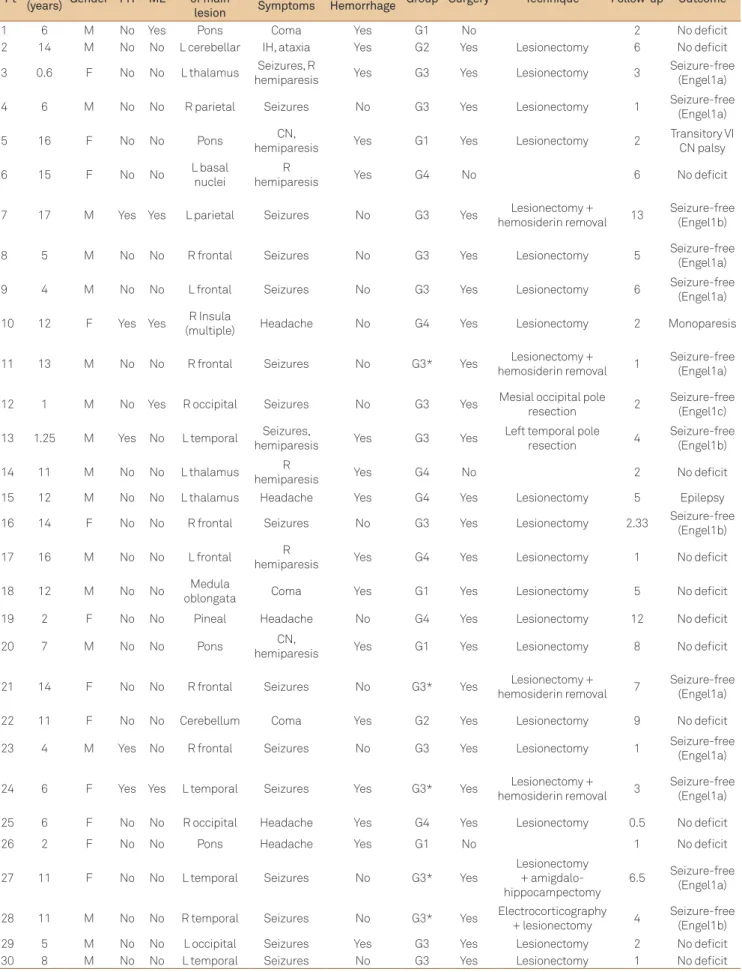
he Study Group included 18 males and 12 females with a male-female ratio of 1.5:1. he mean age at onset ranged from 6 months to 17 years-old (mean 8.7 years-old). Patient’s age and gender, CM location, clinical aspects, and outcome are summarized in Table 1.
he most common presenting symptoms were seizures in 16/30 (53.3%), headaches in 15/30 (50%), followed by fo-cal neurologifo-cal deicits in 11/30 (36.6%), and behavior dis-turbance in 1/30 (3.3%). Acute intracranial hemorrhage was presented in 16/30 (53.3%).
Single CMs were found in 25/30 (83%), while ive pa-tients (17%) had multiple ones. Regarding only symptomat-ic lesions, 23 (76.6%) supratentorial and 7 (23.3%) infraten-torial, location was as follows: frontal lobe (7/30, 23.3%), temporal lobe (6/30, 20.0%), occipital lobe (3/30, 10.0%), pa-rietal lobe (1/30, 3.3%), insula (1, 3.3%), thalamus and basal nuclei (4/30, 13.3%), brainstem (5/30, 16.6%), and cerebellar hemisphere (2/30, 6.6%).
A total of 26 CMs undergone surgery and four patients were managed conservatively. here were no postoperative deaths or signiicant complications in this series. No child was asymptomatic prior to the resection.
In the G1 (5/30), three patients were submitted to sur-gery (Fig 1). Two patients that showed spontaneous total improvement did not undergone surgery. In the G2 (2/30), microsurgical resection was performed after cerebellar hem-orrhage in both patients (Fig 2).
In G3 (16/30), 11 patients underwent simple lesionecto-my without further neurophisiological evaluation. Five pa-tients had long-lasting symptoms (more than one year) or high frequency of seizures and were therefore deeply inves-tigated and treated (Table 1). In cases of epilepsy, the sur-rounding hemosiderin-stained tissue had also been removed. Of 15 children followed-up with preoperative seizures, all of them were rendered seizure-free (Engel’s class 1) after the CM removal.
Seven patients composed G4. Five cases among them were submitted to surgery (Fig 3), and two were managed conservatively (thalamic and basal ganglia CM).
Outcome/follow-up
he mean follow-up period was 4.1 years (range 6 months to 13 years). he overall post-treatment results were positive. Major morbidity and mortality from surgical procedures were absent. One patient (case 10) showed a permanent monopa-resis after surgery.
Pt Age
(years) Gender FH ML
Localization of main
lesion
Main Symptoms
Clinical
Hemorrhage Group Surgery Technique Follow-up Outcome
1 6 M No Yes Pons Coma Yes G1 No 2 No deficit 2 14 M No No L cerebellar IH, ataxia Yes G2 Yes Lesionectomy 6 No deficit 3 0.6 F No No L thalamus Seizures, R
hemiparesis Yes G3 Yes Lesionectomy 3
Seizure-free (Engel1a) 4 6 M No No R parietal Seizures No G3 Yes Lesionectomy 1 Seizure-free
(Engel1a) 5 16 F No No Pons CN,
hemiparesis Yes G1 Yes Lesionectomy 2
Transitory VI CN palsy 6 15 F No No L basal
nuclei
R
hemiparesis Yes G4 No 6 No deficit 7 17 M Yes Yes L parietal Seizures No G3 Yes Lesionectomy +
hemosiderin removal 13
Seizure-free (Engel1b) 8 5 M No No R frontal Seizures No G3 Yes Lesionectomy 5 Seizure-free
(Engel1a) 9 4 M No No L frontal Seizures No G3 Yes Lesionectomy 6 Seizure-free
(Engel1a) 10 12 F Yes Yes R Insula
(multiple) Headache No G4 Yes Lesionectomy 2 Monoparesis 11 13 M No No R frontal Seizures No G3* Yes Lesionectomy +
hemosiderin removal 1
Seizure-free (Engel1a) 12 1 M No Yes R occipital Seizures No G3 Yes Mesial occipital pole
resection 2
Seizure-free (Engel1c) 13 1.25 M Yes No L temporal Seizures,
hemiparesis Yes G3 Yes
Left temporal pole resection 4
Seizure-free (Engel1b) 14 11 M No No L thalamus R
hemiparesis Yes G4 No 2 No deficit 15 12 M No No L thalamus Headache Yes G4 Yes Lesionectomy 5 Epilepsy 16 14 F No No R frontal Seizures No G3 Yes Lesionectomy 2.33 Seizure-free
(Engel1b) 17 16 M No No L frontal R
hemiparesis Yes G4 Yes Lesionectomy 1 No deficit 18 12 M No No Medula
oblongata Coma Yes G1 Yes Lesionectomy 5 No deficit 19 2 F No No Pineal Headache No G4 Yes Lesionectomy 12 No deficit 20 7 M No No Pons CN,
hemiparesis Yes G1 Yes Lesionectomy 8 No deficit 21 14 F No No R frontal Seizures No G3* Yes Lesionectomy +
hemosiderin removal 7
Seizure-free (Engel1a) 22 11 F No No Cerebellum Coma Yes G2 Yes Lesionectomy 9 No deficit 23 4 M Yes No R frontal Seizures No G3 Yes Lesionectomy 1 Seizure-free
(Engel1a) 24 6 F Yes Yes L temporal Seizures Yes G3* Yes Lesionectomy +
hemosiderin removal 3
Seizure-free (Engel1a) 25 6 F No No R occipital Headache Yes G4 Yes Lesionectomy 0.5 No deficit 26 2 F No No Pons Headache Yes G1 No 1 No deficit 27 11 F No No L temporal Seizures No G3* Yes
Lesionectomy + amigdalo-hippocampectomy
6.5 Seizure-free (Engel1a) 28 11 M No No R temporal Seizures No G3* Yes Electrocorticography
+ lesionectomy 4
Seizure-free (Engel1b) 29 5 M No No L occipital Seizures Yes G3 Yes Lesionectomy 2 No deficit 30 8 M No No L temporal Seizures No G3 Yes Lesionectomy 1 No deficit
Among the 15 patients with seizures, eight are now sei-zure — and drug-free; in seven patients, however, antiepilep-tic medications were not discontinued, although these chil-dren also scored Engel’s class 1.
Four patients that were not operated improved sponta-neously and remained asymptomatic in the follow-up.
During the follow-up period, no new lesions or recur-rence on MRI images were seen in all children except for a boy (case 15) with a CM in the thalamus that had to be re-oper-ated. He presented seizures due to severe hyponatremy after surgery and remained with antiepileptic drugs afterwards. he remaining lesions were stable in children with multiple CMs.
DISCUSSION
CM is one of the congenital vascular malformations af-fecting the central nervous system (CNS) that may remain asymptomatic for a long time or may produce clinical manifes-tations12. he pathogenesis of CM remains unclear, and little is known regarding the molecular pathogenesis of the disease3.
here are two types of CMs: sporadic and familial. he fa-milial type is characterized by a multiplicity of lesions13-15 and the possibility of de novo CM formation16. In this series, ive patients (16.6%) had multiple lesions, three of them had also positive familial history. One subject with familial history (total of 4–13.3%) did not have multiple lesions. Among the literature review, incidence of familial history and multiple lesions both varies from 0.0 to 26.3%2,3,6-10,17-19.
Table 2 summarizes previous studies on pediatric CM. Note that only ten series were according to the selection cri-teria and therefore included in this review2,3,6-10,12,17,19.
In our series, the mean age at clinical manifestation was 8.7 years-old, which is similar to the literature. he ratio of male to female was 1.5:1. In addition to the present study, there seems to be a slightly predominance of boys in most an-alyzed studies (Table 2). Since these are patient-based stud-ies, the deinitive predominance is hard to deine.
he majority of CMs come to medical attention after chil-dren experience any number of clinical symptoms, and up to 20% are discovered incidentally19-21. In pediatric patients, CM symptoms in the CNS are mainly present as hemorrhage, sei-zure, or focal neurological deicits3. Xia et al. also observed acute intracranial hemorrhage in 20% of children with CMs, according to CT and MRI studies. However, a higher pro-portion of hemorrhage was conirmed histopathologically in 52.5% (32/61) of operated children with CMs in their se-ries19. Acute intracranial hemorrhage was observed in 53.3% (16/30) in our series.
In the current study, 56.6% of patients sufered from seizures, which is signiicantly higher than in earlier stud-ies2,6-8,10,17,19, although Acciarri et al.9 reported 70% of seizures in a series of 42 children with CMs.
Presentation of symptoms has some correlation with lesion locations19. Patients with lesions located supericial-ly in the cerebral hemispheres more frequentsupericial-ly are pres-ent with seizures. On the other hand, patipres-ents with lesions
Fig 1. Pre (A) and postoperative (B) images of a brainstem cavernous malformation. This patient presented with tetraparesis and 11 points at the Glasgow Coma Scale. Three years after surgery she was asymptomatic. (A): axial T1-weighted magnetic resonance imaging revealed a typical cavernous malformation, characterized by heterogenic mass and hypointense halo signal around it; (B): sagittal T1-weighted gadolinium-enhanced magnetic resonance imaging revealed total removal of the cavernous angioma and decompression of brainstem (Patient 5).
A
B
Fig 3. Axial T2-weighted magnetic resonance imaging (A) revealed a right occipital lesion with a hypointense halo around it, and other lesions can also be seen (arrows). The gradient-echo sequence (B) confirmed multiple cavernous malformations (Patient 12).
A
B
Fig 2. (A) brain computed tomography showed a hemorrhagic lesion on the left cerebellum hemisphere and incipient dilatation of ventricular temporal horns. Axial T1 (B) and T2-weighted (C) magnetic resonance imaging revealed heterogenic lesion with mass effect and fourth ventricle compression. Histopathology confirmed the diagnosis of a cavernous angioma (Patient 2).
n: number of patients; ML: multiple lesions; FH: family history; NA: not available; IH: intracranial hypertension; RS: radiosurgery; >: at least. Data were taken from the text and tables presented in the articles. Symptoms and post-surgical results excluded sometimes the spinal cord cavernomas and/or the patients without enough follow-up, correct amounts of patients are shown.
R ef er ência n A ge Mal e: F emal e FH ML Sit e Main symp toms at diagnosis P os t-sur gical results Supr a:infr a-tent orial
Thalamus and Basal Gnaglia Br
ains
tem
Cer
ebellum Spine Seizur
es Headac he Focal neur ol ogical de ficits none Hemor rhag e Sur gery Foll ow-up Impr ov ed Unc hang ed W or sened Mor tality
6 17 18 m to 16 y 1.12:1 4/17
(23.5%) 2/17
(11.7%) 15:2 2
2/17
(11.7%) 0 0
4
(23.5%) NA NA 0
12 (70.5%) 15 (88.2%) NA 11/15 (73.3%) 1/15 (6.6%) 3/15 (20%) 0
14 19 (mean=9,1)7 m to 17 y 1.11:1 (26.3%)5/19 (26.3%)5/19 12:6 2 (21%)4/19 (10.5%)2/19 1 (27.7%)5/18 (38.8%)7/18 (72.2%)13/18 0 (66.6%)12/18 all 6 m to 9 y (89.4%)17/19 0 (10.5%)2/19 0
18 18 10 m to 17 y 1:1.25 0 0 15:2 NA 0 2/18
(11.1%) 1 11/17 (64.7%) 1/17 (5.8%) 5/17
(29.4%) 0 NA all 1 to 16 y
14/17 (82.3%)
3/17
(17.6%) 0 0
7 24 (mean=8 y)6 m to 15 y NA 0 (8.3%)2/24 20:4 NA NA NA 0 (54.1%)13 NA (9 IH)(41.6%)10 0 (79.1%)19 all (mean=4 y)6 m to 14 y (95.8%)23/24 0 0 (4.1%)1/24
8 36 (mean=8.6)9 m to 17 y 1.4:1 (11.1%)4/36 NA 23:12 6 (19.4%)7/36 (13.8%)5/36 1 (45.7%)16/35 (2.8%)1/35 (28.5%)10/35 (13.8%)5/36 (52.7%)19/36 all NA (85.7%)30/35 (8.5%)3/35 (5.7%)2/35 0
3 33 1 to 20 y
(mean=11.6 y) 1.2:1 0 0 27:6 3
5/33 (15.1%)
1/33
(3%) 0
19
(57.6%) NA NA 0
25 (75.7%)
25 and RS on 8
2 to 17 y (mean=5.8 y) 29/33 (87.9%) 4/33 (12.1%) 0 1/33 (3%)
9 42 10 m to 17 y 1:1 (2.3%)1/42 (11.9%)5/42 35:5 0 (4.7%)2/42 (7.1%)3/42 2 28/40 (70%) (27.5%)11/40 16/40 (40%) 0 (42.5%)17/40 all 1 to 16 y 29/42 (69%) (23.8%)10/42 (7.14%)3/42 0
19 6615 m to 17.8 y
(mean=11.6 y)1.53:1
1/66 (1.5%) 7/66 (10.6%) 55:6 (both:4) 1 2/66 (3%) 4/66 (6%) 1 31/65 (47,7%) 30/65 (46,2%) 8/65 (12,3%) 2/65 (3,1%) 13/65 (20%) 62/66 (93.9%)
5 m to 9.3 y (mean=3.2 y) 43/46 (93.5%) 1/46 (2.2%) 2/46 (4.3%) 0
10 32 (mean=7.1)2 d to 17 y 1.13:1 (9.3%)3/32 (25%)8/32 24:08 1 (18.7%)6/32 (6.2%)2/32 0 (40.6%)13 (18.8%)6 7 (21.8%) 0 (65.6%)21 (87.5%)28 mean=4.43 y(96.4%)27/28 (3.5%)1/28 0 0
2 79 (mean=9.7 y)4 m to 17 y 1.07:1 (1.2%)1/79 (3.8%)3/79 >74:NA NA NA NA 0 41 (51%) 8 (10%) 14 (18%) 0 18 (23%) all (mean=3 y)1 m to 16 y 39/54 (72%) (22.2%) 12/54 (1.8%)1/54 0
Table 2. Summary of literature review.
located in deep structures such as the brainstem generally present with focal neurological deicits. In this series, fo-cal neurologifo-cal deicits (36.6%, 11/30) happened mostly in deep-seated and infratentorial lesions. Only two cases with focal neurological deicits did not result from deep-seated lesions, an acute hemorrhage of a temporal (case 13) and a frontal CM (case 17).
CMs have been observed throughout the CNS, and their locations in children are comparable to those in adults8,19,22. Supratentorial location accounts for about 80.0%, while the other 20.0% are located in the posterior cerebral fossa3,8,19,22.
he proportion of supratentorial CMs in the descending or-der is frontal, temporal, occipital, and parietal lobe. A deep location in the basal ganglia, hypothalamus, or ventricular system is rare8,19,22. In this series, regarding only the sympto-matic lesions, 23 patients (76.6%) presented supratentorial CM and seven (23.3%) infratentorial.
Management
G1: brain-steam; G2: cerebellum; MRI: magnetic resonance imaging.
without prior hemorrhage4. However, without a docu-mented episode of hemorrhage, treatment should be based on the precise location of the cavernoma, patient’s age, symptoms, and outcome expectation23,24.
Additional data of natural history also favor surgical treatment. Seizures seem to happen precociously when CMs are located in the frontal or temporal lobes25, and apparently their risk of development varies from 1.5 to 4.8% per year4,21. Focal deicits and intracranial hypertension may be caused not only by acute macro-hemorrhage, but also due to pro-gressive growing secondary to recurrent micro-hemorrhage24. Figs 4 and 5 summarize management strategies for each following group.
Brain steam cavernomas (G1)
Brainstem CMs constitute 0 to 21% of all cavernomas in different pediatric series3,6,8-10,17-19. Clinical presenta-tion is generally acute and severe due to peculiar local-ization, despite frequent small amount of hemorrhage. Neurological improvement often happens a few days later. This apparently benign course is due to slow flow
and pressure inside the CMs, consequently hemorrhage usually displaces the neural tissue instead of its destruc-tion7. Two of our patients were not submitted to surgery and improved totally spontaneously.
Surgical resection is indicated for those brainstem le-sions that appear in the pial surface23. Asymptomatic patients or with good neurological recover should be observed, mainly if the CMs are located deep-ly in the brain steam4,17. Some authors believe that the risk of not operating on a patient that had al-ready suffered hemorrhage is very high, especially if he/she is young with high life expectancy, independent of the lesions depth, because of the possible catastrophic evaluation if it rebleeds or grows3,26. Neurosurgical team experience and skills, as well as neurophysiologic moni-toring, should be considered to treat such patients19,23.
Cerebellar cavernomas (G2)
Cerebellar location is less frequent, and clinical presen-tation is generally due to great hemorrhage inside the cer-ebellar parenchyma that may cause mass efect and occlude
Infratentorial Cavernomas
(G1 and G2)
Brain-steam cavernoma
Symptomatic
Next to the 4. ventricle or
cistems
Next to the 4. ventricle or
cistems Far from the 4.
ventricle or cistems
Far from the 4. ventricle or
cistems One
bleeding
Multiple bleedings
Next to the 4. ventricle or
cistems
Far from the 4. ventricle or
cistems
Progressive neurologic deterioration
clinical stability
No bleeding
Acidental asymptomatic
finding
Bleeding at the MRI
Cerebellar
Cavernoma SURGERY
SURGERY SURGERY SURGERY
Conservative treatment
Conservative treatment
Conservative treatment
Conservative treatment Young
patient
G teto o tG teto A vernous angiomas; MRI: magnetic resonance imaging.
the fourth ventricle resulting in cerebellar dysfunction and hydrocephalus27.
Patients with cerebellar cavernoma (G2) should be treated with surgery even in the absence of hemorrhage. Surgery is usually safe and successful while a irst or re-current hemorrhage in the posterior fossa represents great threat. Exception is made for multiple asymptomatic cer-ebellar CMs4. Cerebellar cavernomas represent 0.0 to 13.8% in earlier studies3,6,8-10,17-19, in our series there were two pa-tients in this group (6.6%).
Supratentorial cavernomas associated with seizure (G3)
Surgical treatment should be indicated to the majority of patients that sufered hemorrhage and to those which surgical risk was considered low4,9.
Lesionectomy alone showed favorable results28. In a se-ries of patients submitted to lesionectomy of supratentori-al CMs, a strict relationship was found between the dura-tion of seizure history before the surgical treatment and the cure of epilepsy with the surgery. Cohen et al.28 showed that all patients that presented one preoperative seizure or less
Supratentorial Cavernomas
(G3 and G4)
Associated with seizure
Eloquent area
Non-eloquent area
Focal deficit
Headache
Thalamus or basal
nuclei
Controversy Eloquent area
Surgery Surgery
Conservative Conservative treatment Conservative treatment Lesionectomy
Lesionectomy
Extended resection
Electrocorticography guided resection
New Symptom, recurrent or hemorrhage at MRI
Consider as the treatment of brain-steam CAs
<1 year of seizures <5 seizures
>1 year of seizures >5 seizures
Fig 5. Therapeutic guideline for G3 and G4.
than two months of history did not have any seizures after surgery; 75.0 to 80.0% of patients with two to ive preopera-tive seizures or 2 to 12 months of history also had not any seizure after surgery. In addition, 50% of patients with more than ive seizures or more than 12 months of history were postoperatively seizure free28. Although resection of the he-mosiderin stained tissue that surrounds the lesion was not proved to be of better efectiveness4,8-10,29, it was adopted by several studies when CMs are placed in noncritical areas2,3,9, including the present one. Eleven patients of this study un-derwent simple lesionectomy.
Longer seizure history and increased number of preo-perative seizures are consistent with more diiculty in treating epilepsy with simple lesionectomy surgery28. We believe these patients should have a complete epilepsy evaluation to a possible extended surgery guided by elec-trocorticography. In fact, ive patients with refractory sei-zures were further evaluated, and two of them needed an extended resection.
1. Del Curling O, Kelly DL, Elster AD, Craven TE. An analysis of the natural history of cavernous angiomas. J Neurosurg 1991;75:702-708.
2. Hugelshofer M, Acciarri N, Sure U, et al. Effective surgical treatment of
cerebral cavernous malformations: a multicenter study of 79 pediatric patients. J Neurosurg Pediatr 2011;8:522-525.
3. Lee JW, Kim DS, Shim KW, et al. Management of intracranial cavernous
malformation in pediatric patients. Childs Nerv Syst 2008;24:321-327.
4. Amin-Hanjani S, Ojemann RG, Ogilvy CS. Surgical management of
cavernous malformations of the nervous system. In: Schmidek HH (Ed). Operative neurosurgical techniques: indications, methods and results. Philadelphia: Elsevier, Inc; 2006. p. 1307-1324.
5. Faria MP, Fagundes-Pereyra WJ, Carvalho GT, Sousa AA. Cavernous
malformations: surgical management in Belo Horizonte Santa Casa Hospital. Arq Neuropsiquiatr 2004;62:1079-1084.
6. Mazza C, Scienza R, Beltramello A, Da Pian R. Cerebral cavernous
malformations (cavernomas) in the pediatric age-group. Childs Nerv Syst 1991;7:139-146.
7. Di Rocco C, Iannelli A, Tamburrini G. Surgical management of paediatric
cerebral cavernomas. J Neurosurg Sci 1997;41:343-347.
8. Mottolese C, Hermier M, Stan H, et al. Central nervous system
cavernomas in the pediatric age group. Neurosurg Rev 2001;24:55-71.
9. Acciarri N, Galassi E, Giulioni M, et al. Cavernous malformations of the
central nervous system in the pediatric age group. Pediatr Neurosurg 2009;45:81-104.
10. Consales A, Piatelli G, Ravegnani M, et al. Treatment and outcome of children with cerebral cavernomas: a survey on 32 patients. Neurol Sci 2010;31:117-123.
11. Engel JJ, Van Ness PC, Rasmussen TB, Ojemann LM. Outcome with respect to epileptic seizures. In: Engel JJ (Ed). Surgical treatment of the epilepsies. New York: Raven Press; 1993. p. 609-621.
12. Giulioni M, Acciarri N, Padovani R, Galassi E. Results of surgery in children with cerebral cavernous angiomas causing epilepsy. Br J Neurosurg 1995;9:135-141.
13. Rigamonti D, Spetzler RF. The association of venous and cavernous malformations. Report of four cases and discussion of the pathophysiological, diagnostic, and therapeutic implications. Acta Neurochir (Wien) 1988;92:100-105.
14. Scott RM. Brain stem cavernous angiomas in children. Pediatr Neurosurg 1990;16:281-286.
15. Zabramski JM, Wascher TM, Spetzler RF, et al. The natural history of familial cavernous malformations: results of an ongoing study. J Neurosurg 1994;80:422-432.
16. Tekkök IH, Ventureyra EC. De novo familial cavernous malformation presenting with hemorrhage 12.5 years after the initial hemorrhagic Ictus: natural history of an infantile form. Pediatr Neurosurg 1996;25:151-155.
17. Scott RM, Barnes P, Kupsky W, Adelman LS. Cavernous angiomas of the
central nervous system in children. J Neurosurg 1992;76:38-46. 18. Giulioni M, Acciarri N, Padovani R, Frank F, Galassi E, Gaist G. Surgical
management of cavernous angiomas in children. Surg Neurol 1994;42:194-199.
19. Xia C, Zhang R, Mao Y, Zhou L. Pediatric cavernous malformation in the central nervous system: report of 66 cases. Pediatr Neurosurg 2009;45:105-113.
20. Robinson JR, Awad IA, Little JR. Natural history of the cavernous angioma. J Neurosurg 1991;75:709-714.
References
Supratentorial cavernomas not associated with seizure (G4)
his group represented 23.3% of patients (7/30). he man-agement of thalamic and basal ganglia CMs is controversial4. In selected cases, surgery is considered when the CM is next to the ventricle surface or in the insular lobe through to the Sylvian issure approach10. Indeed, among three patients, two were conservatively treated with a remarkable neurological outcome.
Recurrent headache seems to be attributed to the CM. Surgery is indicated to prevent future neurologic deicit, ex-cept for patients with unacex-ceptable risk due to critical lesion location4,9. In this series, one patient (case 19) with a pine-al lesion had insidious onset of headache and somnolence. Another patient (case 10) with multiple lesions had history of recurrent headache and behavior abnormality, the insular le-sion was operated through a pterional approach and a mono-paresis persisted in the follow-up.
If a focal neurologic deicit is present, there is usually an associated hematoma, and the surgical removal should be performed in order to avoid further deicit and to help clinical recovery. he management of CM in eloquent brain areas (motor, sensitive, speech, or visual) should be tailored for each case4.
In conclusion, we found a favorable outcome for surgi-cally treated children with symptomatic CMs, and we sug-gest that resection should be the golden-standard therapy for patients with lesions that do not cause an excessive surgical risk, such as localization in a highly eloquent area.
Natural history is particularly important in children and should be individualized, considering besides bleeding also CM location, symptoms, surgical treatability, and life expec-tancy. Surgical removal of CMs in eloquent areas, such as brainstem and thalamus/basal nuclei, can prevent neurolog-ical deterioration secondary to re-bleeding or more seldom lesion growth, but these are still challenging and conser-vative treatment is still an option, particularly in those ca- ses with considerable clinical improvement. Cerebellar cavernoma should be generally treated with surgery. Recent record of seizures is associated with high rate of success after lesionectomy. Extended surgery associated with CM lesionectomy for long-term epilepsy or high frequency of seizures should be considered after proper investigation and failure of drug therapy.
21. P*zz. /-<,=.>*vani R, Morrone B, Finizio F, Gaist G. Cerebral cavernous angiomas in children. J Neurosurg 1980;53:826-832.
22. Lena G, Ternier J, Paz-Paredes A, Scavarda D. Central nervous system cavernomas in children. Neurochirurgie 2007;53:223-237.
23. Samii M, Eghbal R, Carvalho GA, Matthies C. Surgical management of brainstem cavernomas. J Neurosurg 2001;95:825-832.
24. Detwiler PW, Porter RW, Zabramski JM, Spetzler RF. De novo formation of a central nervous system cavernous malformation: implications for predicting risk of hemorrhage. Case report and review of the literature. J Neurosurg 1997;87:629-632.
25. Requena I, Arias M, López-Ibor L, et al. Cavernomas of the central nervous system: clinical and neuroimaging manifestations in 47
patients. J Neurol Neurosurg Psychiatry 1991;54:590-594.
26. Di Rocco C, Iannelli A, Tamburrini G. Cavernous angiomas of the brain stem in children. Pediatr Neurosurg 1997;27:92-99.
27. Cavalheiro S, Braga FM. Cavernous Hemangioma. In: Choux M, Di Rocco
C, Hockley A, Walker M (Eds). Pediatric neurosurgery. London: Churchill Livingstone; 1999. p. 691-701.
28. Cohen DS, Zubay GP, Goodman RR. Seizure outcome after lesionectomy for cavernous malformations. J Neurosurg 1995;83:237-242.