DEPARTAMENTO DE BIOLOGIA ANIMAL
Can Evolution of Gut Microbiota alter C. elegans Longevity?
Ana Cristina Pereira Gavião Laranjeira
Mestrado em Biologia Evolutiva e do Desenvolvimento
Dissertação orientada por:
Doutor Ivo Chelo, IGC
Doutora Sara Magalhães, FCUL
the same. I am deeply grateful for all your support throughout this chapter of my academic journey. First of all, I would like to express my most sincere gratitude to my main supervisor – Ivo Chelo – for giving me the opportunity to work in the Eco-evolutionary Genetics group. I am genuinely thankful for everything you taught me, for the relentless support during this last year and for all the discussions and problem-solving we did together. Thank you for always having time for me, listening to what I had to say and taking my ideas into consideration, even when they made little sense. I learned a lot with you and I am confident when I say that you helped me grow as a scientist. I sincerely believe that I could have not found a better supervisor for my Masters and for that, I will always be grateful.
I would like to give a special thank you to Ana Paula Marques for all your support during this year. I learned a lot with you and your help during this journey was, without a doubt, extremely important. Thank you for always having time for me and explaining even the most basic things, without ever losing patience. Finally, thank you for all the times that you kindly did something for me and for my work.
I would also like to thank professor Sara Magalhães for generously accepting me as her external student and for teaching me evolutionary concepts which were most important for my work.
I owe a big thank you to my professors Solveig Thorsteinsdóttir and Élio Sucena, for the opportunity to be in this wonderful masters and for being extremely good teachers that inspire their students to always push forward. Furthermore, thanks for always being available to talk to and to help by giving me advice in some of my decisions.
I would like to thank the whole IGC community for creating the most amazing environment to work, which stimulates any scientist, regardless of their age.
I would like to thank my entire family, specially my mom and dad for all the support and patience during this year. Thank you for understanding my passion and for all your interest in my work.
A special thanks to all my friends who shared this adventure with me.
Finally, I would like to thank to Filipe. Without you, this work would not be the same. Thank you for all the discussions, ideas that you gave me and for seeing my work as your own. Thank you for always believing in me and telling me everything was going to be better, especially when it did not seem that way. Most of all, thank you for just being there.
ecológicas que influenciam processos evolutivos. Este tipo de interacções tem vindo a ser descrito e estudado como feedbacks entre processos ecológicos e evolutivos. Um exemplo particular é a interacção entre hospedeiros e microorganismos, uma vez que os microorganismos são fundamentais para a fisiologia e ecologia dos seus hospedeiros. Dentro da comunidade microbiológica, o microbiota intestinal tem sido largamente estudado uma vez que pode influenciar o desenvolvimento embrionário, bem como a saúde em indivíduos adultos ao providenciar nutrientes e metabolizar compostos não digestíveis.
A interacção entre hospedeiro e microbiota intestinal pode ser estudada em organismos modelo, como é o caso de C. elegans. Este nemátode tem sido reconhecido como fundamental para ecologia dos solos e as suas bactérias intestinais influenciam processos como resposta imunitária, fecundidade e longevidade. Na natureza, o microbiota de C. elegans é bastante complexo, sendo composto por diversas espécies de bactérias. Contudo, em ambiente laboratorial o microbiota do nemátode pode ser estudado com uma única espécie de bactéria.
Diversos estudos têm abordado a interacção entre C. elegans e bactérias intestinais. Hoje em dia sabe-se que diferentes estirpes de bactérias influenciam de modo diferente o seu hospedeiro, nomeadamente a sua longevidade. Adicionalmente, a relação do nemátode com as bactérias intestinais muda ao longo da vida do mesmo, passando de uma relação predador-presa para uma relação hospedeiro-agente patogénico. Esta alteração na interacção dos dois organismos está directamente relacionada com a acumulação de bactérias no intestino, que por sua vez está directamente relacionada com níveis de patogenicidade.
Para além deste efeito de acumulação bacteriana, as bactérias também podem influenciar a longevidade dos organismos através de outros mecanismos, nomeadamente existem diversas bactérias que são patogénicas para C. elegans. Um exemplo é a bactéria Serratia marcescens, conhecida por ser patogénica oportunista para humanos e patogénica para o nemátode. O estudo de bactérias patogénicas como microbiota intestinal em C. elegans é bastante vantajoso, uma vez que estas são capazes de colonizar o intestino do nemátode.
Atendendo a esta interacção entre hospedeiro e bactérias patogénicas, o principal objectivo deste trabalho foi investigar se a evolução do microbiota intestinal está directamente relacionada com alterações no hospedeiro, nomeadamente, alterações na longevidade do mesmo. Para tal, foi utilizado um protocolo de evolução experimental onde o microbiota intestinal do nemátode foi seleccionado através da longevidade de C. elegans.
Adicionalmente, foram utilizadas duas estirpes diferentes de C. elegans, a N2 (wild-type) e a mutante derivada PS3551 que possui uma mutação no gene hsf-1. Este gene está relacionado com respostas a stress e tem sido associado a alterações na longevidade de C. elegans, uma vez que a perda de função deste gene leva a uma diminuição da longevidade.
Neste trabalho foi também desenvolvido um novo protocolo que permite criar condições experimentais de modo a testar o impacto de diferentes níveis de selecção na evolução de bactérias, uma vez que a selecção pode actuar em diferentes níveis biológicos. Estudos anteriores demonstraram a importância de uma análise com diferentes níveis de selecção na interpretação de certos processos evolutivos, nomeadamente na evolução de organismos patogénicos.
Por fim, neste trabalho é apresentado um novo protocolo que permite transferir nemátodes adultos de forma muito rápida e com pouco esforço. Neste protocolo, os indivíduos são transferidos com um filtro que permite separar os adultos de ovos e larvas e transferir cerca de 300 indivíduos de uma só vez. Isto permite reduzir o tempo dedicado à transferência de indivíduos, possibilitando o aumento do número de populações experimentais e consequentemente o número de condições experimentais em teste.
Os nossos resultados indicam que a selecção indirecta do microbiota intestinal influencia de modo claro a longevidade do hospedeiro, uma vez que após apenas um ciclo de selecção, nemátodes expostos a microbiota seleccionado tinham uma maior longevidade. Até à data, a evolução do microbiota intestinal através da selecção de uma característica do hospedeiro nunca tinha sido aplicada num hospedeiro animal e os nossos resultados abrem a porta para a possibilidade de seleccionar o microbiota de modo a melhor as características do hospedeiro. Apesar de ter sido demonstrado que a longevidade de C. elegans é alterada em função da selecção do seu microbiota, não foi possível seleccionar diferentes bactérias capazes de diminuir ou aumentar a longevidade. Contudo, este resultado provavelmente deve-se ao facto de se ter feito apenas um ciclo de selecção e provavelmente com mais ciclos de selecção será possível seleccionar diferentes bactérias, uma vez que se sabe que existem estirpes específicas de bactérias capazes de aumentar ou diminuir a longevidade de C. elegans.
Os nossos resultados também demonstram que o protocolo utilizado para criar diferentes condições experimentais com diferentes níveis de selecção funciona e que esta abordagem é importante para a evolução das bactérias. De facto, o aumento na longevidade de C. elegans foi apenas visto quando os animais se encontravam numa condição em que a selecção individual a nível das bactérias é menor.
directa uma bactéria mais patogénica deverá ter um maior fitness. Uma vez mais, estes resultados mostram que a evolução da interacção entre hospedeiro e microbiota do intestino é um processo complexo e futuros estudos poderão ter em consideração diferentes níveis de selecção.
Neste trabalho, também foi demonstrado que a competição entre bactérias pode, por si só, influenciar a longevidade de C. elegans, uma vez que a longevidade do nemátode quando exposto a uma única estirpe bacteriana é diferente da longevidade quando expostos a uma mistura das mesmas estirpes. Este resultado é bastante interessante tendo em conta que na natureza o microbiota de C. elegans não é composto por apenas uma espécie de bactéria, mas sim por diversas espécies que poderão competir entre si. Finalmente, C. elegans expostos a bactérias com novas resistências a antibióticos têm uma maior longevidade, o que evidencia a importância da resistência a antibióticos no estudo da interacção entre hospedeiros e microorganismos.
Palavras-chave: Interacções hospedeiro-agente patogénico; Microbiota intestinal; Longevidade; Evolução
evolutionary processes. One of the most prevalent ecological interactions is the relationship between host and microorganisms, namely the gut microbiota. This kind of interaction can be studied in model organisms such as C. elegans, since the nematode has been acknowledged as a major player in soil ecology and the worms’ gut bacteria can influence processes such as immune response, fecundity and longevity. C. elegans gut microbiota studies can be done with single bacterial species. The possibility of using pathogenic bacteria capable of colonizing the gut, such as S. marcescens, provides an excellent model to study host-microorganism interactions. Given this interplay between host and pathogenic bacteria, we aimed to investigate if gut microbiota evolution is directly related with changes in the host, particularly its longevity. For that, the worms’ gut microbiota was indirectly selected via C. elegans’ longevity. Moreover, we developed a protocol to create experimental conditions that allow us to test the impact of different levels of selection, since this theoretical perspective can be relevant in explaining the evolutionary processes of pathogen evolution. Our results show that by selecting gut microbiota it is possible to alter host longevity, which has never been reported in an animal host. Moreover, results indicate that our protocol creates different environments which allow for different levels of selection, which in turn are important for bacterial evolution. In fact, an increment in longevity was only seen when bacterial individual selection was minimized. In this work, we also found that bacterial competition can have an impact on C. elegans’ longevity, which is interesting since in nature worms are exposed to diverse bacteria that can compete with each other. Finally, we found that the gain of antibiotic resistance can influence the interaction between host and microorganisms.
Keywords: Host-pathogen interaction; Gut microbiota; Longevity; Experimental evolution; Levels of
Sumário ... II
Abstract ... V
I.
Introduction ... 1
1. Species interactions ... 1
2. Host-gut microbiota interactions ... 1
3. C. elegans as a model organism... 3
4. C. elegans and its gut microbiota ... 4
4.1 Bacteria as a food source ... 5
4.2 C. elegans core gut microbiota ... 6
4.3 Bacteria accumulation is deleterious for C. elegans ... 6
5. C. elegans’ longevity is influenced by multiple factors ... 7
5.1 Pathogenic bacteria ... 7
6. C. elegans’ longevity and the insulin/IGF signaling pathway ... 8
6.1 The heat-shock factor-1 ... 9
7. Selection of gut microbiota ... 10
7.1 Indirect selection ... 10
7.2 Multilevel selection ... 10
8. Main questions ... 11
II. Material and methods ... 12
1. Strains and maintenance ... 12
2. Survival assays ... 13
2.1 Transferring C. elegans with filters ... 13
2.2 Survival in Peptone-Free NGM ... 14 2.3 Statistical analysis ... 14 3. Gut colonization ... 14 4. Mutagenesis ... 14 5. Levels of selection ... 15 6. Experimental Evolution ... 15
6.4 Preparation for the next round of selection ... 17
III. Results ... 18
1. C. elegans fed on different pathogenic bacteria have different lifespans ... 18
2. S. marcescens is capable of colonizing C. elegans’ gut ... 19
3. Experimental Evolution ... 22
3.1 Higher genetic variability causes lifespan differences upon decreased individual selection ... 23
3.2 One round of selection in mutant bacteria is sufficient to increase C. elegans’ longevity . 24 4. C. elegans’ lifespan decreases when feeding on a mixture of bacteria ... 25
4.1 C. elegans decrease in lifespan can be associated to S. marcescens db11 when in competition with E. coli OP50 ... 26
5. Kanamycin resistance in S. marcescens is associated with trade-offs ... 27
5.1 S. marcescens kanamycin resistance has an impact on C. elegans’ longevity ... 30
IV. Discussion ... 31
1. C. elegans survival on different bacteria ... 31
2. Gut colonization ... 35
3. Different levels of selection ... 35
4. Experimental Evolution ... 36
5. Antibiotic resistance and trade-offs ... 37
6. Final remarks ... 38
V. Bibliography ... 39
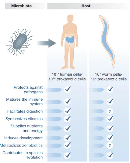
Figure 1 - Functions of gut microbiota in human and nematode hosts ... 2
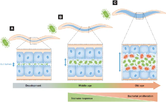
Figure 2 – Changes in C. elegans-gut microbiota interactions during the course of life ... 4
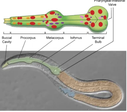
Figure 3 – C. elegans digestive tract ... 5
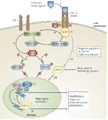
Figure 4 – Insulin/IGF signaling pathway ... 8
Figure 5 – Scheme of experimental evolution protocol ... 16
Figure 6 – Survival of C. elegans fed on different pathogenic bacteria ... 18
Figure 7 – Gut bacteria recovered from C. elegans fed on S. marcescens and E. coli IAI1 ... 20
Figure 8 – Bacteria recovered on day 7 from individuals fed on S. marcescens and E. coli IAI1 ... 21
Figure 9 – Frequency of selectable C. elegans for each time-point ... 22
Figure 10 – Survival of N2 individuals fed on wild-type and mutant S. marcescens ... 23
Figure 11 – Survival of N2 individuals fed on derived S. marcescens ... 24
Figure 12 – Survival of C. elegans fed on different S. marcescens strains and E. coli OP50 ... 25
Figure 13 – Survival of C. elegans fed on a mixture of S. marcescens db10 and db11 ... 26
Figure 14 – Over time frequency of S. marcescens db10 inside C. elegans’ gut ... 27
Figure 15 – S. marcescens from LB-tetracycline plates ... 29
I. Introduction
1. Species interactions
In nature, organisms are part of ecosystems where one species co-occurs and interacts with other species along with the environment, creating a network of biotic and abiotic interactions[1]. The interplay
between natural selection and ecology has been gaining relevance, giving rise to the Eco-Evolutionary Feedback field[2,3]. In light of this concept, selection mediated by ecological dynamics drives evolutionary
changes which in turn alter the form of ecological interactions[3]. For instance, bacterial species adapting to
a new abiotic environment had enhanced growth rate when in polyculture, highlighting the impact species interactions has on adaption to a novel environment[4].
Taking into consideration that an ecosystem usually has an exceptionally complex network, eco-evolutionary feedbacks can be studied as relationships between one or more species and their environment. A particular case is the interaction between hosts and microorganisms, which could be seen as an ecological community[5]. Healthy animals and plants are colonized by a large and diverse number of microorganisms[5]
where bacteria are considered key players in multiple aspects of organisms’ biology[1]. Additionally,
previous works have shown that host-parasite interactions can lead to extremely rapid evolutionary changes[6]. One major focus of host-microorganism interactions is the relationship between a host and its
gut microbiota.
2. Host-gut microbiota interactions
The intestinal tract of animals is a favorable niche for microorganisms where these form a complex and dynamic community that can be seen as an ecosystem[5]. It is estimated that the human gut microbiota
includes 1014 bacteria[7], which is about the number of stars in 1000 Milky Way galaxies.
In recent years, the importance of gut bacteria in host health has gained relevance since bacteria were seen to affect both development and adult-health by acting as stimuli for morphogenesis or by providing essential nutrients and metabolizing indigestible compounds[8]. Indeed, 10% of the metabolites in
the mammalian blood flow are from bacterial origin[9].
Gut epithelia are constantly exposed to hostile bacteria[10] and resident microbes can provide strong
protection against opportunistic pathogens[11]. This protection can be mediated by producing toxins, limiting
resource availability or by directly modulating host immune response[8,12].
Alternatively, a disruption in the gut microbiota could result in pathological outcomes, such as obesity, diabetes, autoimmune disorders, inflammatory bowel disease or even some forms of cancer[13,14].
Besides its importance for host health, the large population size and short generation time of microbes create the potential for rapid adaptation, making the gut microbiota the perfect model for eco-evolutionary studies.
However, the number and nature of questions that can be answered by directly studying mammalian gut microbiota are limited. For instance, host generation time restricts experimental evolution assays concerning host-gut microbiota interactions. A possible solution is to use model organisms, such as
Caenorhabditis elegans. C. elegans is an excellent model organism to study gut microbiota interactions,
given that the intestine is their largest somatic organ and it is typically full of microbes[13]. Moreover, the
gut microbiota in C. elegans performs many of the same functions of human gut microbiota[13] (Figure1).
Figure 1 – Functions of gut microbiota in human and nematode hosts.
Effects of intestinal microbiota on the host.
3. C. elegans as a model organism
The nematode Caenorhabditis elegans was established as a model organism by Sydney Brenner in 1974[16]. Today, C. elegans is a very well established model in several fields such as development, (innate) immunity, apoptosis and aging[16].
C. elegans is a small free-living nematode that is found in rotting fruit, vegetation and soils of
temperate regions[17,18]. Populations are mainly formed by self-fertilizing hermaphrodites and a small
percentage of males[18]. The worm has a short generation time (4 days), which includes 4 larval stages and a
large progeny – each hermaphrodite can lay between 250-300 eggs[17].
In laboratory conditions, C. elegans normally grows at 20-25°C on solid agar medium and is easily maintained in monoxenic cultures of Escherichia coli, strain OP50 (an uracil auxotroph bacteria derivative of E. coli B)[15]. Under those conditions, wild-type C. elegans has a lifespan of 2-3 weeks[17,18]. Furthermore,
worms can be frozen and they are fully transparent, allowing the use of fluorescent reporters in living animals[17]. C. elegans also present a powerful set of genetics tools, with approximately 3000 different
mutants strains publicly available and its whole genome sequenced[17].
Finally, the worm possesses evolutionarily conserved signaling pathways for innate immunity, such as the insulin/IGF signaling pathway, the p38 MAPK and the transforming growth factor (TGF-β)[13,18,19].
4. C. elegans and its gut microbiota
The nematode has been recognized as a major player in soil ecology, where it encounters a wide variety of different bacteria[20]. The interaction between C. elegans and gut bacteria is very complex and
changes throughout the worm’s life course (Figure2)[15]. Accordingly, during development their interaction
with bacteria is exclusively predatory and bacteria found within the intestinal lumen are never intact[15]. In
contrast, live bacteria can only be seen in young adults (day 4-5 of adulthood) where they can form symbiotic or commensal communities in the gut[15]. As worms age, they are more sensitive to bacterial infections and
bacteria proliferation within the intestinal lumen becomes detrimental to the host[21].
This kind of interaction has also been reported in Drosophila, since the presence of bacteria during the first week of adulthood enhances longevity and in the last stage of life causes a decrease in lifespan[21].
Figure 2 – Changes in C. elegans–gut microbiota interactions during the course of life. A – During development, bacteria serve as a source of food;
B – In young adults, bacteria escape the grinders action and establish a community in the intestinal lumen; C – As the worm ages, bacterial accumulation within the lumen become detrimental to the host.
4.1 Bacteria as a food source
As mentioned above, bacteria serve exclusively as a food source in the initial phases of C. elegans life (Figure2A). When C. elegans feed, bacteria pass through the pharynx to the terminal bulb, where there is an organ that destroys bacterial cells. This organ – the grinder – is a tripartite array of interlocking tooth-like structures that process all food[15,22] (Figure3). After passing through the pharynx, bacterial cells reach
the intestine. The worm intestine is a nonrenewable monolayer of 20 epithelial cell arranged to form a tube with a central lumen[15].
The quantity of live bacteria in the nematode intestine is influenced by pharyngeal pumping rate, grinder integrity and digestive system efficiency[23]. Additionally, host immune system and bacterial
proliferative capacity are also important features in controlling gut microbiota[13]. Peculiarly, despite the fact
that C. elegans kills bacteria, the worm has a nutritional requirement for live, metabolically active bacteria, since animals fed on non-viable bacteria appear ill and have diminished fecundity[24].
Figure 3 – C. elegans digestive tract.
The digestive tract is an epithelial tube consisting of the buccal cavity (yellow), pharynx (green) intestine (orange) and hindgut (blue).
4.2 C. elegans core gut microbiota
In young adults, it is possible to see bacterial accumulation and proliferation inside C. elegans intestinal lumen[25]. During young adulthood, bacteria can form a symbiotic or commensal community within
the intestine (Figure2B). In fact, recent studies have reported that C. elegans possesses a species-rich core gut microbiota dominated by Proteobacteria phyla, namely the Enterobacteriaceae family and the
Pseudomonas genera[1,8,26]. In addition, C. elegans microbiome is distinct from its substrate environment and
at least from the congeneric C. remanei[8,26]. A core gut microbiota has also be described in apes, zebrafish,
termites, bees and Drosophila[7](where the core microbiota varies between and within different species,
habitats and even laboratories)[27].
4.3 Bacteria accumulation is deleterious for C. elegans
Even though a healthy core microbiota has been described for C. elegans, as the worm ages they lose the capacity to control the number of live bacteria that reach the intestine. This loss is related with a decline in intestinal functions, such as ingestion and defecation[29] as well as with a decline in innate
immunity[28]. In this phase, even non-pathogenic bacteria like E. coli OP50 are able to escape grinder
function and reach the intestine alive, where they become opportunistic[25].
Accumulation of undigested bacteria is associated with symptoms of pathology, which include increased variability in intestinal shape and size and distention of the pharynx and intestinal lumen[30]
(Figure2C). In addition to the morphological changes, bacteria accumulation within the gut has been correlated with higher mortality rates, since C. elegans grown on bacteria unable to proliferate had a longer lifespan[30]. In fact, an inverse correlation between bacterial accumulation and C. elegans lifespan was
recently described, which suggests that bacteria accumulation contributes to aging[25,31].
However, bacterial accumulation is not the only factor that contributes to increased mortality. Grinder-defect mutants only show increased mortality associated with a compromised immune system[31]
and bacteria such as Enteroccocus faecium are capable of colonizing the gut without causing significant mortality[32]. This suggest that non-pathogenic bacterial accumulation may only increase mortality when the
immune system deteriorates with age.
In summary, bacterial accumulation early in adulthood seems to be controlled by gut immunity and with aging there is a decline in immune response and a deregulation in controlling bacteria proliferation which is strongly and inversely correlated with longevity[25].
5. C. elegans’ longevity is influenced by multiple factors
Along with the effect of bacteria accumulation, bacteria can affect host longevity by other mechanisms. First, bacteria are the food source for C. elegans, therefore, nutritional quality and bacterial metabolites may influence host aging[33]. Moreover, dietary restriction has been reported to extend the
lifespan of worms, flies and mice[34]. Second, bacteria may cause pathogenic infections by creating a
persistent infection or by producing toxins[33,35].
5.1 Pathogenic bacteria
C. elegans possess a grinder that destroys all bacterial cells[15,22], however, pathogenic bacteria are
able to pass through the grinder intact, even in young C. elegans. Once in the intestine bacteria are capable of proliferating and killing the nematode[36]. Bacteria such as Pseudomonas aeruginosa[37], Serratia
marcescens[38,39], Salmonella enterica and Enterococcus faecalis are known pathogenic bacteria for C.
elegans[35].
S. marcescens is a Gram-negative enterobacteriaceae extracellular pathogen that causes disease in
plants and in a wide range of both invertebrates and vertebrates hosts[38]. In humans, it is an opportunistic
pathogen associated with hospital infections and nosocomial infections[18]. Given its pathogenic proprieties,
this bacteria were widely adopted as a model to study the genetic basis of virulence[6,17].
S. marcescens is also a pathogen for C. elegans, however it is unknown if the two species co-exist
in nature, even though this is strongly suggested by the fact that both are common in soils[6]. S. marcescens
is capable of establishing a persistent intestinal infection that kills worms in 6-9 days[18,39]. In the first 6 hours
after infection it is already possible to find bacteria within the intestinal lumen and after 24 hours, a clear distension of the intestinal lumen is visible. 48 hours after infection, there is a strong drop in the rate of egg laying and worms start do die after 72 hours of contact[39].
In addition to bacterial interactions, C. elegans longevity is also influenced by the rate of mitochondrial respiration and by the insulin/IGF signaling pathway – a “master key” in immune response and longevity regulation[34].
6. C. elegans’ longevity and the insulin/IGF signaling pathway
C. elegans possesses a conserved innate immune system that is pathogen-specific[40,19] and decline
as worms age[25,41]. One extremely important signaling pathway in the insulin/IGF signaling pathway (IIS
pathway).
The IIS pathway is one of the most studied and well-characterized pathways in C. elegans (Figure4), since it is an important stress-resistance pathway with implications in processes such as dauer formation[16,42],
stress-resistance and longevity[43]. In general, the pathway consists of the receptor tyrosine kinase DAF-2
(an insulin/IGF-1-like receptor) that controls the fork-head-family transcription factor DAF-16 (the sole orthologue of FOXO transcription factor)[44]. When the receptor DAF-2 is activated, it initiates a kinase
cascade that results in phosphorylation and repression of DAF-16. DAF-16 phosphorylation prevents it from entering the nucleus where it is necessary for the transcription of numerous genes related with innate immunity and stress response[19,22,30,45](Figure4).
Figure 4 – Insulin/IGF signaling pathway
The pathway is activated with the DAF-2 receptor that maintains DAF-16 inactivated in the cytoplasm. In the absence of DAF-2, DAF-16 enters the nucleus where activates genes related with immune response and stress resistance. HSF-1 is also a key player that regulates gene expression longevity and stress resistance genes.
daf-2 mutants have an increased resistance to heat, hypoxia, heavy metals and bacterial
pathogens[42]. This increased resistance requires the activation of DAF-16, which in the absence of DAF-2
is capable of entering the nucleus[42]. Previous studies demonstrated that activation of DAF-16 triggers the
expression of antimicrobial genes, such as the lysozyme lys-7[19]. This gene is upregulated when C. elegans
is exposed to Serratia marcescens.
The lifespan of C. elegans is also regulated by the IIS pathway. Mutations in daf-2 gene are related with an increased lifespan, whereas mutations in daf-16 shorten lifespan. In that way, the activation of DAF-16 is necessary to activate the expression of genes related with aging[22].
Long-lived daf-2 mutants also have lower levels of bacterial accumulation in their gut, however it is not clear whether this is a cause or an effect of daf-2 longevity[25,31]. Mutations that diminish insulin-like
signaling also increase lifespan and stress resistance in Drosophila[46] and mice[47], suggesting that the effect
of this pathway on longevity is conserved.
In C. elegans, the increase in lifespan related with the IIS pathway has been associated with its expression in the intestine. Studies show that expressing daf-16 in the intestine increases lifespan by 50-60%, which seems to indicate that the intestine is an important organ in IIS-mediated lifespan extension[43].
In summary, the IIS pathway involves increasing lifespan by regulating the entry of transcription factors such as DAF-16 into the nucleus. Another transcription factor that is involved in this pathway is the heat-shock factor-1 (HSF-1). In normal conditions, DAF-2 is activated and keeps DAF-16 and (probably) HSF-1 inactivated in the cytoplasm[48](Figure 4).
6.1 The Heat-Shock Factor-1
In vertebrates there are four major heat-shock factors (HSF), while in C. elegans only one HSF homolog exists – HSF-1[49]. HSF-1 is the key regulator of the cellular and organismal response to heat stress
and is conserved in all eukaryotes[50]. It is a leucine-zipper-containing transcription factor that controls the
expression of small heat-shock proteins (HSP). One example is the HSP-16 family, which are molecular chaperones that prevent protein and cellular damage following stress[48,51]. In basal conditions, HSF-1 exists
as a monomer in the cytoplasm and nucleus, whereas in stress conditions it becomes a trimer that accumulates in the nucleus where it binds to heat shock elements in the promoter region of HSP genes[52].
Alongside its stress response function, HSF-1 also contributes to processes such as development, growth, aging, immunity, reproduction and has been implicated in protein miss-folding diseases, such as Huntington’s and Alzheimer’s diseases[49]. In C. elegans, mutations in the hsf-1 gene affect heat-shock
In recent years, HSF-1 has been directly implicated in lifespan regulation. In C. elegans, inhibition of HSF-1 leads to decreased lifespan, since hsf-1 mutants live, on average, 7 days less comparing with wild-type animals[30,53]. Conversely, overexpression of hsf-1 is related to lifespan extension[54].
Even though hsf-1 expression is associated with longevity regulation, the exact mechanism by which HSF-1 influences lifespan is not totally clear. Several studies have linked HSF-1 function with the IIS pathway, since the role of HSF-1 seems to be correlated with DAF-16[54].
One evidence is the fact that daf-16 is required for hsf-1 overexpression, although both genes operate independently from one another[54]. Additionally, it seems DAF-16 and HSF-1 increase longevity,
at least in part, by increasing small heat-shock protein (sHSP) levels which prevent aggregations of unfolded proteins[54].
Another mechanism by which HSF-1 could increase lifespan is correlated with collagen regulation[52]. Collagen has been shown to have cytoprotective proprieties that are related with longevity. In
a recent study, authors show that multiple genes involved in cuticle structure, including collagen genes, are enriched upon HSF-1 activation[52].
7. Selection of gut microbiota
7.1 Indirect selection
Numerous studies have proposed that manipulation of gut microbiota can increase host lifespan, suggesting a possible evolutionary-based strategy to extend longevity[21]. In a recent study, interaction
between a host and its gut microbiota was analyzed from a new angle, where the performance of the hosts is improved by altering their microbiota[55]. Artificial selection can be applied on the gut microbiota in an
indirect manner. The idea is to select gut microbiota by choosing a host trait that is directly influenced by its microbiota. The host trait should be easily measured and must have a strong correlation with the microbiota and with host fitness[55]. Such a trait can be, for example, the longevity of the host, since the
microbiota have a large impact on host longevity.
7.2 Multilevel selection
Selection can act at multiple biological levels[56]and this interaction between different levels can be
very important for microorganism evolution, mostly pathogenic ones. Multilevel selection is often studied in a context of antagonistic relationships[56] which can be interpreted as a trade-off between virulence and
Theories of virulence suggest that pathogens should evolve to a less-virulent state, since harming the host would be detrimental to a long-term survival strategy[57]. However, competition between pathogens
should favor the one with higher proliferative and invasive rates, which translates in higher virulence levels[57]. In that way, within an infected host, individual fitness is central, while at the population level,
efficient transmission between hosts is the critical component[58].
This kind of paradox can be overcome with a theoretical multilevel selection perspective, where traits that are costly at lower levels (individual level) can be beneficial at higher levels (host level)[59,60].
Virus evolution is one of the most studied examples, with selection occurring both within infected hosts and between hosts via transmission. Within the host, individual selection is prevalent since a fast-replication virus will outcompete a slower strain. However, if rapid viral replication incapacitates the host, the fast-replicating virus may not be transmitted as frequently as the slower strain, meaning that at a higher level, selection favors the less virulent strain[56,61].
In summary, high virulence should have a higher benefit at lower levels, due to individual competition, while low virulence should only be selected at higher levels of selection, where individual selection is minimized.
Nonetheless, multilevel selection theory still has some conceptual problems, namely, there is an ambiguity regarding the definition of a “higher level trait”, as well as a lack of a precise definition of “higher level fitness”[62].
8. Main questions
The main goal of this work was to investigate if gut microbiota evolution is directly related with changes in host traits, namely in host longevity. To do this, C. elegans was used as a model host and S.
marcescens as gut microbiota. A second major objective was to explore the role of different levels of
selection in pathogenic bacteria evolution. In order to fulfil these goals, we specifically aimed to:
1. Characterize the survival rate of wild-type N2 and hsf-1 mutant when feeding on different bacteria, mainly pathogenic bacteria;
2. Create experimental conditions favoring high or low levels of individual selection; 3. Correlate different levels of selection with pathogenic bacteria evolution;
4. Select gut microbiota able to colonize C. elegans’ gut and alter its longevity; 5. Prove that the microbiota can be indirectly selected;
II. Material and Methods
1. Strains and maintenance
In this work, two different strains of Caenorhabditis elegans were used: the N2 Bristol strain and the PS3551 strain, which has a mutation in the hsf-1 gene. Both were obtained from the Caenorhabditis Genetics Center – CGC. Populations were maintained, according to standard conditions[63,64], at 20°C in
nematode growth medium (NGM) plated with Escherichia coli OP50.
Synchronized populations were obtained by treating adults with a bleach solution which allows unhatched embryos isolation. Embryos stayed in M9 buffer over-night with shaking, so as to obtain a synchronized population of L1 individuals, that arrested their development due to absence of food[65,66].
Hatched L1 were then seeded (transfer onto NGM plates) to start a new generation.
In order to avoid contaminations, some alterations were applied to the standard protocol, namely M9 buffer was supplemented with gentamicin (10µg/ml). Additionally, all plates with maintenance worms had NGM supplemented with kanamycin (50µg/ml) and every two generations supplemented with ampicillin (100 µg/ml).
Two different strains of E. coli OP50 were used: one with mCherry fluorescence (red) associated with kanamycin resistance and another with GFP fluorescence (green) associated with ampicillin resistance. Both inserts are chromosomic. The use of these strains allows the alternation of antibiotics, as well as easy checking for carrying-over bacteria.
Worms used for experiments were seeded as unhatched embryos (right after bleach treatment), given that development arrest could influence survival of individuals[66]. 4th stage larvae - L4 (approximately 48 hours after seed) worms were used in all experiments. Different bacteria were used during the course of our experiments:
E. coli OP50, both with and without fluorescence; E. coli MG1655;
E. coli IAI1 with mCherry fluorescence (plasmid with ampicillin resistance) and without fluorescence; E. coli aroD;
Serratia marcescens db10 (tetracycline resistant) and db11 strain (tetracycline, kanamycin and streptomycin resistant).
All bacteria came from the CGC and before seeding (bacteria plating), bacteria were grown over-night at 37°C with shaking in liquid lysogeny broth medium (LB) supplemented with antibiotics. Bacteria were stored at 4°C in LB plates supplemented with their respective antibiotic and every two weeks transferred to a new plate.
2. Survival assays
Survival assays started with L4 individuals being transferred onto plates, effectively making L4 stage ‘day 0’. In order to avoid starvation and overcrowding by progeny, worms were transferred daily to new plates during the fertile phase (approximately 6-7 days). Later, individuals were transferred every second day. An individual was considered dead if it failed to either move or respond to touch and did not show any sign of pharyngeal pumping. Worms that show an egg laying defect (bagging) or died from vulva bursting were considered as dead individuals. Worms that disappeared, probably due to crawling off the plate or disintegration after dying, were treated as censored data.
2.1 Transferring C. elegans with filters
In this work, a new method (inspired on the work of K. Lew and J. Miwa[67]) of transferring C.
elegans was developed, which allows the transfer of a large number of individuals and at the same time
separate adults from eggs, L1 and L2 larvae.
In this protocol, a 40µm filter was used to separate and transfer approximately 300 adults at once (see S1.2 for more details). Plates were washed with M9 buffer and C. elegans were filtered and washed two times with M9. Adults did not pass through the filter and were recovered with a glass pipet and transferred in buffer drops to a new petri dish.
To test the efficiency of this new protocol, a survival assay with wild-type N2 and hsf-1 mutants was performed, where individuals were passed daily onto new plates seeded with E. coli OP50 by filtering. With this protocol, it was possible to perform and recover a standard survival curve (FigureS3), were, as expected, N2 individuals had higher survival (hazard ratio=0.515) compared to hsf-1 mutants
2.2 Survival in Peptone-Free NGM
During the experimental evolution protocol, peptone-free NGM (PFN) plates were used in order to guarantee that only S. marcescens from the gut was selected, since this medium was effective in preventing bacterial growth[68]. Given this, survival of N2 individuals fed on S. marcescens db10, db11 and
E. coli OP50 mCherry (used as control) was analyzed. All plates were 90mm PFN plates seeded with a
specific strain of bacteria, which were replicated three times. Before the seed, bacteria grew in an over-night liquid culture with shaking and was concentrated until an OD600=10 in order to form a lawn. Otherwise mentioned, this medium was used in all following experiments.
2.3 Statistical analysis
A Cox Proportional Hazards Regression survival analysis was performed using the coxme function in R, version 3.3.1. This function fits a Cox model containing mixed effects, allowing the use of fixed (e.g. Structured condition) and random effects (e.g. Replicates). A p-value<0.05 was considered to indicate significance of effects. For more details see S7.
3. Gut colonization
During the survival experiment with pathogenic bacteria (S1.3), the ability of those bacteria to colonize C. elegans’ gut was analyzed. Every day, two individuals from each condition were selected at random and crushed in order to recover gut bacteria. Bacteria recovery was done by crushing individuals in 5µl of PBS. Different dilutions were plated in 10µl drops in LB and minimum medium with and without uracil, thus distinguishing between experimental bacteria and E. coli OP50 mCherry. As positive control,
E. coli MG1655 was used (capable of growing in minimum medium with and without uracil) and as negative
control, E. coli OP50 mCherry that only grows in minimum medium with uracil.
4. Mutagenesis
In order to maximize genetic diversity in our bacterial populations a chemical mutagen - ethyl methanesulfonate (EMS) – was used. The protocol was followed according to Parkhomchuk et al., 2009 during different time-points (15, 30, 45, 60 minutes). Control population was subject to the same protocol, however, no EMS was added. E. coli OP50 and S. marcescens db10 were used in this protocol.
Mortality rates of bacteria were evaluated by comparing CFUs of control and mutated bacteria (S2). The 15-minute mutated population of S. marcescens was used in the experimental evolution.
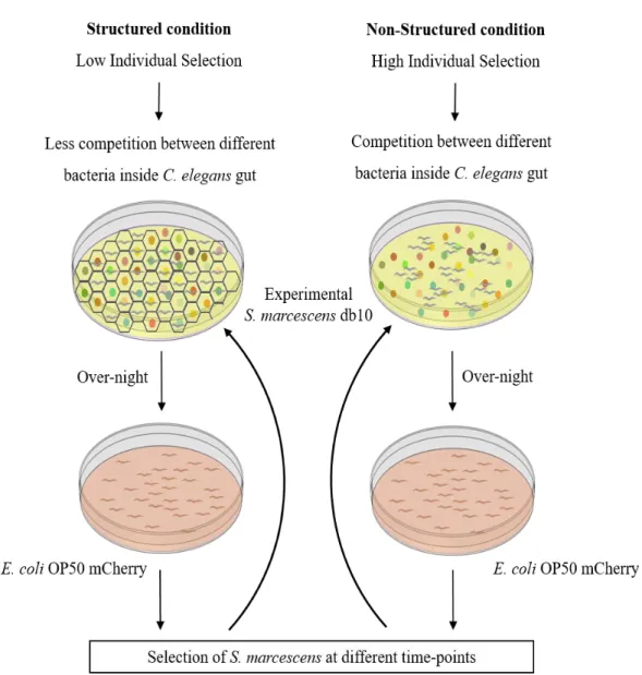
5. Levels of selection
In this work, one objective was to develop experimental conditions to create different levels of selection. For that, in association with the Techinico-scientific Support (TSS) team at the Instituto Gulbenkian de Ciência (IGC), we developed a honeycomb-like 3D scaffold composed by 594 individual cells. This scaffold restricts the movement of individuals by confining then inside a single cell and, ideally each worm has access to a limited number of different bacterial colonies.
Bacteria were placed in 150mm plates in order to obtain approximately 500 individual bacterial colonies per plate. In the structured condition, C. elegans were placed in the petri dish and after 20 minutes the 3D scaffold was added. In contrast, no scaffold was added in the non-structured condition, allowing worms to move freely. The scaffold stayed on over-night and was removed the next morning.
6. Experimental Evolution
For the experimental evolution protocol, N2 individuals were exposed to wild-type and mutant S.
marcescens db10 populations in structured and non-structured conditions. Each treatment was replicated
three times, totaling 24 evolving populations. Individuals were selected in three different time-points and their gut bacteria was recovered in order to start a new round of selection. After one round of selection, survival of individuals exposed to derived bacteria selected at the different conditions was analyzed in the non-structured condition, in order to only test the impact of the initial environment.
6.1 Experimental protocol
Approximately 200 L4 individuals were transferred in M9 buffer drops, onto 150mm NGM-tetracycline plates. These plates had been previously seeded with wild-type or mutant S. marcescens and the 3D scaffold was added in the structured condition plates.
The following day, the 3D scaffold was removed and all plates were washed with M9 buffer. Individuals were transferred onto 90mm PFN plates seeded with E. coli OP50 mCherry, where they were maintained (Figure5). Dead individuals were removed daily in order to guarantee that only individuals which die at specific time-points were considered.
6.2 Selection time-points
Three different selection time-points - T1, T2 and T3 – were defined in order to select bacteria at different stages of C. elegans’ lifespan. T1 was defined as the time when 5% of the total population was dead. T2, which controls for random selection, was defined as the mid-point of worms’ lifespan where dead individuals corresponding to 5% of total population were selected. T3 individuals were the final 10% that stayed alive.
6.3 Selection of individuals
At each time-point, gut bacteria of selected individuals were recovered. Individuals were selected using 5µl droplets of PBS and transferred to an individual well in a 96 well plate. Worms were crushed with a 0,5µm pestle and the solution was diluted in PBS and plated in 5µl drops in both LB and LB-tetracycline. To control for contaminations, a random sample was selected to be plated in both kanamycin and LB-ampicillin. Plates were incubated over-night at 37°C, and on the next day stored at 4°C.
6.4 Preparation for the next round of selection
Since T1 and T3 were separated by one week, a strategy to guarantee that all bacteria were approximately in the same stage had to be developed. Upon T3, samples from all time-points were individually transferred and plated once again in LB-tetracycline. That way, all samples had grown over-night at 37°C before being plated for the next round. 50 colonies from each sample were then selected and placed in liquid LB. OD600 for each sample was measured in order to guarantee that all samples had the same quantity of bacteria. These samples were seeded in 150mm NGM-tetracycline plates to be used in the following round of selection.
III. Results
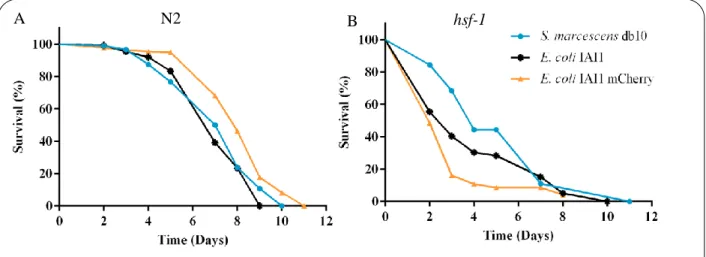
1. C. elegans fed on different pathogenic bacteria have different lifespans
One objective of this work was to characterize C. elegans’ survival when feeding on pathogenic bacteria. To do that, survival of wild-type N2 and hsf-1 mutants fed on S. marcescens db10, E. coli IAI1 and E. coli IAI mCherry was analyzed (S1.3).
There were no differences in survival rates of N2 individuals (Figure6A) feeding on E. coli IAI1 or
S. marcescens (p-value=0.304). Lifespan of individuals fed on E. coli IAI1 or E. coli IAI1 mCherry was
different (p-value<0.0001), being that worms fed on E. coli IAI1 mCherry had a lower probability of death (hazard ratio=0.501).
Unlike wild-type worms, hsf-1 mutants (Figure6B) had different survival rates when feeding on E.
coli IAI1 or S. marcescens (p-value=0.003), being that S. marcescens reduced the risk of death (hazard ratio=0.510). There were also differences between survival rates of hsf-1 worms when feeding on E. coli
IAI1 or E. coli IAI1 mCherry (p-value=0.0004). However, unlike wild-type N2, hsf-1 mutants survived less when feeding on E. coli IAI1 mCherry (hazard ratio=1.608).
A
Figure 6 – Survival of C. elegans fed on different pathogenic bacteria.
A – wild-type N2 fed on S. marcescens db10 (blue) n=285; E. coli IAI1 (black) n=134; E. coli IAI1 mCherry (orange) n=229. B – hsf-1 mutants fed on S. marcescens db10 (blue) n=45; E. coli IAI1 (black) n=130; E. coli IAI1 mCherry (orange) n=161. One replicate per condition.
B
2. S. marcescens is capable of colonizing C. elegans’ gut
The main goal of this work was to evolve gut bacteria, thus it was necessary to verify if bacteria were indeed capable of colonizing C. elegans’ gut. In the previous survival experiment, the ability of bacteria to colonize C. elegans’ gut was analyzed by selecting every day two individuals from each condition and recover its gut bacteria. Note that individuals were feeding on E. coli OP50 mCherry, which is distinguishable from non-fluorescence E. coli IAI1 and S. marcescens colonies.
E. coli IAI1 mCherry lost fluorescence easily, since an over-night growth was enough for
non-fluorescence colonies to appear (data not shown). Thus, this strain was excluded from following experiments.
Concerning the two other bacteria, S. marcescens and E. coli IAI1 were found in the gut 24 hours after infection, since both bacteria grew in LB (Figure7a, d) and in minimum medium plates (Figure7b, c, e, f). Interestingly, results show that individuals fed on S. marcescens did not have E. coli OP50 mCherry in their gut (all colonies were not fluorescent) (Figure7a-c), while individuals fed on E. coli IAI1 had both
E. coli strains in their gut – there were both fluorescent and non-fluorescent colonies (Figure7g-i, n-p). Moreover, non-fluorescent colonies from individuals fed on S. marcescens or E. coli IAI1 in both LB and minimum medium were morphologically different (Figure7a-f, j-m) – S. marcescens colonies were more compact and less translucent.
Figure 7 – Gut bacteria recovered from C. elegans fed on S. marcescens and E. coli IAI1.
First column – LB medium; second column – minimum medium with uracil; third column – minimum medium
without uracil. a-f, j-m: brightfield stereoscope images; g-i, n-p: mCherry fluorescence stereoscope images. Scale bar=1mm (a-i), 250µm (j-l, n-o), 200µm (m,p). Marker lines encircle initial plating area.
On day 6 of the experiment, S. marcescens and E. coli IAI1 could still be found in the gut. However, on day 7, a morphological change in non-fluorescent colonies derived from individuals fed on E. coli IAI1 was visible – colonies became very similar to S. marcescens non-fluorescent ones (Figure8).
To understand this result, a portion of each bacterial colony was recovered and streaked in LB and LB-tetracycline plates. Results show E. coli IAI1 plates were contaminated with S. marcescens (data not shown), consequently it was not possible to conclude if E. coli IAI1 is indeed capable of colonizing C.
elegans’ gut. Moreover, the survival of individuals fed on E. coli IAI1 (Figure6) could be affected by the
presence of S. marcescens.
Given these results, S. marcescens was chosen as experimental bacteria, since it is capable of colonizing C. elegans’ gut.
Figure 8 – Bacteria recovered on day 7 from individuals fed on S. marcescens and E. coli IAI1.
Brightfield stereoscope images, scale bar=1mm. Marker lines encircle initial plating area.
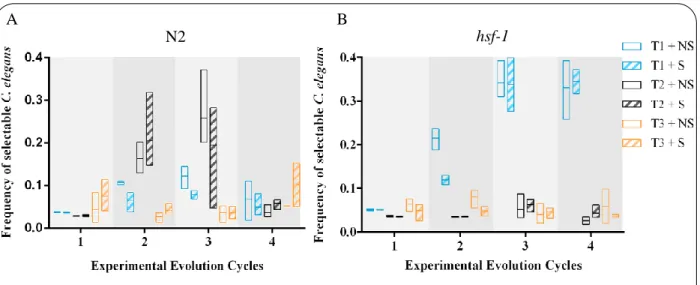
3. Experimental Evolution
In the experimental evolution assays, wild-type N2 and hsf-1 mutant individuals were exposed to a mutant population of S. marcescens db10 in NGM plates with and without structure (S4). During this experiment, survival of individuals was not analyzed and selection time-points were calculated based on a standard curve previously obtained with S. marcescens (Figure6).
Thus, the number of individuals to be selected in each time-point represent a fix percentage (5 or 10%) of the initial population size. Given this, in each time-point there should be a predictable number of individuals to be selected – selectable C. elegans. However, during four rounds of selection, the frequency of selectable individuals in each time-point was not as predicted. Results from N2 individuals (Figure9A) did not present any clear pattern, while hsf-1 mutants showed an increment in the percentage of dead individuals at T1 (Figure9B). Since increased virulence was expected to be selected in T1 (which is in agreement with obtained data), a survival assay for hsf-1 mutants exposed to ancestral and derived S.
marcescens was performed (S1.4) in order to test if this pattern reflected a possible effect of selection. However, no differences in survival were found (FigureS4).
At the end of the fourth round of selection, a possible cross contamination between all experimental populations was found (data not shown), which indicates that all populations were indistinguishable. Consequently, the experiment was abandoned and a new cycle of evolution was started.
A B
Figure 9 – Frequency of selectable C. elegans for each time-point.
Y axis represents the percentage of individuals that could be selected in each time-point. A – wild-type N2; B –
hsf-1 mutants. Non-structure (hollow bars); Structure (striped bars); T1 (blue); T2 (black); T3 (orange); three replicates per condition. Horizontal line represents mean value.
In this new cycle of experimental evolution, only N2 individuals were used and exposed to a new mutant population of S. marcescens. Peptone-free NGM (PFN) plates seeded with E. coli OP50 mCherry were used in order to guarantee that only gut S. marcescens was selected. As before, the structured and non-structured conditions were used and individuals were selected at the three different time-points to recover their gut bacteria.
3.1 Higher genetic variability causes lifespan differences upon decreased individual selection
Before the beginning of this new cycle, the impact of bacterial genetic variability in C. elegans’ survival was analyzed by looking at how survival of N2 individuals was different while feeding on wild-type and mutant S. marcescens with and without structure.
There was no difference in survival between individuals fed on wild-type or mutant bacteria
(p-value=0.32) (Figure10). However, when the structured and non-structured condition was taken into
consideration, a difference in C. elegans survival was detected. Individuals fed on mutant bacteria had lower probability of death when in the structured condition (hazard ratio=0.716 and p-value=0.025). On the contrary, worms fed on wild-type bacteria had no difference in survival in both conditions (p-value=0.34). This result indicates that the two conditions were in fact different and had an impact on mutant bacterial competition, which was reflected in C. elegans’ survival.
Figure 10 – Survival of N2 individuals fed on wild-type and mutant S. marcescens.
Wild-type and non-structure n=668 (full blue); wild-type and structure n=773 (dashed blue); mutant and non-structure n=591 (full orange); mutant and structure n=544 (dashed orange). Three replicates per condition.
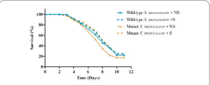
3.2. One round of selection in mutant bacteria is sufficient to increase C. elegans’ longevity
The main goal of this work was to test if it possible to select and evolve gut bacteria capable of altering C. elegans’ longevity. Due to temporal constrains, only one round of selection was performed after which N2 individuals were exposed to bacteria previously selected at the different time-points and conditions. One way by which population evolution can be demonstrated is by comparing the effect of ancestral and derived bacteria on C. elegans’ longevity. Ancestral and derived wild-type S. marcescens were not different (p-value=0.29) (Figure10,11A) and there was no interaction with the structure (p-value=1). However, ancestral and derived mutant S. marcescens (Figure10,11B) were different (p-value<0.0001) and
C. elegans exposed to derived bacteria had a lower probability of death (hazard ratio=0.734). Still, the
presence of structure had no impact on survival of worms fed on mutant S. marcescens (p-value=1). This result shows that one cycle of selection was enough to cause differences in mutant S.
marcescens populations, which translated in an increment of C. elegans longevity.
Besides the comparison between ancestral and derived bacteria, population evolution could eventually be seen in different time-points. However, there was no effect on C. elegans’ survival of selecting bacteria in different time-points for both wild-type and mutant S. marcescens (p-value=0.073 and 0.313, respectively) (Figure11).
A B
Figure 11 – Survival of N2 individuals fed on derived S. marcescens.
A – Wild-type bacteria; T1 non-structure n=620 (full blue); T1 structure n=685 (dashed blue); T2 non-structure n=862 (full black); T2 structure n=719 (dashed black); T3 non-structure n=729 (full orange); T3 structure n=706 (dashed orange). B – mutant bacteria; T1 non-structure n=1035 (full blue); T1 structure n=706 (dashed blue); T2 non-structure n=945 (full black); T2 structure n=794 (dashed black); T3 non-structure n=725 (full orange); T3 structure n=935 (dashed orange). Three replicates per condition.
4. C. elegans’ lifespan decreases when feeding on a mixture of bacteria
Since during experimental evolution PFN plates were used, it was necessary to analyze C. elegans survival in this new medium. Worms were exposed to S. marcescens db10, db11 and E. coli OP50 mCherry (S1.5).
As expected, there were no differences between survival rates of N2 individuals fed on either S.
marcescens db10 or db11 (p-value=0.63) (Figure12). However, individuals fed on S. marcescens db10
survived more (hazard ratio=0.566) when compared with worms fed on E. coli OP50 mCherry
(p-value=0.009). The same happens with S. marcescens db11; worms fed on db11 had lower probability of
death (hazard ratio=0.484) comparing to E. coli OP50 mCherry (p-value=0.001), which was surprising.
Given this unexpected result, individuals from E. coli OP50 plates were picked and their gut bacteria recovered to confirm whether worms were feeding on E. coli. Results indicated a contamination with S.
marcescens db11 (data not shown) giving rise to a new hypothesis where the interaction (most likely
competition) between S. marcescens db11 and E. coli OP50 could be the cause of lifespan decrease[84].
To test this hypothesis, a second experiment was performed where the survival of N2 individuals fed on a mixture of bacteria, was analyzed. Animals were exposed to a mixture of S. marcescens db10 and db11 and then transferred to E. coli OP50 mCherry.
Since the purpose of this experiment was to test the role of bacterial competition in C. elegans’ survival, the experiment was performed in structured and non-structured conditions, which provide different opportunities for bacterial competition to occur.
Figure 12 – Survival of C. elegans fed on different S. marcescens strains and E. coli OP50.
N2 individuals fed on S. marcescens db10 (blue) n=737; S. marcescens db11 (black) n=657; E. coli OP50 mCherry (orange) n=525; three replicates per condition.
To confirm initial bacterial proportions, the mixture was plated in tetracycline and LB-tetracycline-streptomycin. S. marcescens db10 were at higher proportion in initial plates – 98%, which could be explained by a possible higher growth efficiency or simply by the fact that the mixture was not balanced (OD600 results are not an exact measurement).
Once more, C. elegans fed on a mixture of bacteria (Figure13) had higher probability of death than individuals fed on single (Figure12) S. marcescens db10 (hazard ratio=0.346) or db11 (hazard
ratio=0.300) (p-value<0.0001, in both cases). Accordingly, no difference was found when comparing N2
worms fed on either the mixture or on E. coli OP50 mCherry contaminated with S. marcescens db11
(p-value=0.072). Moreover, C. elegans in structured conditions had lower probability of death than individuals
which could freely explore the environment (p-value=0.002).
4.1 C. elegans decrease in lifespan can be associated to S. marcescens db11 when in competition
with E. coli OP50
Taking the previous results into consideration, it was interesting to test if the decrease in lifespan, and consequently higher virulence, was a general effect of bacteria competition or if it was a more specific one associated with one strain in particular.
One way to test this was to recover gut bacteria throughout time and analyze the proportion of different bacteria (S1.5, 6). This idea takes into account the assumption that more virulent bacteria should be in higher proportion in the first individuals to die, and over time it should be replaced by less virulent
Figure 13 – Survival of C. elegans fed on a mixture of S. marcescens db10 and db11.
C. elegans N2 exposed to non-structure (blue) and structure (orange) conditions. 2 replicates per condition; Structure n=172; Non-Structure n=187.
This effect should be stronger in the structured condition, where intra bacterial strain competition is expected to be lower giving an over-time advantage to the less virulent bacteria. If S. marcescens db10 and db11 had different levels of virulence due to competition with E. coli OP50, it should be possible to recover different proportions of the bacteria throughout time.
S. marcescens db10 started out in higher proportions inside C. elegans’ gut and it frequency was
even higher in the non-structured condition (p-value<0.0001) (Figure14). Over time, there was a significant increase in S. marcescens db10 frequency (p-value<0.0001), which was especially pronounced in the structured condition (p-value<0.0001) (Figure14).
5. Kanamycin resistance in S. marcescens is associated with trade-offs
During the initial experimental evolution protocol, NGM plates were supplemented with kanamycin in order to guarantee that only S. marcescens from the gut has selected. However, after four days, well-defined bacterial colonies without fluorescence could be seen on the plates (data not shown). This was an indicator that S. marcescens was growing on plates, since C. elegans were feeding on E. coli OP50 mCherry.
Figure 14 – Over time frequency of S. marcescens db10 inside C. elegans’ gut.
Frequency of recovered bacteria at different time-points and conditions: Non-Structure (blue) and Structure (orange). Each point represents mean value for frequency calculated from 5 CFU measurements for S. marcescens db10 and db11; vertical bars represent standard deviation (SD). Black lines represent predicted model for non-structure (full) and non-structured (dashed) conditions (see S7).