(1) Universidade do Estado de Santa Catarina, Florianópolis, Santa Catarina, Brasil.
(2) Pontifícia Universidade Católica de Campinas, Campinas, São Paulo, Brasil. (3) Universidade Estadual de Campinas,
Campinas, São Paulo, Brasil.
Conflict of interest: Nonexistent
Relationship between oral habits and
spirometry maneuvers, in children
Rafaela Coelho Minsky(1) Tayná Castilho(1) Roseane Rebelo Silva Meira(2) Tatiana Godoy Bobbio(3) Camila Isabel Santos Schivinski(1)
Received on: July 06, 2017 Approved on: November 05, 2017
Mailing address:
Camila Isabel Santos Schivinski Departamento de Fisioterapia da Universidade Estadual de Santa Catarina - UDESC
Rua Pascoal Simone, 358, Coqueiros CEP: 88080-350 - Florianópolis, Santa Catarina, Brasil
E-mail: [email protected]
ABSTRACT
Purpose: to analyze whether deleterious oral habits can influence the number of
attempts of forced spirometry maneuvers performed by healthy children.
Methods: thisobservational and cross-sectional analytical study included 149 healthy
children aged 6-12 years attending public and private schools in Florianópolis, SC, Brazil. A validated protocol was applied for the analysis of deleterious oral habits. The children were grouped according to the number of spirometry maneuvers needed to achieve successful spirometry results, as follows: G1) children who needed 3 maneu
-vers; G2) 4 maneu-vers; G3) 5-8 maneuvers. Data were analyzed with the Kolmogorov-Smirnov test and the Kruskal-Wallis test was applied to compare quantitative variables between the groups. The Chi-square test was used to assess the association between the groups and qualitative variables.
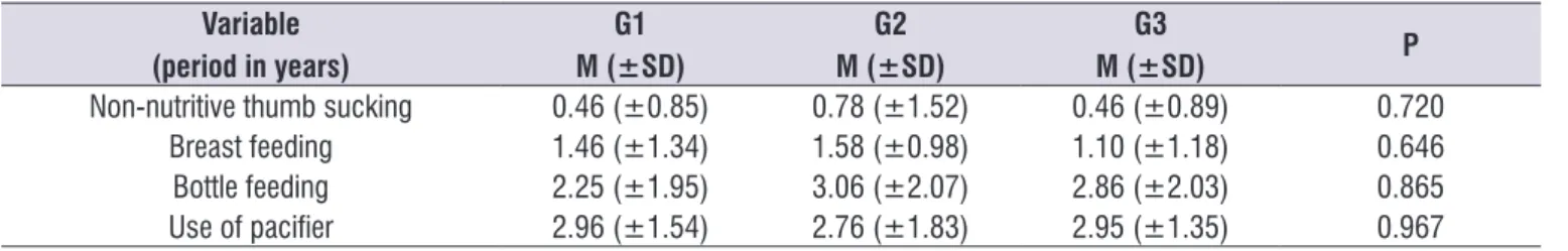
Results: there was no association between the number of attempts and the qualitative
variables evaluated by the protocol. There was also no difference between the groups regarding quantitative variables for breastfeeding time, breastfeeding occurrence, use of pacifiers, and thumb sucking.
Conclusion: the presence of DOH did not influence the number of forced spirometry
maneuvers, performed by the healthy children in this study.
INTRODUCTION
Orofacial function is the result of the integration between the central nervous system and the neuro-muscular system, which includes breathing, chewing, sucking and swallowing1,2. These actions are performed by the stomatognathic system (SS), which is composed of craniofacial structures such as bones of the head, mandible, hyoid, sternum, chewing and swallowing muscles3.
Some oral habits performed by children are considered deleterious to the SS. These include
non-nutritive thumb sucking, mouth breathing, pacifiers,
lower lip interposition/suction, tongue thrusting, nail biting, and jaw propulsion4,5. The persistence of delete-rious oral habits (DOH) plays a key role in causing malocclusions, orthodontic abnormalities and phonetic disorders, since they affect the growth and devel-opment of the muscles and bones of the mandible5-7. Thus, DOH may even interfere in facial aesthetics.
Spirometry is a pulmonary function test indicated for the diagnosis and monitoring of respiratory diseases,
widely used in scientific research and public health8,9. When performing spirometry, the individual must place the mouthpiece on his or her tongue and seal the lips so that air does not escape during the maneuvers, which require forced expiration, muscle strength and motor coordination. For this reason, children with a history of DOH and possible impairment of oral functions may
show difficulties in performing the test, requiring a
higher number of maneuvers and possible changes in their results.
Therefore, this study aims to analyze the relationship between the presence of DOH and the number of attempts of forced spirometry maneuvers performed by healthy children.
METHODS
This study has been approved by the Ethics Committee of the State University of Santa Catarina under Resolution Number 1.006.003 - (CAAE: 38770314.1.0000.0118), registered in the National Research Ethics Committee involving Human Beings (CONEP). All participants provided a minor’s assent document and a parent or guardian authorized their participation by signing an Informed Consent Document.
This is an observational and analytical cross-sectional study, including healthy children between 6 and 12 years of age from public and private schools
in the metropolitan area of Florianópolis - Santa Catarina/Brazil. Data was collected in the school site and the following individuals were selected: healthy non-athletes (children not enrolled in high-performance sports federations), non-obese children (body mass index above 25 kg/m²) and non-undernourished (mass index lower than 18.5kg/m2)10, children with no diagnosis or history of cardiorespiratory, musculo-skeletal, rheumatic, neurological diseases, auditory
or visual deficits, and children with normal spirometry
parameters.
Schoolchildren´s health was checked according to the International Study of Asthma and Allergies in Childhood (ISAAC)11 and the analysis of health history, previously completed by a parent or tutor. Spirometry showing forced expiratory volume in one second (FEV1) and forced vital capacity (FVC) above 80% of predicted, according to Polgar et al. (1971)12 was also considered. A JAEGER™ MasterScope IOS™ spirometer was used to record the parameters of FVC, FEV1 and peak
expiratory flow (PEF). The examination was conducted
according to the American Thoracic Society´s guidelines13, and forced spirometry maneuvers were performed until the values and curves were acceptable and reproducible, on a maximum of eight attempts. Children who failed to perform a valid test in this number of maneuvers were excluded.
An assessment tool based on the Orofacial Myofunctional Evaluation protocol (MBGR)14 was used to identify the presence of DOH. This tool considers elements including time of breastfeeding and the use
of the bottle; difficulties in introducing solid foods;
characteristics of chewing; frequency of episodes
such as choking, gastroesophageal reflux (GERD) and
coughing during meal. It also controls oral habits such
as use of pacifier or non-nutritive thumb sucking and
speech acquisition time. These items were answered by a parent or guardian. Ultimately, it includes the participants´ physical observation (made by a trained examiner) relating to lip positioning, saliva control, mobility and oral musculature tonus by means of a manual resistance (with emphasis on tongue, bucci-nators/orbicularis lips).
spirometry maneuvers needed to achieve successful spirometry results, as follows: G1) children who needed 3 maneuvers; G2) 4 maneuvers; G3) 5-8 maneuvers.
For the analyses, the variables were divided into qualitative and quantitative. Qualitative variables
included: labial posture, saliva control, feeding difficulty
(introduction of the cup, transition from liquid to pasty and from pasty to solid foods), characteristic of chewing (time with food inside the mouth), presence of GERD and coughing, history of non-nutritive thumb sucking or
use of pacifier, bottle feeding, history of breastfeeding
and speech quality. Quantitative variables included: breastfeeding period, whether the child was breastfed,
used a pacifier and sucked their thumbs.
Initially, statistical analysis was conducted by evalu-ating distribution of data using the Kolmogorov-Smirnov test. Then, the Kruskal-Wallis test was used to compare the quantitative variables between the 3 groups. An association between the groups and the qualitative variables was observed by means of the Chi-square
test. For all analyses, the significance level was set at
5% (p <0.05).
RESULTS
A total of 149 children participated in this study. They were allocated into 3 groups, whose age, gender and anthropometric measurements are described in Table 1.
Table 1. Distribution of gender, age and anthropometric data of the participants of each group
G1 M (±SD)
G2 M (±SD)
G3 M (±SD)
Sample
(prevailing gender)
59 (36 girls)
53 (30 boys)
37 (21 girls)
Age 9.24 (±1.89) 9.58 (±1.90) 8.59 (±1.87)
Weight 34.25 (±10.97) 37.96 (±11.54) 34.78 (±12.08)
Height 1.41 (±0.12) 1.44 (±0.13) 1.40 (±0.12)
Note: G1: group 1; G2: group 2; G3: group 3; Age: in years; Weight: in kilograms; Height: in centimeters; M: mean; SD: standard deviation.
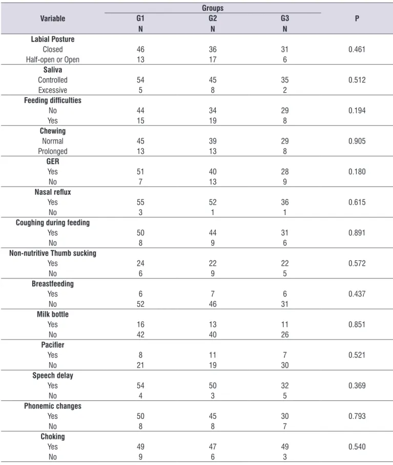
The results of the analyses of associations between the qualitative variables of orofacial function and the group are presented in Table 2. No statistically
significant difference could be observed, indicating no
association between the qualitative variables and the number of spirometry maneuvers.
Table 2. Result of Chi-square test for the comparison between quantitative variables of orofacial function and allocation into 3 groups
Variable
Groups
P G1
N
G2 N
G3 N Labial Posture
Closed 46 36 31 0.461
Half-open or Open 13 17 6
Saliva
Controlled 54 45 35 0.512
Excessive 5 8 2
Feeding difficulties
No 44 34 29 0.194
Yes 15 19 8
Chewing
Normal 45 39 29 0.905
Prolonged 13 13 8
GER
Yes 51 40 28 0.180
No 7 13 9
Nasal reflux
Yes 55 52 36 0.615
No 3 1 1
Coughing during feeding
Yes 50 44 31 0.891
No 8 9 6
Non-nutritive Thumb sucking
Yes 24 22 22 0.572
No 6 9 5
Breastfeeding
Yes 6 7 6 0.437
No 52 46 31
Milk bottle
Yes 16 13 11 0.851
No 42 40 26
Pacifier
Yes 8 11 7 0.521
No 21 19 30
Speech delay
Yes 54 50 32 0.369
No 4 3 5
Phonemic changes
Yes 50 45 30 0.793
No 8 8 7
Choking
Yes 49 47 49 0.540
No 9 6 3
Airflow leaks may be related to dentition. The correct
positioning of the teeth in the mouthpiece ensures that leaks will not occur. But, especially in the age range in which the primary teeth will be replaced, the presence of spaces between the teeth can allow air to escape.
Among the various DOH, oral breathing is a frequent habit among children, as reported in a study conducted by Melo and Pontes, showing occurrence of 48.60%. They also suggest that the habit of breathing through the mouth can interfere with the growth of the facial skull, favoring some physical characteristics, namely,
flaccidness face muscles, half-open lips, drooping
cheeks, hypotonic tongue in the lower position or between teeth and dental malocclusion19.
Dental malocclusion, which may make the mouth-piece of the spirometer unstable, is often caused by DOH7. Its presence may be determinant for an oral respiratory pattern in some individuals, which may lead to alterations in the orofacial musculature, such as hypotonia and hypofunction of the mandibular elevator muscles20.
Isometric contractions are mostly related to the alter-ations in the muscular strength (MS) and, therefore, to muscle fatigue21,22. They may interfere with the perfor-mance of a spirometry maneuver, since muscular action of the stomatognathic system is needed to seal the mouthpiece spirometer. A study conducted by Busanello-Stella et al. found a decrease in the muscular strength of the orbicularis oris muscles in children with nasal and oral breathing when sustaining isometric contraction against resistance. The group with mouth breathers reported earlier muscle fatigue21.
GER was one of the variables that, although not
statistically significant, was very frequent among
children who participated in this study. The literature shows that a GER´s complication includes tooth erosion and it is associated with bruxism23,24. Thus, GER may alter SS functions and consequently interfere in the spirometry test.
DISCUSSION
This is the first study to observe whether the
presence of DOH can directly interfere with the pulmonary function test. Notably, the maintenance of these habits can impair the structures and functions of the stomatognathic system, which are necessary for the performance of spirometry15. And although a direct relationship between the number of forced expiratory maneuvers and the history of DOH performed by the
MBGR protocol has not been identified, this may have
happened for some reasons that caused limitations in the study.
A first limitation of the study was the use of part
of the MBGR protocol. Therefore, the entire protocol should be administered. A second limitation was the use of part of the protocol that consisted mostly of qualitatively evaluated items. However, in the literature, the effects of DOH have been evaluated mainly qualita-tively with the MBGR protocol14,16.
Thus, specific SS assessment mechanisms are
relevant, including the strength and tone of the facial musculature, as well as their respective functions. This analysis enables the understanding of the real contribution of each element of the SS in examinations involving correct placement and maintenance of a
mouthpiece. In this context, electromyography verifies
the activation of certain facial muscles that are used during the spirometry testings17 and could be included in further studies on the subject.
Some factors should be considered in order to achieve an acceptable spirometry maneuver: the examiner´s adequate practical training, command voice, properly calibrated good quality equipment and control of the environment. In addition to these factors related to the technical aspects of the test, the under-standing and cooperation of the patient to perform the maneuver adequately should also be considered, in order to avoid early glottis closure, early termination,
glottal noise, buccal obstruction and airflow leak18.
Table 3. Result of Kruskal-Wallis test for the comparison between quantitative variables of orofacial function and allocation into 3 groups
Variable (period in years)
G1 M (±SD)
G2 M (±SD)
G3
M (±SD) P
Non-nutritive thumb sucking 0.46 (±0.85) 0.78 (±1.52) 0.46 (±0.89) 0.720
Breast feeding 1.46 (±1.34) 1.58 (±0.98) 1.10 (±1.18) 0.646
Bottle feeding 2.25 (±1.95) 3.06 (±2.07) 2.86 (±2.03) 0.865
Use of pacifier 2.96 (±1.54) 2.76 (±1.83) 2.95 (±1.35) 0.967
This study investigated the relationship between spirometry performance and DOH given the common
difficulty faced by the pediatric age group to perform
spirometry maneuvers adequately. This encouraged
further researches that identified possible factors
responsible for this event. In clinical practice, it is common for children to have spirometry curves with artifacts and parameters with values lower than those predicted for their age and gender. Coughing during
the testing, mouthpiece obstruction, airflow leak,
Valsalva maneuver and even unsatisfactory onset of forced spirometry maneuvers, with no evidence of effort and with inadequate termination, are some of the usual factors responsible for not meeting the criteria required for a valid the test. The relationship between these elements and DOH, i.e., the hypothesis formulated
in this study, has not been confirmed, and should be
further investigated.
Comprehensive research should also be conducted on the relationship between history of DOH and
specific evaluation of the orofacial musculature and
the performance of spirometry maneuvers in children with respiratory impairment, such as asthma, allergic
rhinitis and cystic fibrosis. In the study conducted by
Carvalho-Oliveira et al., although on asthmatic adults, there was a high frequency of SS changes, which had an association with the severity of asthma observed by
the forced expiratory volume in the first second (FEV1) of spirometry test25. Campanha et al. performed speech therapy treatment and observed clinical and functional improvement in asthmatic children, evidenced by the increase in the percentage values of the spirometry parameters26.
CONCLUSION
There was no relationship between the presence of DOH and the number of forced spirometry maneuvers performed by healthy children in the study population. However, a comprehensive investigation of the SS is required. Furthermore, this study should be conducted in a pediatric population with respiratory impairment.
REFERENCES
1. Strini PJ, Barbosa TD, Gavião MB. Assessment of orofacial dysfunctions, salivary cortisol levels and oral health related quality of life (ORHQoL) in young adults. Arch Oral Biol. 2011;56(12):1521-7.
2. Bakke M, Bergendal B, McAllister A, Sjogreen L, Asten P. Development and evaluation of a
comprehensive screening for orofacial dysfunction. Swed Dent J. 2007;31(2):75-84.
3. Cielo CA, Ribeiro VV, Christmann MK, Lima JPM, Pacheco-Rubim AB, Hoffmann CF et al. Stomatognatic system changes in dysphonic individuals. Rev. CEFAC. 2016;18(3):613-25.
4. Garde JB, Suryavanshi RK, Jawale BA, Deshmukh V, Dadhe DP, Suryavanshi MK. An epidemiological study to know the prevalence of deleterious oral habits among 6 to 12 year old children. J Int Oral Health. 2014;6(1):39-43.
5. Suhani RD, Suhani MF, Muntean A, Mesaros M, Badea ME. Deleterious oral habits in children with hearing impairment. Clujul Medical. 2015;88(3):403. 6. Prado DGA, Sovinski SRP, Nary Filho H, Brasolotto
AG, Berretin-Felix G. Oral motor control and orofacial functions in individuals with dentofacial deformity. Audiol Commun Res. 2015;20(1):76-83. 7. Sharma S, Bansal A, Asopa K. Prevalence of oral
habits among eleven to thirteen years old children in Jaipur. Int J Clin Pediatr Dent. 2015;8(3):208. 8. Miller MR, Hankinson JA, Brusasco V, Burgos F,
Casaburi R, Coates A et al. Standardisation of spirometry. Eur Respir J. 2005;26(2):319-38.
9. Brazzale D, Hall G, Swanney MP. Reference values for spirometry and their use in test interpretation: A position statement from the Australian and New Zealand Society of Respiratory Science. Respirology. 2016;21(7):1201-9.
10. World Health Organization. The World Health Organization Quality of Life assessment (WHOQOL): position paper from the World Health Organization. Soc Sci Med. 1995;41:1403-9.
11. Solé D, Vanna AT, Yamada E, Rizzo MCV, Naspitz CK. International study of asthma and allergies in childhood (ISAAC) written questionnaire: validation of the asthma component among Brazilian children. Invest Allergol Clin Immunol. 1998;8(6):376-82. 12. Polgar G, Weng TR. The functional development of
the respiratory system from the period of gestatin to adulthood. Am Rev Respir Dis. 1979;120(3):625-95. 13. American Thoracic Society. Standardization of
spirometry – 2005. Eur Respir J. 2005;26:319-38. 14. Genaro KF, Berretin-Felix G, Rehder MIBC,
Marchesan IQ. Avaliação Miofuncional Orofacial – Protocolo MBGR. Rev. CEFAC. 2009;11(2):237-55. 15. Pereira TS, Oliveira F, Cardoso MCAF. Association
2017 [cited 2017 Nov 05] ; 29(3):e20150301. Available from: http://www.scielo.br/scielo. p h p ? s c r i p t = s c i _ a r t t e x t & p i d = S 2 3 1 7 -17822017000300302&lng=en. Epub May 15, 2017. http://dx.doi.org/10.1590/2317-1782/20172015301. 16. Johanns CM, Silvério K, Furkim AM, Marchesan
I. Há relação de hábitos orais deletérios com a tipologia facial e a oclusão dentária? Rev. CEFAC. 2011;13(6):1095-102.
17. Prates LS, Gois M, Berwig LC, Blanco-Dutra AP, Busanello-Stella AR, Silva AMT. Clinical and electromyographic evaluation of mastication within different facial growth patterns. Rev. CEFAC. 2016;18(1):104-12.
18. Pereira CAC. Espirometria. J bras pneumol. 2002;28(3):1-82.
19. Melo PED, Pontes JRS. Deleterious oral habits in a group of children from a public school in Sao Paulo city. Rev. CEFAC. 2014;16(6):1945-52.
20. Emmerich A, Fonseca L, Elias AM, Medeiros UV. The relationship between oral habits, oronasopharyngeal alterations, and malocclusion in preschool children in Vitória, Espírito Santo, Brazil. Cad Saúde Pública. 2004;20(3):689-97. 21. Busanello-Stella AR, Blanco-Dutra AP, Corrêa ECR,
Silva AMT. Electromyographic fatigue of orbicular oris muscles during exercises in mouth and nasal breathing children. Codas. 2015;27(1):80-8.
22. Buzinelli RV, Bérzin F. Electromyographic analysis of fatigue in temporalis and masseter muscles during continuous chewing. J Oral Rehabil. 2001;28:1165-7.
23. Ranjitkar S, Smales RJ, Kaidonis JA. Oral
manifestations of gastroesophageal reflux
disease. J Gastroenterol Hepatol. 2012;27(1):21-7. 24. Mengatto CM, Dalberto Cda S, Scheeren B, Barros
SG. Association between sleep bruxism and
gastroesophageal reflux disease. J Prosthet Dent.
2013;110(5):349-55.
25. Carvalho-Oliveira M, Salles C, Terse R, D’Oliveira Júnior A. Association between severe asthma and changes in the stomatognathic system. J Bras Pneumol. 2016;42(6):423-8.