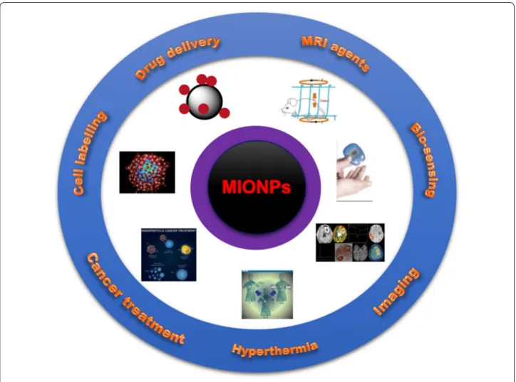
Multifunctional magnetic iron oxide nanoparticles: diverse synthetic approaches, surface modifications, cytotoxicity towards biomedical and industrial applications
Texto
Imagem


Documentos relacionados
Methods: Two translations and their respective back-translations were performed, as well as the evaluation of the semantic equivalence, the prepara- tion of the synthetic version,
E eu que- ria dizer que o Brasil somente ofertará esse setor de serviços educacionais se vocês, aqui, da Associação, chegarem a um consenso: “Queremos que o setor seja incluído
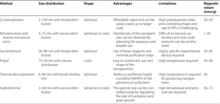
Source: Recent progress on magnetic iron oxide nanoparticles: synthesis, surface functional strategies, and biomedical applications (13), and In situ doxorubicin
Por fim, selecionei como ângulo de ataque inicial as narrativas temáticas do espetáculo, considerando os atravessamentos interpretativos/estéticos diversos que poderiam
descrever as alterações dermatológicas ocorrida nos animas que desenvolveram cisto folicular durante o ciclo reprodutivo, não sendo possível realizar a repdrodução
gradual, e não instantânea, do organismo humano, que se processa durante os curtos meses que medeiam entre a fecundação do óvulo e o nascimento da criança. Uma vez que a
The original tabletop - Exact IGRT Couch - presents higher attenuation values, followed by kVue TM Universal Couch (carbon fiber) and, finally, the Kvue TM Calypso
A avaliação diagnóstica das competências sociais foi organizada segundo quatro dimensões 4 : i) respeitar as regras de aula/trabalhar de forma cooperativa; ii)
