Joana Raimundo Pimenta
ACCUMULATION, RESPONSES AND GENOTOXICITY OF TRACE ELEMENTS IN
OCTOPUS VULGARIS
“Dissertação apresentada para obtenção do Grau de Doutor em Bioquímica pela Universidade Nova de Lisboa, Faculdade de Ciências e Tecnologia.”
I - Nº de arquivo
III Dedico este trabalho a duas mulheres fantásticas: A minha Mãe e a minha Avó
V
Agradecimentos
Ao Engenheiro Carlos Vale, por toda a sua disponibilidade, ajuda e comentários ao longo da realização do trabalho. Por todo o seu apoio nos momentos críticos e por acreditar em mim!!!! Os meus sinceros agradecimentos. À Professora Doutora Isabel Moura, por me ter permitido trabalhar no seu laboratório, e sem quaisquer objecções, aceitar o meu Doutoramento. Obrigada pela confiança transmitida pelos dois!
Depois de 11 anos a trabalhar no laboratório de metais do IPIMAR, sinto-me, sem dúvida, uma pessoa com muita sorte. Um obrigada muito grande à Patrícia, João, Hilda, Miguel, Rute, Pedro, Vasco, Marta, Engª Ana, Cristina, Isabelina, Rute, Miguel Nuno, Nuno, Pereira e Rui por todo o apoio e amizade ao longo destes anos. À Maria João, Pedro e Teresa, que embora “mais recentes”, têm sido incansáveis no seu apoio!
Os meus agradecimentos ao Professor Doutor José Moura por manter a porta do seu laboratório sempre aberta, e pelo seu constante interesse no trabalho. Obrigada!
A todos do 4º e 6º piso do Departamento de Química da Universidade Nova por me terem recebido sempre bem, apesar do “mau cheiro”! Um obrigada especial à Gabi, ao Pablo e à Sofia. Gostaria ainda de transmitir o meu profundo agradecimento à Ana Teresa, Marta, Raquel, Célia, Gabi, Rui, Rui, Filipe e muitos outros. Obrigada, pelos almoços, opiniões, sugestões e, principalmente, pela vossa paciência!
Gostaria, igualmente, de agradecer à Professora Doutora Maria Helena Costa por me ter disponibilisado o seu laboratório no decorrer do trabalho. Em partícular um obrigada ao Pedro por todos os momentos bem passados e por mergulhos parasidíacos, e ao Jorge e Mário pela sempre boa disposição.
Ao João, obrigada por seres tudo aquilo que eu preciso. Pela tua boa disposição, e apoio incondicional. E pela minha Matilde!! Obrigada!
Aos meus pais, sem o apoio dos quais teria sido impossivel atingir tal meta. Obrigada pela vossa constante presença, perseverança e incentivo.
À minha Avó, mulher sem igual! Ao meu irmão e à Sílvia, aos meus primos, tios e família somente por existirem e estarem sempre ao meu lado. A todos os meus amigos pela constante presença.
VI
À Fundação para a Ciência e a Tecnologia (FCT) pelo apoio concedido através da atribuição de uma bolsa de doutoramento (SFRH/BD/37730/2007).
VII
Sumário
IX
Summary
XI
Table of Contents
Acknowledgments V
Sumário VII
Summary IX
List of Figures XV
List of Tables XVII
Lista de Figuras XIX
Lista de Tabelas XXI
Chapter 1. General Introduction 1
General introduction 3
Coastal environment 3
Trace elements in marine organisms 3
Detoxification of accumulated trace elements 5
Sub-lethal effects 8
Octopus vulgaris (common octopus) 9
Cephalopods metal contamination 11
Portuguese coast as study area 13
Aims and Structure of the Thesis 15
Chapter 2. Elemental concentration and partitioning 27
2.1 Partitioning of Fe, Cu, Zn, Cd and Pb concentrations among eleven tissues of Octopus vulgaris from the Portuguese
coast 30
Abstract 31
Introduction 31
Material and Methods 32
Samples 32
Analytical procedure 32
Statistical analysis 33
Results 33
Metal concentrations 33
Effect of size/weight and sex on metal concentration 35
Metal–metal correlations 35
Differences of Fe, Cu, Zn, Cd and Pb among tissues 35
Discussion 35
Comparison with other works 35
Accumulated metals in organs/tissues 48
References 40
2.1 Total lead and its stable isotopes in digestive gland of Octopus vulgaris as a fingerprint 43
Abstract 45
Introduction 45
Material and Methods 46
Samples 46
Analytical methodology 57
Sample pre-treatment 47
Methods 48
Statistical analysis 49
Results 49
Biologic parameters in octopus 49
Lead concentrations and isotopic ratios in digestive gland 49
Aluminium and lead concentrations in sediments 50
XII
Discussion 51
References 54
2.3 Relations between mercury, methyl-mercury and selenium in tissues of Octopus vulgaris from the Portuguese
Coast 57
Abstract 59
Introduction 59
Material and Methods 60
Samples 60
Analytical methodology 61
Statistical analysis 62
Results 62
Biological data 62
Metal concentrations in digestive gland and mantle 63
Differences between areas of capture 64
Discussion 64
Effect of size and gender on Hg accumulation 64
Comparison of metal levels with the literature 64
Elevated concentration of Hg in octopus from SE Portuguese coast 66
Relationships between levels in digestive gland and mantle 66
Selenium and Mercury 67
Octopus as a source of Hg in human consumption 68
References 69
3. Sub-cellular responses to elemental concentrations 73
3.1 Sub-cellular partitioning of Zn, Cu, Cd and Pb in the digestive gland of native Octopus vulgaris exposed to different
metal concentrations (Portugal) 75
Abstract 77
Introduction 77
Material and Methods 78
Samples 78
Sub-cellular fractionation 79
Metal analyses 79
Statistical analysis 80
Results 80
Whole digestive gland 81
Insoluble fraction 82
Discussion 82
References 87
3.2 Sub-cellular partitioning of trace elements in digestive gland, kidney and gills of native Octopus vulgaris (Portugal) 91
Abstract 93
Introduction 93
Material and Methods 94
Samples 94
Analytical methodology 94
Sub-cellular fractionation 94
Trace elements 95
Statistical analysis 95
Results 96
Biological data 96
Influence of tissue and sampling area on trace element variability 96
Trace element concentrations in tissues 96
Trace element concentrations in sub-cellular fractions 98
XIII
Discussion 100
Metal content in the cytosolic fraction 100
Metals in organelle fractions 101
Relationships of trace element concentrations between sub-cellular fractions and whole tissue 101
References 105
3.3 Association of Zn, Cu, Cd and Pb with protein fractions and sub-cellular partitioning in the digestive gland of
Octopus vulgaris living in different metal exposure 111
Abstract 113
Introduction 113
Material and Methods 114
Composite samples 114
Protein purification 115
Metal analyses 115
Statistical analysis 116
Results and Discussion 116
Metal concentrations 116
Cytosolic fraction 117
Chromatographic analysis 118
Metal association with LMW proteins 119
Metal-metal relationships 120
References 122
3.4 Metallothioneins and trace elements in digestive gland, gills, kidney and gonads of Octopus vulgaris 125
Abstract 127
Introduction 127
Material and Methods 128
Study areas 128
Sampling 129
Metal determinations 129
Quantification of metallothionein (MT) 129
Statistical analysis 130
Results 130
Metal concentrations in tissues 130
Levels of metallothioneins-like proteins (MT) 132
Discussion 133
Relation between MT and Metals 134
References 138
4. Genotoxic effects 143
4.1 DNA damage and metal accumulation in digestive gland, gills, kidney and mantle of wild Octopus vulgaris
(Portugal) 145
Abstract 147
Introduction 147
Material and Methods 148
Samples 148
Analytical methodology 149
Metals 149
DNA Strand Breaks 149
Statistical analysis 150
Results 151
Influence of size/weight and gender 151
Metal partitioning 151
Differences of metal concentrations between areas of capture 152
XIV
Discussion 153
References 156
5. General Discussion 159
General discussion 161
Elemental concentrations and partitioning 161
Sub-cellular responses to elemental concentrations 163
Genotoxic effects 164
Final Remarks 165
References 166
Appendix – Methodologies 169
Methodologies 171
Biological samples 171
Metal analyses 171
Biological samples 171
Sediment samples 171
Analytical methods 172
Sub-cellular fractionation 172
Protein purification 174
Metallothionein analyses 175
DNA strand breakages 175
XV
List of Figures
Figure 1.1 - Mechanisms occurring in organisms: uptake, storage and detoxification of contaminants. 5
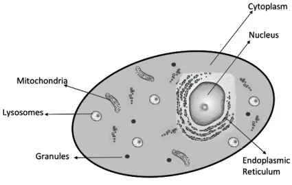
Figure 1.2 - Schematic example of a eukaryotic cell, with the various components/organelles. 8
Figure 1.3 - Schematic representation of processes leading to DNA damages from exposure to effects on populations (Me –
metals). 9
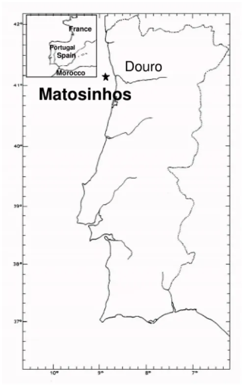
Figure 2.1.1 - Location of the sampling area of O. vulgaris in the Portuguese coast: Matosinhos. 32
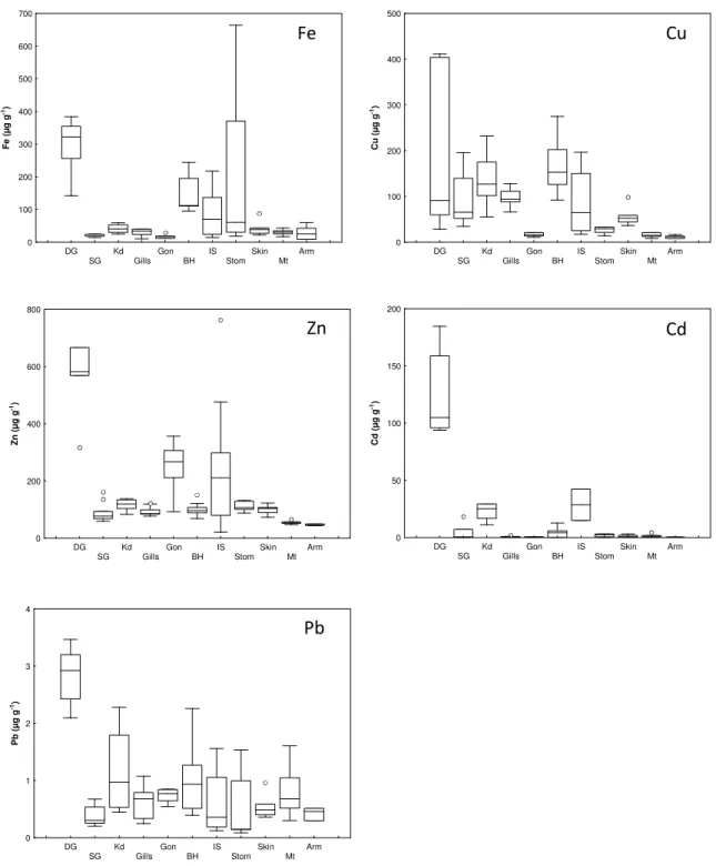
Figure 2.1.2 - Median, 25 and 75% percentile, minimum and maximum, and the extreme values ( ) and outliers (•), of Fe, Cu, Zn, Cd and Pb concentrations (µg g-1, dry weight) in the digestive gland (DG), posterior salivary glands (SG), kidneys (Kd), gills, gonads (Gon), branchial hearts (BH), ink sac (IS), stomach (Stom), skin, mantle (Mt) and arm of common octopus, O. vulgaris.
34
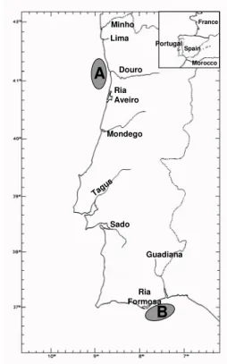
Figure 2.2.1 - Octopus vulgaris. Location of the two sampling sites in the Portuguese coast: A (Matosinhos) and B (Olhão). 47
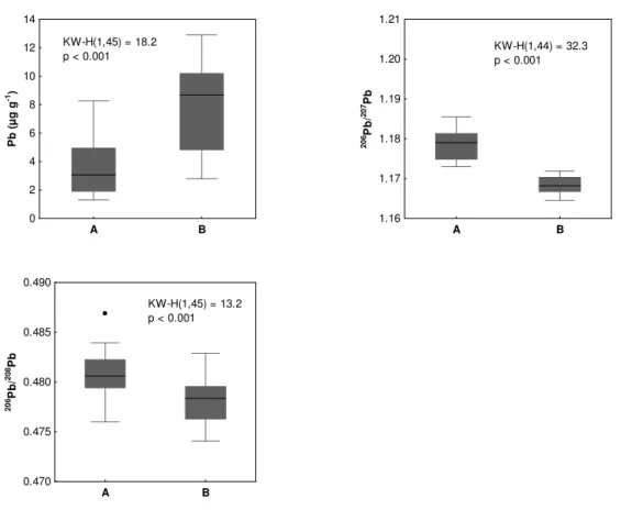
Figure 2.2.2 - Octopus vulgaris. Median, 25% and 75% percentiles, minimum and maximum, outliers (•), Kruskal-Wallis test (KW-H) and p-values of Pb concentrations (µg g-1, dry weight) and 206Pb/207Pb and 206Pb/208Pb ratios in the digestive gland of common octopus collected in two areas of the Portuguese coast (A and B).
50
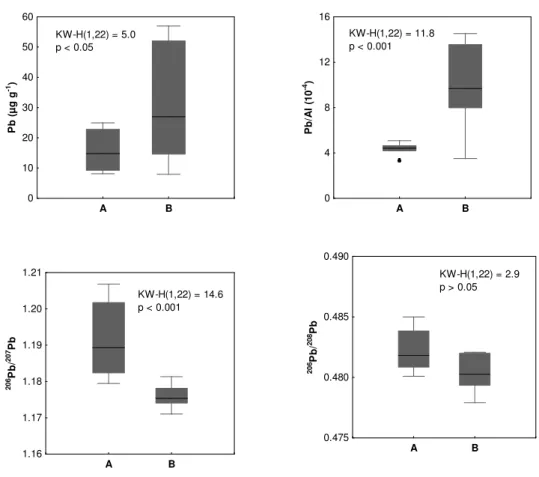
Figure 2.2.3 - Median, 25% and 75% percentiles, minimum and maximum, outliers (•), Kruskal-Wallis test (KW-H) and p-values of Pb concentrations (µg g-1, dry weight), Pb/Al (10-4) and 206Pb/207Pb and 206Pb/208Pb ratios in the surface sediments collected in two areas of the Portuguese coast (A and B).
51
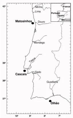
Figure 2.3.1 – Location of the three areas of capture of Octopus vulgaris in the Portuguese Coast: Matosinhos, Cascais and
Olhão. 60
Figure 2.3.2 - Median, 25 and 75% percentile, minimum and maximum, and the extreme values ( ) and outliers (•), of Hg, MeHg and Se concentrations (µg g-1, dry weight) and MeHg (%) in the digestive gland (black boxes) and mantle (white boxes) of common octopus, O. vulgaris from the three areas of capture.
63
Figure 2.3.3 – Relationships between concentrations of Hg and MeHg (µg g-1, dry weight) in mantle and digestive gland of O.
vulgaris from Matosinhos (♦), Cascais ( ) and Olhão ( ). 67
Figure 2.3.4 – Relationships between concentrations of Hg and MeHg (µg g-1, dry weight) for the digestive gland and mantle
of O. vulgaris from Matosinhos (♦), Cascais ( ) and Olhão ( ). 67
Figure 3.1.1 – Location of the two sampling sites of O. vulgaris in the Portuguese coast: Matosinhos and Olhão. 78
Figure 3.1.2 – Relationships between levels of Zn, Cu, Cd and Pb (µg g-1, dry weight) in: nuclei, mitochondria, lysosomes and micrososmes and the whole digestive gland of O. vulgaris from Matosinhos (♦) and Olhão ( ). 85
Figure 3.2.1 – Location of the three areas of capture of Octopus vulgaris in the Portuguese Coast: Matosinhos (A), Cascais (B)
and Olhão (C). 94
Figure 3.2.2 – Principal component analysis of metals in tissues of common octopus, O. vulgaris from the three capture areas.
DG - digestive gland, K - kidney and G – gills. 97
Figure 3.2.3 - Median, 25 and 75% percentile, minimum and maximum, of Log V, Co, Zn, Cu, As, Cd and Pb concentrations (µg g-1, dry weight) in the digestive gland (black boxes), kidney (grey boxes) and gills (white boxes) of common octopus, O. vulgaris from the three areas of capture.
98
Figure 3.2.4 – Median, 25 and 75% percentile, minimum and maximum, of Log V, Co, Zn, Cu, As, Cd and Pb concentrations (µg g-1, dry weight) in the digestive gland (black boxes), kidney (grey boxes) and gills (white boxes) of common octopus, O. vulgaris from the three areas of capture. a – Granules; b – Mitochondria; c – Lysosomes plus microsomes; d – HDP; e – HSP.
99
Figure 3.2.5 - Relationships between Log levels of Co and Cd (µg g-1, dry weight) in: granules (Gran), mitochondria (Mit), lysosomes plus micrososmes (Lys+Mic), HDP and HSP and the whole digestive gland (♦), kidney ( ) and gills ( ) of O. vulgaris.
XVI
Figure 3.3.1 – Location of the two sampling areas of O. vulgaris in the Portuguese coast: Matosinhos and Olhão. 114 Figure 3.3.2 – Median, 25 and 75% percentile, minimum and maximum of Zn, Cu, Cd and Pb (µmol g-1, dry weight)
concentrations in composite samples of Octopus vulgaris digestive gland captured in Matosinhos and Olhão. 117
Figure 3.3.3 - Sephadex G-75 profiles of Zn, Cu, Cd and Pb concentrations (nmol L-1) (♦) in Octopus vulgaris digestive gland cytosol of samples from Matosinhos and Olhão and 280 nm absorbance (-). 119
Figure 3.3.4 – Cadmium concentrations (nmol L-1) (♦) in the octopus digestive gland of samples from Matosinhos and Olhão
and 254:280 nm ratio in the chromatographic profile (-). 120
Figure 3.3.5 – Relations between Cd and Cu and Zn (nmol L-1) in the peak with low molecular weight of the cytosol from the
digestive gland of octopus from Matosinhos and Olhão. 121
Figure 3.4.1 – Location of the three areas of capture of Octopus vulgaris in the Portuguese Coast: Matosinhos (A, 41º 09.0’ N08º 41.1’ W), Cascais (B, 38º 36.0’ N; 09º 27.2’ W) and Olhão (C, 36º 55.0’ N; 07º 52.7’ W). 128
Figure 3.4.2 - Median, 25 and 75% percentile, minimum and maximum of Co, Zn, Cu and Cd logarithmic concentrations (µg g -1
, dry mass) in the digestive gland (Dig Gland), Gills, Kidney and Gonads of common octopus, O. vulgaris from areas A (black boxes), B (grey boxes) and C (white boxes).
131
Figure 3.4.3 – Median, 25 and 75% percentile, minimum and maximum, of V, Cr, Ni and As concentrations (µg g-1, dry mass) in the digestive gland (Dig Gland), Gills, Kidney and Gonads of common octopus, O. vulgaris from areas A (black boxes), B (grey boxes) and C (white boxes).
132
Figure 3.4.4 – Median, 25 and 75% percentile, minimum and maximum, of metallothionei (MT) concentrations (mg g-1, dry mass) in the digestive gland (Dig Gland), Gills, Kidney and Gonads of common octopus, O. vulgaris from areas A (black boxes), B (grey boxes) and C (white boxes).
133
Figure 3.4.5 – Principal component analysis of metals and metallothionein (MT) concentrations in the (a) digestive gland (Dig
Gland), (b) Gills, (c) Kidney and (d) Gonads of common octopus, O. vulgaris from areas A, B and C. 135/136
Figure 4.1.1 – Location of the two areas of capture of Octopus vulgaris in the Portuguese Coast: Matosinhos and Olhão. 149
Figure 4.1.2 – Comet examples of DNA-SB from Octopus vulgaris: ≅ 0% (A, gonads), ≅ 27% (B, kidney), ≅ 68% (C, gills) and
≅74% (D, digestive gland). 150
Figure 4.1.3 - Median, 25 and 75% percentile, minimum and maximum, and the extreme values ( ) and outliers (•), of Zn, Cu, Cd and Pb concentrations (µg g-1, dry weight) in the digestive gland (Dig Gland), Gills, Kidney and Gonads of common octopus, O. vulgaris from Matosinhos (black boxes) and Olhão (white boxes).
152
Figure 4.1.4 – Median, 25 and 75% percentile, minimum and maximum, and the extreme values ( ) and outliers (•), of DNA strand breakage (DNA-SB) (%) in the digestive gland (Dig Gland), Gills, Kidney and Gonads of common octopus, O. vulgaris
from Matosinhos (black boxes) and Olhão (white boxes).
153
Figure Ap.1– Schematic representation of mantle length measurement. 171
Figure Ap.2 – Schematic procedure of the sub-cellular fractionation by sequential centrifugation (adapted from Campbell et
al., 2005). 173
Figure Ap.3 – Schematic procedure of the sub-cellular fractionation by sequential centrifugation (adapted from Wallace et al.,
XVII
List of Tables
Table 2.1.1 – Metal-metal correlations (r) and associated probabilities (a – 0.05; b – 0.01; c – 0.001) in the digestive gland, salivary glands, ink sac and stomach of the O. vulgaris captured in Matosinhos. 35
Table 2.1.2 – Comparison of Fe, Zn, Cu, Cd and Pb levels (µg g-1, dry weight) in the digestive gland, branchial hearts, gills,
gonads, posterior salivary glands, mantle, arm and mantle skin of O. vulgaris from Matosinhos (present study) with data from the literature.
37
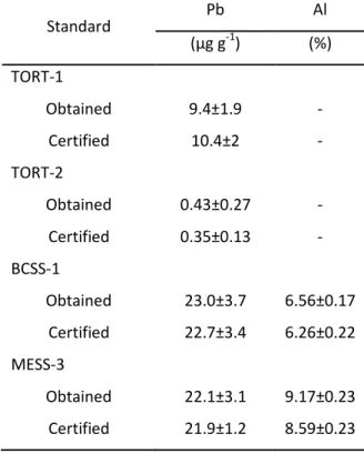
Table 2.2.1 – Lead (µg g-1, dry weight) and Al (%, dry weight) concentrations of lobster hepatopancreas (TORT-1 and TORT-2) and marine sediments (BCSS-1 and MESS-3) (NRCC) obtained in the present study and certified values. 48
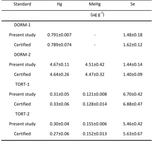
Table 2.3.1 – Mercury, MeHg and Se concentrations (µg g-1, dry wt) of dogfish muscle (DORM-1 and DORM-2) and lobster hepatopancreas (TORT-1 and TORT-2) (NRCC) determined in the present study and certified values. 62
Table 2.3.2 – Comparison of Hg and Se levels (µg g-1, dry weight) in the digestive gland and mantle of O. vulgaris from Portuguese coast (Matosinhos, Cascais and Olhão) with values in the literature. 65
Table 3.1.1 – Number of individuals (n), and ranges of weight (g) and length (mm) of specimens included in the composite samples of digestive gland of Octopus vulgaris from Matosinhos and Olhão; three sampling periods were considered. 79
Table 3.1.2 - Zinc, Cu, Cd and Pb concentrations (µg g-1, dry wt) of lobster hepatopancreas certificate standards (TORT-1 and
TORT-2) (NRCC) determined in the present study and certified values. 80
Table 3.1.3 – Median and ranges of Zn, Cu, Cd and Pb concentration (µg g-1, dry weight) in whole digestive gland and their insoluble fractions (nuclei, mitochondria, lysosomes and microsomes) of common octopus (n=6 Matosinhos; n=7 Olhão). 81
Table 3.1.4 – Comparison of Zn, Cu, Cd and Pb levels (µg g-1, dry weight) in the digestive gland of
O. vulgaris from Matosinhos
and Olhão with cephalopod data from the literature. 83
Table 3.2.1 - Size (mm), weight (g) and female:male proportion of Octopus vulgaris captured in the three sampling areas along
the Portuguese coast. 96
Table 3.2.2 – Sub-cellular mass (%), V , Co, Zn, Cu, As, Cd and Pb levels (µg g-1, dry weight) and sub-cellular distribution in the
digestive gland, kidney and gills of O. vulgaris. 109
Table 3.3.1 - Number of individuals (n), and the ranges of weight (g) and mantle length (mm) of Octopus vulgaris captured in
the Portuguese coast. 114
Table 3.3.2 - Zinc, Cu, Cd and Pb concentrations (nmol g-1, dry wt) of lobster hepatopancreas (TORT-1 and TORT-2) (NRCC)
XIX
Lista de Figuras
Figure 1.1 - Mecanismos que ocorrem nos organismos: uptake, armazenamento e desintoxicação de contaminantes. 5
Figure 1.2 – Exemplo esquemático de uma célula eucarionte, com os diversos componentes/organelos. 8
Figure 1.3 – Representação esquemática dos processos que favorecem o aparecimento de danos no DNA, desde a exposição
até aos efeitos na população (Me-Metais). 9
Figure 2.1.1 – Localização da area de captura de O. vulgaris na costa Portuguesa: Matosinhos. 32
Figure 2.1.2 – Mediana, percentis 25 e 75%, mínimo e máximo, e valores extremos ( ) e outliers(•), das concentrações de Fe, Cu, Zn, Cd e Pb (µg g-1, peso seco) na glândula digestiva (DG), glândulas salivares posteriores (SG), rim (Kd), brânquias, gónadas (Gon), corações branquiais (BH), saco de tinta (IS), estômago (Stom), pele, manto (Mt) and braço do polvo comum, O. vulgaris.
34
Figure 2.2.1 - Octopus vulgaris. Localização das duas áres de captura: A (Matosinhos) e B (Olhão). 47
Figure 2.2.2 - Octopus vulgaris. Mediana, percentis 25 e 75%, mínimo e máximo, outliers (•), teste Kruskal-Wallis (KW-H) e valores de p das concentrações de Pb (µg g-1, peso seco) e razões 206Pb/207Pb e 206Pb/208Pb na glândula digestive do polvo comum capturado em duas áreas da costa Portuguesa (A e B).
50
Figure 2.2.3 - Mediana, percentis 25 e 75%, mínimo e máximo, outliers (•), teste Kruskal-Wallis (KW-H) e valores de p das concentrações de Pb (µg g-1, peso seco), Pb/Al (10-4) e razões 206Pb/207Pb e 206Pb/208Pb em sedimentos superficiais colhidos em duas áreas da costa Portuguesa (A e B).
51
Figure 2.3.1 – Localização das três áreas de caputra de Octopus vulgaris na costa Portuguesa: Matosinhos, Cascais e Olhão. 60
Figure 2.3.2 - Mediana, percentis 25 e 75%, mínimo e máximo, e valores extremos ( ) e outliers(•), das concentrações de Hg, MeHg e Se (µg g-1, peso seco) e MeHg (%) na glândula digestiva (black boxes) e manto (white boxes) do polvo comum,
O. vulgaris das três áreas de captura.
63
Figure 2.3.3 – Relações entre as concentrações de Hg e MeHg (µg g-1, peso seco) no manto e glândula digestiva de O. vulgaris
de Matosinhos (♦), Cascais ( ) e Olhão ( ). 67
Figure 2.3.4 – Relações entre as concentrações de Hg e MeHg (µg g-1, peso seco) para a glândula digestiva e manto de O.
vulgaris de Matosinhos (♦), Cascais ( ) e Olhão ( ). 67
Figure 3.1.1 – Localização das duas areas de captura de O. vulgaris na costa Portuguesa: Matosinhos e Olhão. 78
Figure 3.1.2 – Realções entre os níveis de Zn, Cu, Cd e Pb (µg g-1, peso seco) no: nucleo, mitocôndria, lisossomas and microssomas e a glândula digestiva de O. vulgaris de Matosinhos (♦) e Olhão ( ). 85
Figure 3.2.1 – Localização das três areas de captura de Octopus vulgaris na costa Portuguesa: Matosinhos (A), Cascais (B) e
Olhão (C). 94
Figure 3.2.2 – Análise de Componentes Principais dos metais nos tecidos do polvo comum, O. vulgaris das três áreas de
captura. DG – glândula digestiva, K - rim e G – brânquias. 97
Figure 3.2.3 - Mediana, percentis 25 e 75%, mínimo e máximo, do Logaritmo das concentrações de V, Co, Zn, Cu, As, Cd e Pb (µg g-1, peso seco) na glândula digestiva (caixas pretas), rim (caixas cinzentas) e brânquias (caixas brancas) do polvo comum, O. vulgaris das três áreas de captura.
98
Figure 3.2.4 – Mediana, percentis 25 e 75%, mínimo e máximo, do Logaritmo das concentrações de V, Co, Zn, Cu, As, Cd e Pb (µg g-1, peso seco) na glândula digestiva (caixas pretas), rim (caixas cinzentas) e brânquias (caixas brancas) do polvo comum, O. vulgaris das três áreas de captura. a – Granulos; b – Mitocôndria; c – Lisossomas e microssomas; d – HDP; e – HSP.
99
Figure 3.2.5 – Relações entre o Logaritmo dos níveis de Co e Cd (µg g-1, peso seco) nos: granulos (Gran), mitocôndria (Mit),
lisossomas e microssomas (Lys+Mic), HDP e HSP e a glândula digestiva (♦), rim ( ) e brânquias ( ) de O. vulgaris. 103/104
XX
Figure 3.3.2 – Mediana, percentis 25 e 75%, mínimo e máximo, das concentrações de Zn, Cu, Cd e Pb (µmol g-1, peso seco) nas amostras compostas de glândula digestiva de Octopus vulgaris capturado em Matosinhos e Olhão. 117
Figure 3.3.3 – Perfis de Sephadex G-75 das concentrações de Zn, Cu, Cd and Pb (nmol L-1) (♦) no citosol da glândula digestive
de Octopus vulgaris de Matosinhos e Olhão e absorvância 280 nm (-). 119
Figure 3.3.4 – Concentração de Cd (nmol L-1) (
♦) nas amostra de glândula digestiva de polvo de Matosinhos e Olhão e razões
254:280 nm do perfil cromatográfico (-). 120
Figure 3.3.5 – Relações entre Cd e Cu e Zn (nmol L-1) no pico de baixo peso molecular do citosol da glândula digestiva do polvo
de Matosinhos e Olhão. 121
Figure 3.4.1 – Localização das três áreas de captura de Octopus vulgaris na costa Portuguesa: Matosinhos (A, 41º 09.0’ N08º 41.1’ W), Cascais (B, 38º 36.0’ N; 09º 27.2’ W) e Olhão (C, 36º 55.0’ N; 07º 52.7’ W). 128
Figure 3.4.2 - Mediana, percentis 25 e 75%, mínimo e máximo, do Logaritmo das concentrações de Co, Zn, Cu e Cd (µg g-1, massa seca) na glândula digestiva (Dig Gland), brânquias, rim e gónadas do polvo comum, O. vulgaris das áreas A (caixas pretas), B (caixas cinzentas) e C (caixas brancas).
131
Figure 3.4.3 – Mediana, percentis 25 e 75%, mínimo e máximo, das concentrações de V, Cr, Ni e As (µg g-1, massa seca) na glândula digestiva (Dig Gland), brânquias, rim e gónadas do polvo comum, O. vulgaris das áreas A (caixas pretas), B (caixas cinzentas) e C (caixas brancas).
132
Figure 3.4.4 – Mediana, percentis 25 e 75%, mínimo e máximo, das concentrações de metalotioninas (MT) (mg g-1, massa seca) na glândula digestiva (Dig Gland), brânquias, rim e gónadas do polvo comum, O. vulgaris das áreas A (caixas pretas), B (caixas cinzentas) e C (caixas brancas).
133
Figure 3.4.5 – Análise de Componentes Principais das concentrações de metais e metalotioninas (MT) na (a) glândula
digestiva (Dig Gland), (b) brânquias, (c) rim e (d) gónadas do polvo comum, O. vulgaris das três áreas A, B e C. 135/136
Figure 4.1.1 – Localização das duas areas de captura de Octopus vulgaris na costa Portuguesa: Matosinhos e Olhão. 149
Figure 4.1.2 – Exemplo de Comets de DNA-SB de Octopus vulgaris: ≅ 0% (A, gónadas), ≅ 27% (B, rim), ≅ 68% (C, brânquias)
and ≅74% (D, glândula digestiva). 150
Figure 4.1.3 - Mediana, percentis 25 e 75%, mínimo e máximo, e valores extremos ( ) e outliers(•), das concentrações de Zn, Cu, Cd e Pb (µg g-1, peso seco) na glândula digestiva (Dig Gland), brânquias, rim e gónadas do polvo comum, O. vulgaris de Matosinhos (caixas pretas) e Olhão (caixas brancas).
152
Figure 4.1.4 – Mediana, percentis 25 e 75%, mínimo e máximo, e valores extremos ( ) e outliers(•) dos danos de DNA (DNA-SB) (%)glândula digestiva (Dig Gland), brânquias, rim e gónadas do polvo comum, O. vulgaris de Matosinhos (caixas pretas) e Olhão (caixas brancas).
153
Figure Ap.1– Representação esquemática da mediçã do comprimento. 171
Figure Ap.2 – Procedimento para fraccionamento sub-celular por centrifugação sequencial (adaptado de Campbell et al.,
2005). 173
Figure Ap.3 – Procedimento para fraccionamento sub-celular por centrifugação sequencia (adaptado de Wallace et al., 2003 e
XXI
Lista de Tabelas
Table 2.1.1 – Correlações metal-metal (r)e probabilidade (a – 0.05; b – 0.01; c – 0.001) na glândula digestive, glândulas salivares, saco de tinta e estômago de O. vulgaris capturado em Matosinhos. 35 Table 2.1.2 – Comparação dos níveis de Fe, Zn, Cu, Cd e Pb (µg g-1, peso seco) na glândula digestive, corações branquiais,
brânquias, gónadas, glândulas salivares posteriors, manto, braço e pele do manto de O. vulgaris de Matosinhos (presente estudo) comdados da literatura.
37
Table 2.2.1 – Concentrações de Pb (µg g-1, peso seco) e Al (%, peso seco) no hepatopâncreas de lagosta (TORT-1 e TORT-2) e sedimentos marinhos (BCSS-1 e MESS-3) (NRCC) obtidos no presente estudo e valores certificados. 48
Table 2.3.1 – Concentrações de Hg, MeHg e Se (µg g-1, peso seco) no músculo de peixe-cão (DORM-1 e DORM-2) e hepatopâncreas de lagosta (TORT-1 e TORT-2) (NRCC) determinados no presente estudo e valores certificados. 62
Table 2.3.2 – Comparação dos níveis de Hg e Se levels (µg g-1, peso seco) na glândula digestiva e manto de O. vulgaris da costa Portuguesa (Matosinhos, Cascais e Olhão) com valores da literatura. 65
Table 3.1.1 – Número de indivíduos (n), e ranges de peso (g) e comprimento (mm) dos especimens incluídos nas amostras compostas da glândula digestiva de Octopus vulgaris de Matosinhos e Olhão; foram considerados três períodos de amostragem.
79
Table 3.1.2 – Concentrações de Zn, Cu, Cd e Pb (µg g-1, peso seco) no hepatopâncreas de lagosta (TORT-1 e TORT-2) (NRCC)
determinados no presente estudo e valores certificados. 80
Table 3.1.3 – Medianas e ranges das concentrações de Zn, Cu, Cd and Pb (µg g-1, peso seco) na glândula digestive e fracções sub-celulares (nucleo, mitocôndria, lisossomas e microssomas) do polvo comum (n=6 Matosinhos; n=7 Olhão). 81
Table 3.1.4 – Comparação dos níveis de Zn, Cu, Cd e Pb (µg g-1, peso seco) na glândula digestiva de O. Vulgaris de Matosinhos e
Olhão com dados da literatura em cefalópodes. 83
Table 3.2.1 - Tamanho (mm), peso (g) e proporção fêmea:macho de Octopus vulgaris capturados em três áreas da costa
Portuguesa. 96
Table 3.2.2 – Mass sub-celulart (%), níveis de V , Co, Zn, Cu, As, Cd e Pb (µg g-1, peso seco) e distribuição sub-celularna glândula
digestiva, rim e brânquias de O. vulgaris. 109
Table 3.3.1 – Número de indivíduos (n), e ranges de peso (g) e tamanho do manto (mm) de Octopus vulgaris capturado na costa
Portuguesa. 114
Table 3.3.2 – Concentrações de Zn, Cu, Cd e Pb (nmol g-1, peso seco) no hepatopâncreas de lagosta (TORT-1 e TORT-2) (NRCC)
Chapter 1
Chapter 1
General introduction
Context
This chapter presents an overview of the issues developed in this thesis. The first part reviews the
accumulation pathways and detoxification mechanisms found in organisms. It also briefly describes the
processes leading to sub-lethal effects, e.g. genotoxic effects. The second part describes the octopus
Chapter 1
3 Introduction
Coastal environment
From an environmental point of view, the coastal environment is a geographic space influenced
by terrestrial inputs and intense processes of bio-production (Fernandes, 1997; Castro et al., 1999). Sixty
percent of the existing large cities, wich comprise more than 2.5 million people, are located near the
coast. A substantial proportion of wastewater generated from human activity reaches directly or through
rivers the coastal environment with little or no treatment (Islam and Tanaka, 2004). Concerns of
long-term adverse effects of contaminants on aquatic ecosystems emerged in the last decades (van der Oost et
al., 2003). In addition, the fate and effects on exposed target organisms have also been extensively
studied in the aquatic ecosystem (e.g. Depledge and Fossi, 1994). In line with the potential risks for the
coastal ecosystem wellbeing the European Water Framework Directive (WFD; 2000/60/EC) establishes a
framework for the protection and improvement of ecological quality in transitional and coastal waters.
The aim is to achieve a good quality water status for all aquatic systems (Muxika et al., 2007). In
particular, the efforts on restoring impacted ecosystems have been widely welcomed by scientists and
environmental managers (Kowalski, 2009, references herein). However, coastal environments are
dynamic and complex ecosystems, and spatial-temporal variability associated from natural processes may
mask the effect of anthropogenic pressures.
Trace elements in marine organisms
Trace elements are widely found in marine organisms, reflecting its availability on the
environment. Some trace elements at certain concentration intervals are important for organisms, playing
an essential role in tissue metabolism and growth (Leland and Kuwabara, 1985). For example, Cu, Zn and
Fe are known as vital components of enzymes, respiratory proteins and certain structural elements of
organisms (Depledge and Rainbow, 1990). Manganese, Se and Co have also important roles in various
cellular components like, pyrovate carboxylase, glutathione peroxidase and vitamin B12, respectively.
While a range of trace metals must be delivered to the tissues of an organism in order to meet the diverse
metabolic requirements, accumulation of potentially toxic metal species may also occur. Indeed, some
elements display high concentrations in tissues of marine organisms, and the question is whether it
results from natural processes or influenced by the increasing availability in contaminated areas. Metals,
such as Cd, Hg and Pb, have been considered as non-essential because they have no known biological
role; these metals become highly toxic when found associated with metabolically active sites, even at
relatively low concentrations (Rainbow, 1985).
Assimilation pathways. The uptake of trace elements by marine organisms can occur from water, including suspended particulate matter, food and sediments. The exposure of organisms to contaminants
via food and water will depend on the ecological lifestyles of the aquatic organisms (Valavanidis et al.,
Chapter 1
4
organisms lacking external shells (e.g. cephalopods) and in all molluscs some absorption of dissolved
metal across the digestive epithelium (Langston et al., 1998). Nevertheless, since the branchial epithelium
represents the primary target for waterborne due to their respiratory/nutritional functions, enhanced
metal levels are often found (Langston et al., 1998; Pan and Zhang, 2006). Food may also be a significant
source of metals, if not the primary source, to organisms. Studies suggested that metal partitioning in
tissues such as digestive gland and muscle, are mostly affected by metals accumulated from food, while
for gills the major vector is water (e.g. Langston et al., 1998). Water-soluble (hydrophilic) elements are
more bioavailable to organisms than water-insoluble (hydrophobic) elements that are strongly sorbed or
bound to suspended particles, dissolved organic matter, or biological systems (Rand and Petrocelli, 1985).
Class B elements such as Cu, Zn and Cd, form a wide range of covalent compounds and are therefore less
likely to exist as free ions in solution (Simkiss and Mason, 1984). Usually they are present in biological
tissues as divalent cations, which are free or complexed to different classes of biological ligands (Soto and
Marigómez, 1995). These elements can be bound to sulphydryl, hydroxyl, carboxyl, amino residues of
proteins, peptides, aminoacids at the amino (–NH) and carbonyl (–C=O) groups of the protein chain
backbone (Soto and Marigómez, 1995). Nevertheless, due to the difference in atomic number and
electronegativity, affinity for the different class of ligands may vary in a great deal (Rainbow, 1993). Due
to this broad affinity, many different uptake processes may be involved and the rate at which the metal
enters the organism is related to the level in the environment (Simkiss et al., 1982).
Bioaccumulation. Bioaccumulation in a tissue is the net balance between uptake and depuration rates of an element (Brown and Depledge, 1998). The different accumulation strategies go from a strong
accumulation and weak depuration to weak accumulation and strong depuration. The subsequent fate of
the element depends on the particular physiology of the organism, as to whether the metal is used for an
essential metabolic purpose, stored in the body, excreted, or even gains access to the “wrong”
biomolecule (Rainbow, 2002). Essential metals may be subject to regulation either by limiting metal
uptake at the level of the body concentrations or by involving organism-specific accumulation strategies
with active excretion from the metal excess pool and/or storage in an inert form and/or excretion of
stored (detoxified) metal (Figure 1). Whereas for non-essential metals, excretion from the metal excess
pool and internal storage without elimination are the major strategies (Rainbow, 2002). Aquatic
organisms take up and accumulate trace metals whether essential or not, all of which have the potential
to cause toxic effects (Rainbow, 2007). Uptake of non-essential elements is almost universally determined
by the degree of exposure. In contrast, body burdens of some essential metals may be less influenced by
Chapter 1
5
Figure 1.1 – Mechanisms occurring in organisms: uptake, storage and detoxification of
contaminants.
Detoxification of accumulated trace elements
Whether the well-being of the organism is eventually affected by the presence of trace elements
at undesirable concentrations may depend upon many factors, some intrinsic (e.g. age, sex, health and
nutritional status of the organism) and others extrinsic (e.g. dose, duration, route of exposure to the
contaminant and the presence of other chemicals) (van der Oost et al., 2003). Organisms may survive
within environments containing toxic chemicals in spite of the tendency to overload the normal
physiological mechanisms of biotransformation or detoxification present in the cell (Moore, 1985). Metals
in excess are potentially toxic and should be removed from the vicinity of important biological molecules
to maintain the regular function of cells (Vijver et al., 2004).
The tolerance of marine organisms is associated with the presence of detoxifying mechanisms to
prevent toxic substances from affecting metabolism or damaging sensitive structures within cells.
Sub-cellular metal partitioning is the basis of internal metal sequestration over different organs and tissues,
depending upon many factors such as metal-type and metal pre-exposure (Vijver et al., 2004).
Detoxification mechanisms include: redox reactions of metals enhancing elimination; once in the
cytoplasm, interaction of metals with high-molecular-weight ligands (HMW, such as metalloenzymes),
low-molecular-weight ligands (LMW, e.g. glutathione) and metallothioneins (MTs) (Di Giulio et al., 1995),
and sequestration of toxicants in less mobile tissues, or organelles, thus limiting the access to the more
sensitive tissues and organelles (Leland and Kuwabara, 1985). Overall, systems at sub-cellular level may
be activated in order to prevent deleterious effects. The onset of toxicity can occur at any total body
concentration if the uptake rate changes such that it exceeds the combined rates of excretion and
detoxification for sufficient time for the concentration of metabolically available metal to exceed a
threshold (Rainbow, 2007).
Chapter 1
6
Metallothioneins. Metallothioneins (MTs) were firstly isolated from equine renal cortex by Margoshed
and Vallee (1957). They are cytosolic proteins characterised by low-molecular-weight (6-7 KDa, 57-75
amino acids), high thiol richness (18-20 cysteines per molecule), heat stability, and lack of aromatic amino
acids (Viarengo, 1989; Viarengo and Nott, 1993; Simes et al., 2003; Vergani et al., 2007 and references
herein). Due to the sulphur atoms of cysteine residues, MTs are able to bind very strongly and specifically
some class B elements such as Cu, Cd, and Zn, forming metal-thiolate complexes (Dallinger, 1995). MTs
are now though to be almost ubiquitous among aquatic organisms being reported for some 50 different
aquatic invertebrates (Langston et al., 1998). They are most abundant in parenchymatous tissues (i.e.,
liver, kidney, pancreas and intestines) but their occurrence and biosynthesis have been document in many
tissues and cell types (Pourang et al., 2004). The most important functions of MTs are: the essential
element homeostasis (e.g. Cu and Zn); metal detoxification by way of induction (e.g. Cd and Hg); radical
scavenging; and gene regulation (Thornalley and Vasak, 1985; Roesijadi, 1992, 1996; Dallinger, 1995;
Langston et al., 1998; Park et al., 2001). It seems clear that most of the MTs functions are related to
cellular stress events. What makes these proteins so significant in the cell is the fact that they may meet
different demands simultaneously. While MTs detoxify excess amounts of Cd, for instance, at the same
time, they have to supply cellular compartments with essential elements (Dalinger, 1995) by donating Cu
and Zn to appropriate receptor molecules (metalloenzymes, respiratory pigments, nuclei acids and
membranes).
Various studies have showed that MTs, in marine invertebrates, are employed as a detoxification
strategy (Bebianno and Langston, 1991; Roesijadi, 1992; Viarengo and Nott, 1993). Experimental works
have proved that trace elements can act, at certain levels, as effective MTs inducers (Bebianno et al.,
1993; Bebianno and Serafim, 1998; Lueng and Furness, 2001; Ng and Wang, 2004; Shi and Wang, 2005).
The MTs production has also been recorded in organisms exposed to complex mixtures of contaminants
under environmental conditions (Geffard et al., 2002; Bebianno and Serafim, 2003; Smaoui-Damak et al.,
2004). Important to the assessment of the degree of toxicity is to determine the amount of metal that is
bound to MTs. The fact that organisms have the capacity to synthesize these metalloproteins that can
sequester and subsequently detoxify metals implies that an increased body burden of metals will not
necessarily result in increased toxic effects (Di Giulio et al., 1995). Inducible metal-binding proteins may
provide an initial high-affinity mechanism for control of metals within the cell, since protein turnover is
relatively rapid (Fowler, 1987).
Organelles. Organelles are probably a stable compartment to storage toxic elements. The cell is deemed to be the most basic structural and functional unit of all living organisms and is often called a “building
block of life” (Chou and Shen, 2007). Organelles, one of the constituents of the cell, are specialized to
carry out different tasks. They are recognised to be sensitive to metal contamination and its examination
Chapter 1
7
Wallace et al., 2003). The partition of metals in these sub-cellular fractions is related to the fact that
storage takes place in compartments that are particularly rich in, or capable of synthesizing relatively
large quantities of metal-binding ligands (Langston and Spence, 1995). The impacts on trophic transfer of
metals may be evaluated according to the fraction where metals are accumulated. Metals associated with
lysosomes, mitochondria, HSP and HDP fractions may be trophically available for transfer to predators,
whereas metals associated with granules and cellular debris may be unavailable for transfer (Wallace et
al., 2003).
Within cells there is an intricate network of organelles that all have unique functions (Figure 2).
These organelles allow the cell to function properly. The nucleus of the eukaryotic cell contains the
genetic material (DNA) governing all functions of the cell (Chou and Shen, 2007). Granules are fairly
ubiquitous in molluscs, though they may serve different functions within distinct cells in relation to the
distribution of metals (Langston et al., 1998). Mitochondria are multifunctional cellular organelles with
both energetic and ion-sequestration functions (Chavez-Rooker et al., 2002; Chou and Shen, 2007). It is
considered as a more metal-sensitive compartment (Bonneris et al., 2005), because metals can bind to
crucial enzymes and respiratory protein complexes. Accumulated metals in the mitochondria fraction
reduce energy conversion efficiency and uncouple oxidative phosphorylation that causes oxidative
damage (Di Giulio et al., 1995). Lysosomes are membrane-bound cell organelles containing hydrolytic
enzymes and involved in intracellular digestion (Cajaraville et al., 1995). They play a role in the normal
turnover of cytosolic proteins such as MTs, providing means for metal accumulation in the internal
lysosome milieu (Fowler, 1987; Dallinger, 1995). They are known to be involved in sequestration functions
reducing the negative effect of high accumulated metal concentrations in other organelles, metals are
precipitated within the lysosome and complexed (Viarengo et al., 1985, 1987). The functional
consequences of metal accumulation in these cellular structures may result in the inhibition of enzyme
activities, disruption of the normal process of lysosomal biogenesis causing functional impairment of this
essential cellular system (Fowler, 1987). Another sub-cellular fraction that should be considered is the
microsome, a vesicle-like artifacts formed from the endoplasmic reticulum after cells broke-up during
centrifugation. It has been proposed that metals in this fraction could point to toxicity due to the
presence of fragmented endoplasmic reticulum, which is generally responsible for synthesis and transport
Chapter 1
8
Figure 1.2 – Schematic example of a eukaryotic cell, with the various components/organelles.
Sub-lethal effects
It is documented that a wide range of cellular activities are involved in the response of organisms
to environmental metals. Once metals entered the cells they undoubtedly become bound to a variety of
ligands and it is the metabolism of these complexes that determine the subsequent fate of metals and the
final body load (Simkiss and Mason, 1984). However, due to surpass of the capacity of detoxification
systems to protect the cell, damages may occur.
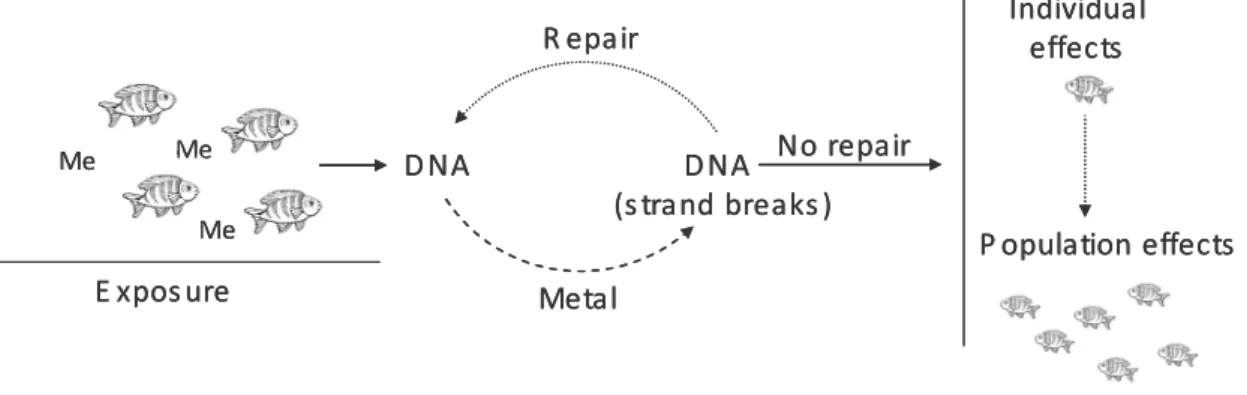
Genotoxicity. Among the molecular components of the cell, DNA is an important target of environmental stress in organisms (Frenzilli et al., 2001). DNA is present in the cell as a functionally stable,
double-stranded polymer without discontinuity (strand breaks) or abnormal modifications and is complexed with
proteins (Shugart, 1995). The exposure of organisms to metal contamination promotes interactions
between metals and DNA (Figure 3). The interactions are manifested primarily by structural alterations in
the DNA molecule and can take the form of adducts (where the chemical or it metabolite becomes
covalently attached to the DNA), of mutations, of strand breakage, or of chemically altered bases
(Shugart, 1995; 2000) and eventually carcinogenesis and other health disorders (Kurelec, 1993). DNA is
the only molecule with capacity for self-repair (Shugart, 1995). However, the ability to repair depends on
exposure. If a DNA lesion induced by a metal can be repaired before fixation, there may be no effect on
DNA. However, this is only true in low-levels exposure where repair enzymes are not saturated by
significant numbers of damaged DNA sites (Shugart, 1995). Because all organisms exhibit this response,
the increased environment contamination leads to an enhancement in the levels of repair indicating DNA
toxicity. Changes in the integrity of DNA have been proposed as useful endpoints for assessing the effects
of environmental pollutants at individual, population and ecosystem level (Klobucar et al., 2003). The
single–cell gel electrophoresis (Comet) assay, has become a widespread technique for detection of DNA
damage induced by xenobiotics (e.g. Cd, by Desai et al., 2006; Fourie et al., 2007; Hg, by Tran et al., 2007;
organic compounds, by Costa et al., 2008). The alkaline version of the assay has proven to be a simple and
Nucleus Cytoplasm
Mitochondria
Lysosomes
Granules Endoplasmic
Reticulum Nucleus Cytoplasm
Mitochondria
Lysosomes
Granules Endoplasmic
Chapter 1
9
reliable method for the quantitation of total DNA fragmentation as a result of the formation of single–
and double–strand breakage, xenobiotic–DNA adducts and alkali–labile sites (e.g. unstable altered
nucleotides) (Singh et al., 1988). Nevertheless, the mechanistic of genotoxicity is still poorly known and
thus the relative potency of contaminants to induce DNA damage and the differential susceptibility of
various organs towards genotoxic damage still need further research. Moreover, the majority of studies
deal with one and/or a limited number or combinations of contaminants, and thus research in aquatic
ecosystems with complex mixtures and interactions of metals and other contaminants is still missing.
Figure 1.3 – Schematic representation of processes leading to DNA damages from exposure to
effects on populations (Me – metals).
Octopus vulgaris (common octopus)
General characteristics. Octopus vulgaris belongs to the class Cephalopoda (Fisher et al., 1987; Mangold, 1983), which is considered as the most active, intelligent and specialized class of molluscs. Octopus have a
soft-bodied that consists of: a muscular mantle that houses the internal organs and represents 8% of body
weight in adults (Trueman and Packard, 1968); and 8 circumoral arms (no tentacles) with bases connected
by a membranous web, and suckers in two series, without chitinous rings or hooks, along the length of
the arms (Jereb et al., 2005). The arms account for 70% of its body weight (Dilly et al., 1964). The mouth
has a pair of chitinous jaws (the beaks) and, as in other molluscs, a chitinous tongue-like radula (band of
teeth) (Jereb et al., 2005). They lack any internal shell which allowed the development of the powerful
muscular mantlethat became the main locomotory organ for fast swimming, via water jetting from the
funnel (Jereb et al., 2005). The circulation of water through the mantle not only produces the power for
swimming, but it provides oxygen for their gills. The surface area of cephalopod gills have been much
increased by a type of folding and are not ciliated as in other mollusks (Gonçalves, 1993). These cilia are
unnecessary since cephalopods are predators not filter feeders. The circulation of water over the gills is
the reverse of what it is in the gastropods, since water leaves the mantle cavity by means of the funnel
(Wells, 1978). Also, the digestive system works with the circulatory system to provide the nutrients
needed to keep the heart pumping. Octopus have a closed circulatory system, with a principal, or
systemic, heart, two branchial hearts and developed arterial, venous and capillary systems supplying
Me Me
Me
E xpos ure
D NA D NA
(s trand breaks ) R epair
Metal
Individual effects
No repair
P opulation effects
Me Me
Me
E xpos ure
D NA D NA
(s trand breaks ) R epair
Metal
Individual effects
No repair
Chapter 1
10
blood to the muscles and organs (Jereb et al., 2005). The nervous systemis highly developed, with a large
brain and peripheral connections. Octopus has the most complex brain of all the invertebrates, just like
vertebrates, they have long term and short-term memories. They are able to change color by using a
complex system of chromatophores under nervous control. The chromatophores are pigment-filled sacs
present in the skin, and capable of remarkable expansion and contraction. This system responds to
current situations in the environment. They produce ink, a dark, viscous fluid also expelled through the
funnel. The ink may take the form of a mucoidal ‘pseudomorph’ (false body) to decoy potential predators,
or of a cloud to obscure the escaping cephalopod. The common octopus is a benthic, neritic species
occurring from the coast line to the outer edge of the continental shelf, in depths from 0-200 m, where it
is found in a variety of habitats (Vaz-Pires et al., 2004). It is normally solitary and territorial, using cavities
or digging a burrow as a home for itself, and leaves it only to feed or reproduce. They often protect and
hide their homes with shells (called the middens), stones and other solid things that they gather. Both
sexes are randomly distributed within patches of different density and are also randomly spaced
(Mangold, 1983). When not travelling in- or offshore, O. vulgaris seems to be a truly sedentary species.
Octopus is a predator being exploratory and opportunistic (Mather and O’Dor, 1991). They feed mainly on
crustaceans, fish and bivalves, leaving the dens at dusk to go for hunting trips, and return at dawn (Wells,
1978). Preys are killed by means of a secretion produced in the posterior salivary glands, cephalotoxin
(Wells, 1978). Octopuses have fast growth rates, up to 13% body weight per day, and food conversion
rates 15-43% (Mangold, 1983; Navarro and Villanueva, 2003). They have been long considered of
cosmopolitan occurrence in temperate and tropical seas (Roper et al., 1984), although a possible
occurrence of cryptic species among O. vulgaris-like octopods is also reported (Guerra et al., 1999). Thus,
the distribution of O. vulgaris in a strict sense may be restricted to the Mediterranean Sea and eastern
Atlantic Ocean (Mangold, 1983). Throughout its distribution range, this species is known to undertake
limited seasonal migrations, usually overwintering in deeper waters and occurring in shallower waters
during summer (Roper et al., 1984).
Birth and offspring. O. vulgaris have a short life span of 12 to 18 months. In the early spring, adult
octopus move closer to the shore for spawning (Mangold and Boletzeky, 1973). They have separate sexes,
and fertilization is internal. Within 2 months after mating, the female releases up to 500,000 eggs
(Mangold, 1983). They are laid in shallow water and are always attached to a substrate, between rocks
and coral reefs. On sandy or muddy bottom, eggs are laid in empty shells or in man-made objects such as
cans, bottles or tires. The female take care of the eggs providing oxygen by squirting them with streams of
water and cleans them with the suckers. She also defends them from predators until they hatch. Soon
after the eggs have hatched the female dies. At hatching, this species has very small hatchlings, paralarvae
(2 mm mantle length) (Boletzky, 1987). Paralarvae are planktonic for 1–3 months, depending on the effect
of temperature on growth rate, and adopt the benthic life mode of the adults at around 7.5 mm ML
Chapter 1
11
(Boletzky, 1977), and it does not imply a total change in feeding behaviour. Growth is very rapid and
juveniles can reach 0.5-0.6 kg within six months of hatching and 1.4-1.8 kg within eight months (Iglesias et
al., 2004). Studies on the Portuguese shelf highlighted the role of temperature and upwelling in
modulating seasonality and distribution of O. vulgaris paralarvae. The influence of the physical
environment was especially pronounced for this specie (Moreno et al., 2009).
Fisheries and Aquaculture. Cephalopods seem to be one of the remaining marine resources, in some
areas, that still experience an increase in landings (Caddy and Rodhouse, 1998). In Portugal the landings
and the economic value of cephalopods have, over the years, maintained a significant growth, indicating
an increasing dependence of the fisheries economy on its landings (Pierce et al., 2010). A study by
Campos et al. (2007) in the Portuguese trawl fleet showed that octopus accounted for a high proportion
of the total landings of these vessels. It is currently ranked third in landings and generates the highest
revenue of all species taken in Portuguese fisheries (Pereira et al., 1997). O. vulgaris is captured by various
methods: hooks and lines, pots and traps in small-scale coastal fisheries in depths of 20-200 m (Pierce et
al., 2010). It is marketed fresh, frozen and dried salted, mostly for human consumption. Despite their
widespread consumption and high market value, commercial aquaculture of cephalopods is very recent
and so far concerns this specie (Vaz-Pires et al., 2004). As mentioned above, octopus presents a series of
characteristics favourable for commercial farming, including: very fast growth rate (Mangold, 1983; Miliou
et al., 2005), high feed conversion efficiencies (Mangold and Boletzky, 1973), high reproductive rate
(Iglesias et al., 1997), a great tolerance to captive conditions (Iglesias et al., 2000), and a high market price
(Vaz-Pires et al., 2004). However, important problems remain regarding the culture of early life stages,
planktonic paralarvae (Iglesias et al., 2007), restricting farming practices to the ongrowing of subadults,
typically obtained from fisheries (Iglesias et al., 2000; Chapela et al., 2006; Rodríguez-Domínguez et al.,
2006).
Cephalopods metal contamination
Cephalopods represent an essential link in marine trophic chains and are eaten by many top
predators. They are known for their ability to accumulate high levels of essential and non-essential
elements to metabolic functions (e.g., Martin and Flegal, 1975; Miramand and Guary, 1980; Mangold,
1983; Finger and Smith, 1987; Miramand and Bentley, 1992; Bustamante et al., 1998a, b; Bustamante et
al., 2000).
Influence of biological parameters. The effect of biological parameters (i.e., size, weight and gender) on the metal accumulation in cephalopods is far from being consensual. Some works reported similar
concentrations in small and large individuals (Barghigiani et al., 1991; Bustamante et al., 1998a; Raimundo
et al., 2004; Seixas et al., 2005a), others indicated correlation between accumulated levels, such as Hg,
and size (Monteiro et al., 1992; Rossi et al., 1993; Pereira et al., 2009). Negative relationships were also
Chapter 1
12
situations were reported with respect to gender: no differences in metal accumulation between males
and females (Miramand and Bentley, 1992; Monteiro et al., 1992; Bustamante et al., 1998a; Barghigiani et
al., 2000; Seixas et al., 2005a, b), lower levels in females (Rossi et al., 1993; Pierce et al., 2008) and higher
levels in females, like Zn (Seixas et al., 2005a; Bustamante et al., 2006). Instead, it appears that
bioaccumulation processes in cephalopod tissues are strongly influenced by metal availability in the
environment (including the food web).
Metal partitioning in tissues. Some studies have evaluated the partition of metals in tissues of
cephalopods (Miramand and Bentley, 1992; Nessim and Riad, 2003; Raimundo et al., 2004, 2005;
Napoleão et al., 2005; Bustamante et al., 2008; Pereira et al., 2009). A general conclusion can be extracted
from all studies: for most of the elements (As is one exception) digestive gland accumulates higher levels.
These findings are related with the intrinsic capacity of this organ to store these elements, which suggests
a major role in detoxification and assimilation processes (e.g., Bustamante et al., 2002; Bustamante et al.,
2006a; Storelli et al., 2006). Cadmium is mainly accumulated in this tissue, reaching 98% of the total body
burden in some species (e.g., Miramand and Guary, 1980; Bustamante, 1998). Additionally, cephalopods
also have the ability to concentrate high metal levels in other tissues. The branchial hearts are known to
accumulate high concentrations of Cu, Fe, Zn, Cd, Ni, V and Mo (Miramand and Bentley, 1992; Nessim and
Riad, 2003; Napoleão et al., 2005). The enhanced levels of Fe in the branchial hearts may be due to the
presence of adenochromes (Ghireti-Magaldi et al., 1958). Gills tend to concentrate high levels of Cu
(Nessim and Riad, 2003) and kidney of Mn, Ni and Pb (Miramand and Bentley, 1992). The elevated
concentrations of Cu found in the branchial hearts and gills are probably associated with the presence of
the heamocyanin (respiratory pigment), in which Cu is one of the main components (Soldevilla, 1987;
Villanueva and Bustamante, 2006; Craig and Overnell, 2003). Metals in branchial hearts and kidney are
probably associated with storage and excretory functions and detoxification mechanisms of these tissues
(Schipp and Hevert, 1978; Guary and Fowler, 1982; Rainbow and Phillips, 1993; Villanueva and
Bustamante, 2006).
Geographical variations. Metal accumulation in cephalopods, mainly in digestive gland, can reflect their
origin (Bustamante et al., 1998b, 2000; Nessim and Riad, 2003; Raimundo et al., 2004; Seixas et al., 2005a,
b). In some cases geographical variations of metal availability can overcome the biological differences
(Nessim and Riad, 2003). Contrasting geographic patterns were observed in digestive gland of specimens
collected in the Portuguese coast, with higher levels of Zn, Pb, Cu and Hg in organisms collected in
Southern areas, while Cd increased drastically in Northern areas (Raimundo et al., 2004). These patterns
were in good relation with water surveys in the Portuguese waters (Caetano and Vale, 2003). Similar
results were obtained for octopus in the same area by Napoleão et al. (2005). Another study with
cephalopods, showed differences between the ones collected in the sub-Artic area (higher Cd levels)
compared with cephalopods from lower latitudes such as along the French Atlantic coast (Bustamante et
Chapter 1
13
vertebrate predators. As a result, cephalopods have been proposed as important vectors of metal
transference to top predators (Bustamante et al., 1998a, b).
Detoxification. These high levels found in the digestive gland would be expected to be toxic unless efficient regulation and detoxification processes are available (Simkiss and Taylor, 1982; Phillips and
Rainbow, 1989; Bustamante et al., 2002). Several studies aimed to assess the mechanisms responsible for
such “absence” of toxicity. Interactions between essential (Zn and Cu) and non-essential elements (Cd)
were assessed in the digestive gland of S. officinalis and O. vulgaris, and explained as a competition for
ligands, and a possible detoxification mechanism (Raimundo et al., 2005; Pereira et al., 2009), since
relationships were more pronounced in specimens captured in areas with enhanced levels of Cd and
positively related with weight. Finger and Smith (1987) and Tanaka et al. (1983) searched for associations
between metals and protein in the squids, Nototodarus gouldi and Ommastrephes bartrami. Proteins with
low, intermediate and high molecular weight were pointed out as potential binding sites for trace metals,
mainly Cu, to a lesser extent Cd and little Zn. Studies have also evaluated relations in wild specimens of
cephalopods to investigate how they mange to tolerate such amounts of Cd looking for the involvement
of MTs and the subcellular distribution, however, relationships to Cd concentrations have not been found
(Bustamante et al., 2002; Bustamante et al., 2006). Instead, they proposed that an alternative mode of
detoxification may be activated as Cd reached a threshold, being the lysosomal fraction involved in this
“new” process. Less data is available for other elements (Bustamante et al., 2006).
Human consumption. Cephalopods are an important food resource being consumed in large quantities in several countries world wide (Amaratunga, 1983). In general, mantle and arm of octopus, which are the
commercial items contain low Cd concentrations, generally below the safety limit established by the
European Commission (1.0 µg g-1, ww of Cd, Journal of EU Communities 2001, EC rule no. 466/2001).
More than 95% of octopus sampled in the Portuguese coast (Raimundo et al., 2005; Raimundo et al.,
2009) presented levels below that limit. According to the joint FAO/WHO expert committee the
Provisional Tolerable Weekly Intake (PTWI) recommended for Cd is approximately 7 µg Kg-1 body weight
(WHO, 2003). On the basis of values registered in the two contrasting areas in Portuguese coast and
assuming an average weight of 60 Kg for humans, the estimated values for PTWI ranged between 2 and 8
kg. Estimated values exceeds largely the weekly average consumption of fishery products in Portugal of
1120 g (FAO, 2007) pointing to a marginal or no risk of its consumption (Raimundo et al., 2009).
Portuguese Coast as study area
The Portuguese coast is extensive with 943 km and it is under pressure, as result of a fast growing
Chapter 1
14
Morphologic features and oceanographic conditions. In the northern west coast, several rivers discharge directly into the sea, like the Minho, Douro and Mondego rivers. The river discharges have a pronounced
seasonal variability, being higher in the winter (http://www.inag.pt) and influencing the stratification of
the coastal waters (Moita, 2001). The Douro estuary, which is the end-member of the largest watershed
of the Iberian Peninsula, is located in one of the most populated zone of Portugal and is subject to
progressive human intervention. The Douro river plume is integrated into larger low salinity waters fed by
the winter-intensified runoff of several rivers on the northwest coast of Portugal and Spain (Peliz et al.,
2002; Alvarez et al., 2006). The plume is detected in a narrow band less than 20 km wide and its extension
is about 100 km (Peliz et al., 2002; Santos et al., 2004). In the south and southwest coasts, major rivers,
like Tagus and Sado are characterised by having large estuaries that tends to trap a great part of the
material transported by low and moderate flows. Tidal currents cause daily suspension of topmost
sediment layers and associated contaminants, and redistribution inside the estuaries according the
morphology (Vale and Sundby, 1987). These systems may receive episodically abrupt quantities of
freshwater and land-derived contaminants (Vale et al., 1990; Martins et al., 2005). In addition, the
Portuguese continental shelf includes several canyons that influence water circulation (Fiuza, 1983).
Water currents and upwelling. Surface waters of the Iberian coast change circulation according to the season (Haynes and Barton, 1990; Pingree, 1993; Wooster et al., 1976), being, in winter, northwards to
the Bay of Biscay in France, and in summer, it becomes weaker and reverses due to the N trade wind
regime (Fiuza, 1983). This southward current promotes cooling and wind-induced upwelling along the
shelf break (Fiuza, 1983; Abrantes and Moita, 1999). North of the Nazaré canyon, the coastal waters are
characterised by a homogeneous upwelling of NACW along the shore (Fiuza, 1983). From Lisbon to Cape
Sines, the upwelling is affected by the presence of coastal protrusions like Cascais, Lisbon and Setúbal
canyons. To South of Cape Sines until Cape S. Vicente, the upwelling structure becomes more regular but
is affected by warmer and saltier offshore surface waters (Moita et al., 2003).
Metals in Portuguese coastal waters. Cotté-Krief et al. (2000) analysed Cd, Cu, Ni and Zn in water off the
continental Portuguese shelf and compared the influence of contaminated rivers (Tinto, Odiel and Tagus)
and the coastal upwelling in defining levels in coastal waters. It was found that upwelling, contrarily to
other systems in non-contaminated areas do not act as a source of trace metal enrichment of the
continental margin waters. Concentrations of dissolved metals reported in that work were lower than
values close to the shoreline in NW of Portugal (Leal et al., 1997). Surface water collected at 1 mile from
the mouth of major estuarine systems in Portugal, pointed to the export of Cd and Cu from the rivers in