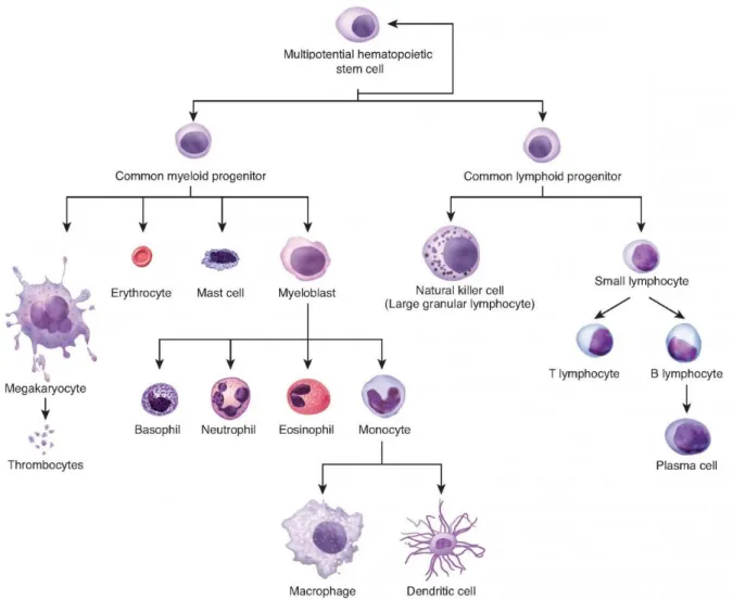
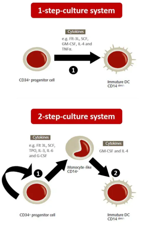
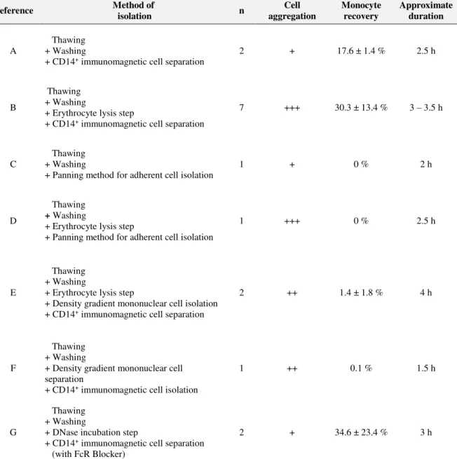
OPTIMIZATION OF PRODUCTION OF ANTI-CANCER VACCINES BASED ON DENDRITIC CELLS
Texto
Imagem


Documentos relacionados
However after being tested on products and services in four cases studies, based on the protocol (as explained in the next chapter), it was possible to propose the
Optimization of this culture medium showed that is possible to increase protease production and reduce the cost of the culture medium with the use of yeast cells.. Yeast
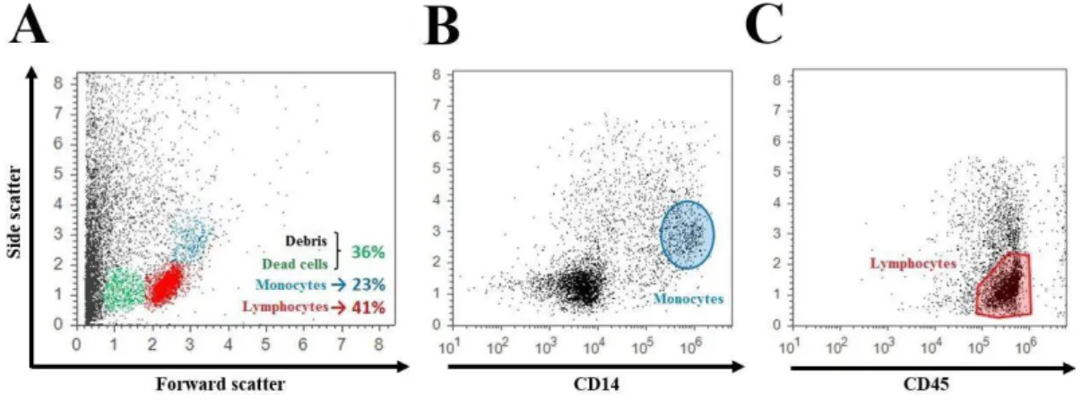
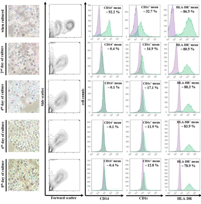
Photomicrographs of cultured monocytes differentiated into macrophages ( M ) and stained with hematoxylin and eosin, at 2–5 days of culture, using peripheral blood mononuclear
(a) Inoculation of shoot tip in initial culture medium supplemented with 5 mg l -1 BA and 0.1 mg l -1 IAA (b) multiple shoot formation on shoot apices in MS medium supplemented
Neste contexto, o artigo pretende avaliar e discutir os impactes resultantes da subida do NMM sobre a população residente, o edificado e as principais infraestruturas
Comparando a constituição dos fatores que foram extraídos na análise entre o período de 2012 a 2016, observa-se para os anos de 2012 e 2013, o F 1 foi composto por indicadores
Ao gerar novos produtos, diferentes dos actualmente produzidos pelo mercado (tanto pela sua forma como pela sua função), o design poderá contribuir para um novo
Em uma revisão sistemática que buscou identificar os mecanismos de governança utilizados pelas redes interorganizacionais, observou-se que esses mecanismos estão ba-
