Selective Bond Cleavage in Potassium Collisions with Pyrimidine Bases of DNA
Diogo Almeida,1Filipe Ferreira da Silva,1Gustavo Garcı´a,2and Paulo Lima˜o-Vieira1,*
1Laborato´rio de Coliso˜es Ato´micas e Moleculares, CEFITEC, Departamento de Fı´sica, Faculdade de Cieˆncias e Tecnologia,
Universidade Nova de Lisboa, 2829-516 Caparica, Portugal
2
Instituto de Fı´sica Fundamental, Consejo Superior de Investigaciones Cientı´ficas, Serrano 113-bis, 28006 Madrid, Spain
(Received 12 October 2012; published 8 January 2013)
Electron transfer in alkali-molecule collisions to gas phase thymine and uracil yieldingH formation
is selectively controlled in the energy range between 5.3 and 66.1 eV. By tuning the collision energy, electron transfer from the alkali to partly deuterated thymine, methylated thymine at the N1 and
methylated uracil at theN3 positions, H loss proceeds not only through the breaking of the (C–H) against (N–H) bonds but also throughN1againstN3sites. Such selectivity, as far as bond and site are concerned, is here reported for the first time by electron transfer induced dissociation experiments in alkali-molecule collisions.
DOI:10.1103/PhysRevLett.110.023201 PACS numbers: 34.20.Gj, 34.50.Ez, 34.50.Lf, 34.80.Ht
Many investigations during the past century have been devoted to the understanding of the alterations induced by high energy radiation in biological systems, particularly within living cells and the DNA/RNA molecule. The biological effects of such radiation are now known to be essentially produced by the secondary species generated along the radiation track and their subsequent reactions within irradiated cells [1]. These species can cause muta-genic, genotoxic, and other potentially lethal DNA lesions [2], such as base and sugar modifications, base release, single strand breaks, and cluster lesions, which include a combination of two single modifications, e.g., double strand breaks and cross-links. Secondary electrons are the most abundant of the secondary species produced by the primary interaction [1,2]. The vast majority of these secondary electrons are created with energies below 20 eV [1], producing, therefore, large quantities of highly reactive radicals, cations, and anions. These species are found to be more efficient producing degradation than the primary radiation; i.e., they are more reactive. As such, studying chemical reactions for biomolecular systems is relevant to understand radiation induced damage at the molecular level with the uttermost need to develop more efficient radiation therapies.
Molecular reaction dynamics and chemical reactivity have been considerably explored by different approaches [3], where laser probing techniques have gained a particu-lar relevance during the last decades. Controlling and inducing selectivity of chemical reactions in molecular collisions have been achieved by mode-selective excita-tion in ultrafast laser pulses [4], by quantum molecular dynamics of photoexcited molecules [5], and, in the case of unimolecular reactions, through coherent quantum ma-nipulation [6,7]. A tunable soft x-ray to stimulate chemical reactions or to selectively break large organic molecules was considered further to site-specific fragmentation of
small molecules such as carbon oxide and acetone [8]. Core-electron excitation inducing selective chemical bond scission due to its special localization and selectivity was also used on thin films of organic polymers by soft x-ray interactions [9]. Additionally, such reaction control and selectivity have been achieved in inelastic electron tunneling microscopy [10] and in the gas phase by site-and bond-specific dissociation through low energy elec-tron interactions [11–14]. Moreover, functional group dependence in the dissociative electron attachment (DEA) process leading to site-selective fragmentation of molecules and the possibility of making use of electron energy as a parameter for control of chemical reactions, have been reported [15]. Steric effects were shown to enhance reactive scattering in molecular beams with ori-ented molecules [16], whereas, at room temperature with random molecular orientation, regarding ion-pair forma-tion, such site- and bond-selective dissociation by tuning the proper collision energy has never been reported.
ionic diabatic potential surfaces). For simplicity, let us consider a diatomic molecule, although for polyatomics hyperdemensional surfaces must be similarly considered. The ionic surface lies above the covalent surface, the endoergicity at large atom-molecule distances being
E¼IEðKÞ EAðABÞ; (1)
where K stands for the potassium atom andABa molecule. However, due to the Coulombic interaction there is a crossing point for which both stationary nonadiabatic po-tential energy surfaces have the same value [22]. During the collision process and near that crossing (Rc), there can
be a perturbation of the stationary states induced by the projectile or target nuclear motion leading to an adiabatic coupling. This leads, after the collision path, to the for-mation of a positive ion Kþ and a molecular temporary negative ion, -TNI, allowing access to parent molecular states which are not accessible in free electron attachment experiments [23,24].
The experiments were performed in a crossed atom-molecule beam arrangement consisting of a potassium source, an oven, and a time-of-flight (TOF) mass analyzer [25]. The components were housed in two high-vacuum chambers at a base pressure of10 5 Pa
. A neutral potas-sium beam at an energy resolution of0:5 eV(FWHM)
generated from a charge exchange chamber intersected orthogonally with an effusive molecular beam consisting of pyrimidine molecules. AtomicKþions, obtained from a potassium ionic source, were accelerated through a cham-ber containing potassium vapor where they resonantly charge exchanged to form a beam of neutral K fast atoms. The energy of the resultant K neutral beam was established by the initial acceleration of the ions. After charge ex-change, the ions that have not been neutralized were removed by electrostatic fields, the resulting neutral K molecular beam was now comprised of two components, a ‘‘hyperthermal’’ beam and an ‘‘effusive thermal energy’’ beam. Since the electron transfer process is endoergic, the thermal beam does not contribute to the formation of anions. The hyperthermal alkali beam entered a high vac-uum chamber where it was monitored by an iridium sur-face ionization detector of the Langmuir-Taylor type. This detector sampled the beam intensity but did not interfere with the beam passing to the collision region. It operates in a temperature regime that only allows detection of the fast beam. The biomolecular target beams were produced in a hot gas cell (oven) and admitted to vacuum by an effusive source through a 1 mm diameter orifice where they were crossed with the neutral hyperthermal potassium beam. At a temperature of about 390 K (measured by a platinum resistance—Pt100) the density of intact molecules was high enough to yield a reasonable negative-ion signal. The negative ions produced in the collision were extracted by a 250 V=cm pulsed electrostatic field towards the
entrance of a TOF where they were analyzed and detected
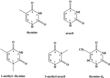
in a single-pulse counting mode. The spectra collected at each collision energy showing the recorded anionic sig-nals, were obtained by subtracting the background signal from the sample signal. In potassium-pyrimidine collision studies (Fig.1), the total energy available in the center-of-mass frame, including potassium ionisation energy (4.34 eV), varies from5up to 66 eV. 1-methyl-thymine (1-meT), 3-methyl-uracil (3-meU) and partly deuterated thymine (thymine-d4) were obtained from Sigma Aldrich
with a stated purity of 98%, respectively. The samples were used as delivered. Time-of-flight mass spectra of the differ-ent anions formed in collisions of neutral potassium atoms with the pyrimidine molecules have been obtained and a typical TOF mass spectrum of 1-methyl-thymine (1-meT) at 66.1 eV collision energy is depicted in Fig. 2. In this Letter we are focusing our attention on theH yield only. FIG. 1. Molecular structure of thymine, uracil, 1-methyl-thymine (1-meT), 3-methyl-uracil (3-meU), and partly deuter-ated thymine (thymine-d4).
However, this anion is a product from many other mole-cules such as water and hydrocarbons that are present in the HV chamber, and in the case of the former, even as moisture in the sample. Thus the spectra in the figures showing the recorded anionic signals were obtained by subtracting the background signal from the sample signal.
The ion yields (relative intensity as a function of the collision energy) ofH from 1-meT are shown in Fig.3(a) at three different collision energies. Methylation at the
N1 position completely supresses H formation in the
low-energy collisions, i.e., at 7.6 eV, whereas at higher energies (9.0 and 66.1 eV) H loss mainly originates from the N3 and the carbon positions. However, at the
available energy of 9.0 eV, accessing the resonance yield-ing H from CH3 is energetically unfavorable [11].
Therefore, we must conclude that at this energy (9.0 eV), the signal mainly originates from the N3 position. In
Fig. 3(b) we show the H yield measured upon
potas-sium collisions with uracil methylated at theN3 position
(3-meU). Here, we clearly see a distinct signal ofH loss
at 5.3 and 7.4 eV, in contrast to the strong suppression of theH signal in 1-meT at 7.6 eV. These findings indicate thatH loss from theN1position is contributing to such a
signal. The signal observed at 64.4 eV is now comprising the contributions fromN3,C6, andCH3. In order to support
these unprecedented results, we have obtained theH and
D yields from thymine deuterated at the C positions (thymine-d4) and the results are shown in Fig.3(c). It is
obvious thatD loss from the C positions is restricted to
collision energies above 7.4 eV, whileH loss from the N sites essentially occurs from the N1 position. This is in
clear agreement with the resonance position in the disso-ciative electron attachment studies [11] even at the present moderate collision energy resolution. In addition to these findings in the potassium-pyrimidine collisions, we can also add that H loss from the C positions is essentially
due to C6, which is particularly relevant in the case of
1-meT.
In DEA to thymine the minimum electron energy required to break a particular bond (N1–H,N3–H,C6-H,
andCH2–H) lies between 4 to 5 eV [26], so bond and site
selectivity [12] to the gas phase of methylated and deuter-ated pyrimidines yielding H formation does not result
from any particular energy constraint [11]. Since energy constraints cannot explain site selectivity, the electronic structure of the associated transient precursor ions accessed by electrons of different energies (either shape or core excited resonances) has been suggested as the main effect responsible for such an achievement. The dissocia-tion mechanism in the potassium collisions yielding a neutral dehydrogenated molecule and H loss, can be
regarded as a pseudodiatomic behavior. In this context, we recall Eq. (1) and for large potassium-molecule values the van der Waals and induction forces can be neglected
and consequently the covalent potential is zero and the ionic potential is purely Coulombic. If this approximation holds, Rc is given by 14:41=EðAÞ [23], when E is
expressed in eV. Taking the adiabatic electron affinities of 1-meT, 3-meU, and thymine-d4, asð0:0250:010Þ eV
[27], ð0:0350:010Þ eV [27] and ð0:0690:007Þ eV
[28], the values for Rc are found for the three molecular
FIG. 3. H =D ion yield as a function of the collision energy.
The vertical dotted lines indicate the mean values of theH =D
position of the centre of the different resonances forN1,N3,C6,
and CH3 positions obtained in DEA studies (from Fig. 3 in
Ref. [11]). (a)H formation from thymine methylated at theN1
position (1-meT) at 7.6, 9.0 and 66.1 eV; (b)H formation from
uracil methylated at the N3 position (3-meU) at 5.3, 7.4, and
64.4 eV; (c) H and D formation from partly deuterated
targets at 3:3 A. The corresponding total cross sections
for ion-pair formation will be of the order ofRc 2
, which is much larger than the corresponding gas kinetic cross sections. Contributions to ion-pair formation through elec-tron transfer to excited states of molecular negative ions have been observed in other polyatomic molecules, such as benzene and fluorobenzene [24]. In the case of thymine and uracil [29], the dehydrogenated parent anion fraction (not shown here) is attributed to excited states and at low collision velocities accounts for 10%–20%of the total cross section. The velocity dependence has shown that above 2104 ms 1
(>40 eV), the contribution of the
excited states is negligible. This is a remarkable finding since similar behavior was observed in diatomics [23] and this result can be used here for the present molecular targets. Therefore, if an excited state of the negative ion is involved, such a contribution may be reached via a smaller crossing distance. To reach such a crossing no electron transfer should occur at the first crossing and the excitation may occur at the inner crossing. When the collision energy is increased the diabatic probabilities controlling the electron transfer process at the first crossing as well as the inner crossing increase and the effect of these excited states will be reduced.
Charge transfer deposited on gas-phase thymine and uracil by an electron harpooning mechanism in atom-molecule collisions [29] induces the loss of hydrogen which exclusively takes place from the N positions. The bond selectivity can also be made site selective by proper adjustment of the collision energy. While at 5.3 eV collision energy results in the loss of hydrogen fromN1in
3-methyl-uracil, the reaction can be suppressed fromN3by
tuning the collision energy to 7.6 eV as is in 1-methyl-thymine. Moreover, D formation from thymine
deuter-ated in the C positions is suppressed at 7.4 eV showing that
H formation in 3-methyl-uracil proceeds only through the
N1 position. Here, we find that energy and charge transfer
are completely inactivated when theN1-Hbond is replaced
byN1-CH3. These findings point to a new achievement in
controlling chemical reactions that may have particular relevance for the investigation of early molecular processes in the nascent stages of DNA damage by secondary elec-trons, especially those related to strand breaks. Such charge transfer processes may also play an important role in low-temperature plasmas as well as in the regions of planetary atmospheres.
D. A. and F. F. S. acknowledge the Portuguese Foundation for Science and Technology (FCT-MEC) for a post-graduate Grant No. SFRH/BD/61645/2009 and post-doctoral Grant No. SFRH/BPD/68979/2010, respectively. We also acknowledge partial funding from the Portuguese research Grant No. PEst-OE/FIS/UI0068/2011 through FCT-MEC, and from the Spanish Ministerio de Economı´a y Competitividad (Project No. FIS 2009-10245). Some of this work forms part of the EU/ESF COST Actions
Nano-IBCT-MP1002 and The Chemical Cosmos-CM0805. The authors are extremely grateful to Professor Eugen Illenberger from the Free University Berlin, Germany, for fruitful and lively discussions.
*Corresponding author. [email protected]
[1] B. Boudaı¨ffa, P. Cloutier, D. Hunting, M. A. Huels, and L. Sanche,Science287, 1658 (2000).
[2] C. von Sonntag,Free-Radical-Induced DNA Damage and Its Repair(Springer, New York, 2005).
[3] R. D. Levine and R. B. Bernstein, Molecular Reaction Dynamics and Chemical Reactivity (Oxford University Press, New York, 1987).
[4] E. D. Potter, J. L. Herek, S. Pedersen, Q. Liu, and A. H. Zewail,Nature (London)355, 66 (1992).
[5] H. Rabitz, R. de Vivie-Riedle, M. Motzkus, and K. Kompa,Science288, 824 (2000).
[6] H. H. Fielding and M. A. Robb,Phys. Chem. Chem. Phys.
12, 15 569 (2010).
[7] L. Ratschbacher, C. Zipkes, C. Sias, and M. Ko¨hl, Nat. Phys.8, 649 (2012).
[8] W. Eberhardt, T. K. Sham, R. Carr, S. Krummacher, M. Strongin, S. L. Weng, and D. Wesner,Phys. Rev. Lett.50, 1038 (1983).
[9] S. Wada, R. Sumii, K. Isari, S. Waki, E. O. Salo, T. Sekiguchi, T. Sekitani, and K. Tanaka, Surf. Sci. 528, 242 (2003).
[10] P. A. Sloan and R. E. Palmer,Nature (London)434, 367 (2005).
[11] S. Ptasin´ska, S. Denifl, V. Grill, T. D. Ma¨rk, E. Illenberger, and P. Scheier, Phys. Rev. Lett.95, 093201 (2005).
[12] H. Abdoul-Carime, S. Gohlke, and E. Illenberger,Phys. Rev. Lett.92, 168103 (2004).
[13] V. S. Prabhudesai, A. H. Kelkar, D. Nandi, and E. Krishnakumar,Phys. Rev. Lett.95, 143202 (2005). [14] I. Bald, J. Kopyra, and E. Illenberger,Angew. Chem., Int.
Ed.45, 4851 (2006).
[15] R. Balog and E. Illenberger,Phys. Rev. Lett.91, 213201 (2003).
[16] P. R. Brooks,Science193, 11 (1976).
[17] V. Vuitton, P. Lavvas, R. V. Yelle, M. Galand, A. Wellbrock, G. R. Lewis, A. J. Coates, and J.-E. Wahlund, Planet. Space Sci.57, 1558 (2009).
[18] D. Almeida, R. Antunes, G. Martins, G. Garcia, R. W. McCullough, S. Eden, and P. Lima˜o-Vieira, Int. J. Mass Spectrom.311, 7 (2012).
[19] F. Gobet, S. Eden, B. Coupier, J. Tabet, B. Farizon, M. Farizon, M. J. Gaillard, S. Ouaskit, M. Carre´, and T. D. Ma¨rk,Chem. Phys. Lett.421, 68 (2006).
[20] H. Tanaka and M. Inokuti,Adv. At. Mol. Opt. Phys.43, 1 (2000).
[21] M. Larsson, W. D. Geppert, and G. Nyman, Rep. Prog. Phys.75, 066901 (2012).
[23] A. W. Kleyn and A. M. C. Moutinho, J. Phys. B 34, R1 (2001).
[24] P. Lima˜o-Vieira, A. M. C. Moutinho, and J. Los,J. Chem. Phys.124, 054306 (2006).
[25] R. Antunes, D Almeida, G Martins, N. J. Mason, G. Garcı´a, M. P. J. Maneira, Y. Nunes, and P. Lima˜o-Vieira, Phys. Chem. Chem. Phys.12, 12 513 (2010).
[26] S. Denifl, S. Ptasin´ska, M. Probst, J. Hrusˇa´k, P. Scheier, and T. D. Ma¨rk,J. Phys. Chem. A108, 6562 (2004).
[27] C. Desfrancois, H. Abdoul-Carime, S. Carles, V. Pe´riquet, J. P. Schermann, D. M. A. Smith, and L. Adamowicz, J. Chem. Phys.110, 11 876 (1999).
[28] J. H. Hendricks, S. A. Lyapustina, H. L. de Clercq, J. T. Snodgrass, and K. H. Bowen, J. Chem. Phys.104, 7788 (1996).