Anatomical Variations of the Middle Turbinate
Concha Bullosa and its Relationship with Chronic
Sinusitis: A Prospective Radiologic Study
Raja Kalaiarasi
1Venkataramanan Ramakrishnan
1Santhosh Poyyamoli
21Department of ENT, Sri Lakshmi Narayana Institute of Medical
Sciences, Puducherry, India
2Department of Diagnostic and Interventional Radiology, Kovai
Medical Centre and Hospital, Coimbatore, Tamil Nadu, India Int Arch Otorhinolaryngol 2018;22:297–302.
Address for correspondence Dr. Raja Kalaiarasi, MS, DNB, Department of ENT, Sri Lakshmi Narayana Institute of Medical Sciences, Osudu, Agaram Village, Villianur Commune Kudupakkam Post, Puducherry, 605502, India (e-mail: [email protected]).
Introduction
A concha bullosa (CB) represents the presence of air cell in the turbinates, and the middle turbinate (MT) concha bullosa is a common nasal cavity anatomical variation. Pneumatiza-tion of the MT happens due to variaPneumatiza-tion in the ethmoidal air cell system development. The incidence rates for pneumati-zation of the MT is between 13 and 53.6%.1–4Concha bullosa
is generally asymptomatic and diagnosed incidentally by
computed tomography. Sometimes, an over-pneumatized MT can lead to nasal obstruction, contact headache, deviated nasal septum and chronic sinusitis. Concha bullosa can be unilateral or bilateral and can be classified into three types according to the site of pneumatization. They are lamellar-type (vertical lamella of MT pneumatization), bulbous-lamellar-type (inferior portion of MT pneumatization) and extensive/large type (vertical lamella and inferior portion of the MT Keywords
►
sinusitis
►
turbinates
►
mucocele
Abstract
Introduction
A pneumatized turbinate, also called concha bullosa, is a normal anatomical
variant of the paranasal sinus region. Depending on the site of pneumatization, the concha
is classi
fi
ed into extensive, bulbous or lamellar type. The middle turbinate concha bullosa
has been implicated as a possible etiological factor in chronic sinusitis.
Objectives
The aim of this study was to investigate the anatomical variations of the
concha bullosa, based on paranasal sinus imaging, and its possible association with
sinusitis.
Methods
This prospective descriptive study was performed at the Department of ENT
and Head Neck Surgery over a period of one year, from 2016 to 2017. We studied the
computed tomography scans of the nose and paranasal sinuses
—
in axial, coronal and
sagittal planes
—
of patients who had symptoms of nasal obstruction, or headache and
features of chronic sinusitis.
Results
Out of the 202 scans studied, the prevalence of concha bullosa was 31.7%. The
concha was bilateral in 35 (54.7%) patients and unilateral in 29 (45.3%) patients. Out of 99
conchae, 54 were on the right side and 45 were on left side. Ipsilateral sinusitis was found in
40.4% of the sides in the scans of subjects with concha. There was no statistically signi
fi
cant
association between any type of middle turbinate concha with sinusitis, but sinusitis was
more predominant with the extensive type of concha (
p
>
0.05).
Conclusion
Multiple air cells, mucocele, pyocele and in
fl
ammatory mucosal
thicken-ings in the concha are relatively rare. Detailed knowledge of anatomic variations of the
concha bullosa is imperative for the radiologists and the operating surgeons.
received June 29, 2017 accepted
December 21, 2017 published online March 13, 2018
DOIhttps://doi.org/ 10.1055/s-0038-1625978. ISSN 1809-9777.
Copyright © 2018 by Thieme Revinter Publicações Ltda, Rio de Janeiro, Brazil
pneumatization). A disease process of the paranasal sinuses can affect the CB, resulting in mucosal thickening, retention of mucous secretion, mucocele and pyocele within the CB. The CB itself can cause mechanical obstruction, affecting the drainage pathway and leading to sinusitis. Understanding the anatomical variations of the CB make it possible to plan for appropriate management. In this article, we described some of the rare anatomical variations of MT CB, such as extensive mucosal thickening within the CB, air cells in the CB, and mucopyocele of the CB, which can cause orbital complications. All these conditions and their clinical impor-tance are discussed. The role of the MT CB in predisposition to chronic sinusitis is in question, and there is dissensus on the relationship between the CB and sinus pathology. Very few research works have investigated the pneumatization and anatomical variation of the CB and its possible associa-tion to chronic sinusitis.
Objectives
The objectives of this study were to investigate the anato-mical variation of the MT concha bullosa based on para-nasal sinus imaging, and its possible association with rhinosinusitis.
Methods
A prospective descriptive study was performed at the Department of ENT and Head Neck Surgery over a period of 1 year, from 1st May 2016 to 30th April 2017, to determine the prevalence of CB in the paranasal sinus imaging of patients who had symptoms of nasal obstruction or head-ache and features of chronic sinusitis. Demographic data, clinical presentation, computed tomography (CT) of nose and paranasal sinuses 5-mm-thick imaging scans in the axial, coronal and sagittal planes were recorded. The inclusion
criterion was the presence of any type of pneumatization of the MT in CTs of the nose and paranasal sinuses. Any mucosal thickening of 4 mm or more in the sinus cavity wall was taken as positive for sinusitis.5 Informed consent forms signed by the patients as well as the institute ethical com-mittee’s clearance were obtained prior to this study.
In this study, CT scans of the nose and paranasal sinus of 202 patients were studied, out of which 64 patients (37 males, 27 females; mean age 30.3 years; range 16 to 60 years) had concha bullosa in their imaging study. The CB was classified as lamellar, bulbous or extensive type, according to the classifi ca-tion developed by Bolger et al.6
Patients who underwent any nasal surgery, had any congenital abnormalities of the nose or had been in accidents involving the faciomaxillary region before taking the CT scan were excluded from this study. A total of 42 patients (65.6%) underwent surgery to treat symptomatic CB.
Results
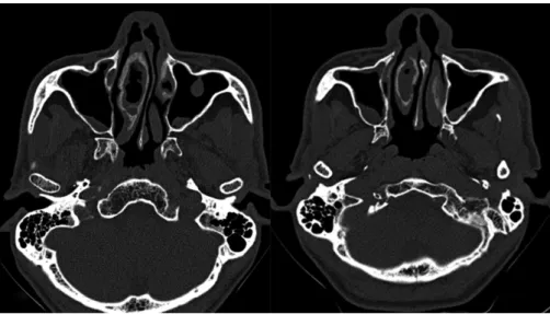
Out of the CT scans studied of the nose and paranasal sinus of 202 patients, the prevalence of CB was 31.7%. Concha bullosa was bilateral in 35 (54.7%) patients, and unilateral in 29 (45.3%) patients. Out of 99 cases of CB, 54 (54.5%) were on right side and 45 (45.5%) were on left side. Two patients had mucopyocele of the CB, in its lamellar part, and four patients had extensive mucosal thickening within the CB (►Fig. 1a and1b). The distribution of anatomical variation of CB is shown in►Table 1. Computed tomography of mucocele or pyocele within a CB shows a prominent well circumscribed soft tissue density with a thin bony framework at the margin (►Fig. 2aand2b). Migrating ethmoidal air cells within the CB is a rare finding (►Fig. 3a and 3b) seen in one patient, bilaterally.
The most common incidental pathology accompanying CB was nasal septal deviation (n¼49) (►Fig. 4a). Ipsilateral
Fig. 1 (a and b) Axial sections of computed tomography of the paranasal sinuses showing mucosal thickening within the right concha bullosa,
sinusitis was found in only 40 (40.4%) of the sides in scans of subjects with CB. Out of the 49 extensive conchae, 11 were on the right side (22.4%), which had associated ipsilateral osteomeatal complex blockage and mucosal thickening within the sinuses, and 16 (32.6%) had free ipsilateral osteomeatal complex without mucosal thickening of the sinuses (►Fig. 5). In our study, the maxillary sinus was the most commonly involved sinus, followed by the ethmoid and frontal sinuses. Two proportion tests were used for the statistical analysis using STATA version 12.0 (Stata Corp, College Station, TX, USA). A p value<0.05 was taken as statistically significant. There was no statistically significant association between any type of CB with sinusitis, but sinusitis was more predominant with the extensive type than with any other type of CB (►Table 2).
Discussion
The ethmoturbinal and maxilloturbinal are the embryologi-cal precursors of the nasal turbinates, which appear between the eighth and tenth weeks of gestation. The ethmoturbinal gives rise to the uncinate process, the MT and the superior
turbinate. The MT is formed by the medial part of the ethmoid bone. The ethmoidal air cells extending into the frontal, maxillary, and the sphenoid paranasal sinus bones retain their ostia at the site of initial evagination.7The ostia remain as their drainage pathway. Anterior ethmoidal cells originating from the middle meatus pneumatize the MT in55% of cases, which usually drain into the frontal recess.8Posterior ethmoid cells originated from the superior meatus pneumatize in45% of the cases, and they usually drain into the retrobulbar recess. Most commonly, the drai-nage occurs through the conchal ostium present near the frontal recess region into which the frontal sinus drains. The CB becomes apparent after 7–8 years of age and continues its development even after the period of adolescence.9 The degree of pneumatization and the inflammatory changes that occur within the CB may correlate with the presentation and the severity of symptoms. The mean age (30.3 years) of this study’s participants with CB was consistent with other studies on the same topic.10,11The proportion of males was higher than that of females in our study, in contrast with other studies.4,10,12,13The prevalence of CB was 31.7% in our study. Aramani et al and Koo SK et al reported a prevalence Table 1 Anatomical variations of middle turbinate concha bullosa
S.No Anatomical variation of concha bullosa Number (%) Total number (%)
1. Pure extensive type 44 (44.5%) 49 (49.5%)
2. Mucosal thickening within the extensive type 4 (4%)
3. Polyp within extensive type 1 (1%)
4. Pure bulbous type 26 (26.3%) 28 (28.3%)
5. Air cells within bulbous type 2 (4%)
6. Pure lamellar type 20 (20.2%) 22 (22.2%)
7. Mucopyocele within lamellar type 2 (2%)
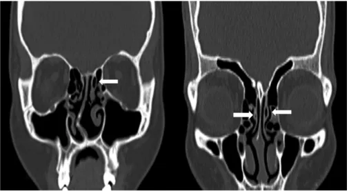
Fig. 2 (a) Coronal and (b) axial sections of non-contrast computed tomography of paranasal sinuses show expansile soft tissue density within
rate of 53.7% in their studies.11,14In our study, the MT CB prevalences of extensive, bulbous and lamellar types were 49 (49.5%), 28 (28.3%) and 22 (22.2%), respectively, which are the same as noted in a study by Tonai et al.15Various other studies report different incidences, which could be due to the differences in the target population and racial variation.4,5,16 The CB, rarely whenfilled with fluid and pus results in mucopyocele. Concha bullosa mucopyocele happens due to chronic obstruction of the CB ostium, which prevents the optimal air current flow between the CB cavity and the surrounding structures, such as the frontal recess, ethmoidal cells or middle meatus.17–19 For this reason, the epithelial
lining in a CB mucocele/pyocele remains intact, lacking an epithelial covering in other types of mucocele.18,19It is impor-tant to differentiate between CB mucocele/pyocele and
eth-moidal pyocele. Concha bullosa pyocele shows an enlarged tip or body of the MT that touches the nasal septum medially and bulges into the lateral wall of the nose laterally or into the medial wall of the orbit. Mucocele of the ethmoid sinus usually displaces the MT inferiorly, against the septum. The MT is seen distinctly as an intact structure, but compressed. Any secre-tions within a CB will have a mucoid attenuation of 10–18 HU in the CT scan. The presence of a bony shell around the CB on a CT scan evaluation allows a conchal mucocele to be differen-tiated from other nasal masses, but the bony rim may some-times be absent or extremely thinned out due to bone remodeling in the pathogenesis of mucocele (►Fig. 2). Concha bullosa mucocele/pyocele can masquerade as an intranasal tumor, and it is very important to consider CB mucocele/ pyocele in the differential diagnosis.
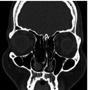
Fig. 3 (a) Coronal and (b) axial sections of computed tomography of the paranasal sinuses show the presence of multiple air cells within the
bilateral concha bullosae. Deviation of the nasal septum to the left side is noted. Also seen is a small mucosal polyp in the inferior wall of the left maxillary sinus.
Fig. 4 (a and b) Coronal sections of non-contrast computed tomography scan of the paranasal sinuses showing pneumatized vertical lamella of
Mucosal thickening and polyp formation within a CB is also uncommon. The inner surface of the CB is lined with mucous membrane and any inflammatory process will incite mucosal hypertrophy and polyp formation.20Usually, the CB contains only a single air cell. Multiple air cells are relatively rare. Ceylan S et al described a giant complex CB, in which the ethmoid bulla invaginated into the MT CB.21In our study, we found ethmoidal air cells in the CB; however, the clinical importance of such discovery is still unclear. Extensive pneumatization of the turbinates with mucosal contact can lead to headache, even in the absence sinonasal inflammation.22
In our study, there was no significant association between any type of CB with ipsilateral rhinosinusitis, but, clinically, we found that sinusitis was more predominant in the exten-sive type of CB, compared with other types; however, this finding was not statistically significant. We also found that patients with mucopyocele of the CB always associated with
ipsilateral sinusitis. Lee et al, Yasan et al, and Armengot et al also reported similarfindings in their case studies that CB mucocele/pyocele cause ipsilateral sinusitis.3,18,23Aktas D et al studied the CT paranasal scans of 54 patients with CB and found no relationship between unilateral and bilateral CB with sinusitis.24
Conclusion
Radiologists and surgeons should be aware of anatomical variants of the CB present in the paranasal sinus imaging. Our findings show that the CB does not appear to give rise to chronic sinusitis, but the extensive type was more sympto-matic than the bulbous or lamellar types. Certain anatomical variations, like air cells in a MT CB and extensive mucosal thickening within a MT CB, are herein defined for thefirst time in the literature.
References
1 Joe JK, Ho SY, Yanagisawa E. Documentation of variations in sinonasal anatomy by intraoperative nasal endoscopy. Laryngo-scope 2000;110(2 Pt 1):229–235
2 Al-Sebeih KH, Bu-Abbas MH. Concha bullosa mucocele and muco-pyocele: a series of 4 cases. Ear Nose Throat J 2014;93(01):28–31 3 Lee JH, Hong SL, Roh HJ, Cho KS. Concha bullosa mucocele with
orbital invasion and secondary frontal sinusitis: a case report. BMC Res Notes 2013;6(06):501
4 Unlü HH, Akyar S, Caylan R, Nalça Y. Concha bullosa. J Otolaryngol 1994;23(01):23–27
5 Rak KM, Newell JD II, Yakes WF, Damiano MA, Luethke JM. Paranasal sinuses on MR images of the brain: significance of mucosal thickening. AJR Am J Roentgenol 1991;156(02):381–384 6 Bolger WE, Butzin CA, Parsons DS. Paranasal sinus bony anatomic variations and mucosal abnormalities: CT analysis for endoscopic sinus surgery. Laryngoscope 1991;101(1 Pt 1):56–64
7 Zinreich SJ, Mattox DE, Kennedy DW, Chisholm HL, Diffley DM, Rosenbaum AE. Concha bullosa: CT evaluation. J Comput Assist Tomogr 1988;12(05):778–784
8 Van Alyea OE. Nasal sinuses: an anatomic and clinical considera-tion. Baltimore: Williams and Wilkins; 1951
9 Cohen SD, Matthews BL. Large concha bullosa mucopyocele replacing the anterior ethmoid sinuses and contiguous with the frontal sinus. Ann Otol Rhinol Laryngol 2008;117(01):15–17 10 Stallman JS, Lobo JN, Som PM. The incidence of concha bullosa and its relationship to nasal septal deviation and paranasal sinus disease. AJNR Am J Neuroradiol 2004;25(09):1613–1618 11 Aramani A, Karadi RN, Kumar S. A study of anatomical variations
of osteomeatal complex in chronic rhinosinusitis patients—CT
findings. J Clin Diagn Res 2014;8(10):KC01–KC04
12 Nadas S, Duvoisin B, Landry M, Schnyder P. Concha bullosa: frequency and appearances on CT and correlations with sinus disease in 308 patients with chronic sinusitis. Neuroradiology 1995;37(03):234–237
13 Lam WWM, Liang EY, Woo JK, Van Hasselt A, Metreweli C. The etiological role of concha bullosa in chronic sinusitis. Eur Radiol 1996;6(04):550–552
14 Koo SK, Kim JD, Moon JS, Jung SH, Lee SH. The incidence of concha bullosa, unusual anatomic variation and its relationship to nasal septal deviation: A retrospective radiologic study. Auris Nasus Larynx 2017;44(05):561–570
15 Tonai A, Baba S. Anatomic variations of the bone in sinonasal CT. Acta Otolaryngol Suppl 1996;525:9–13
16 Uygur K, Tüz M, Doğru H. The correlation between septal deviation and concha bullosa. Otolaryngol Head Neck Surg 2003;129(01):33–36
Fig. 5 Coronal section of computed tomography of the paranasal
sinuses shows extensive pneumatisation of the inferior portion and vertical lamella of the bilateral middle turbinates, more on the right side, suggestive of extensive/large type of concha bullosa.
Table 2 Association of different types of middle turbinate concha bullosa with sinusitis
S. No Concha bullosa type and laterality Associated with sinusitis Not associated with sinusitis Pvalue
1. Right extensive 11 16 0.965
2. Left extensive 10 12 0.5834
3. Right bulbous 4 10 0.332
4. Left bulbous 6 8 0.839
5. Right lamellar 5 8 0.8793
17 Lee JS, Ko IJ, Kang HD, Lee HS. Massive concha bullosa with secondary maxillary sinusitis. Clin Exp Otorhinolaryngol 2008; 1(04):221–223
18 Yasan H, Doğru H, Tüz M, Yeşildağ A, Karahan N. Polypoid degeneration of the middle turbinate mucocele mimicking intra-nasal neoplasia. J Otolaryngol 2005;34(03):207–209
19 Pinto JA, Cintra PP, de Marqui AC, Perfeito DJ, Ferreira RD, da Silva RH. [Mucopyocele of the middle turbinate: a case report]. Rev Bras Otorrinolaringol (Engl Ed) 2005;71(03):378–381
20 Erkan AN, Canbolat T, Ozer C, Yilmaz I, Ozluoglu LN. Polyp in concha bullosa: a case report and review of the literature. Head Face Med 2006;2(02):11
21 Ceylan S, Bora F, Batmaz T, Atalay B. Complex Concha Bullosa. Otolaryngol (Sunnyvale) 2012;2:109
22 Kantarci M, Karasen RM, Alper F, Onbas O, Okur A, Karaman A. Remarkable anatomic variations in paranasal sinus region and their clinical importance. Eur J Radiol 2004;50(03):296– 302
23 Armengot M, Ruiz N, Carda C, Hostalet P, Basterra J. Concha bullosa mucocele with invasion of the orbit. Otolaryngol Head Neck Surg 1999;121(05):650–652