ABSTRACT | Purpose: To compare postoperative changes in retinal nerve fiber layer thickness in patients with macular holes treated with vitrectomy with Brilliant Blue-assisted internal limiting membrane peeling. Methods: Twenty-two eyes of 20 patients with macular holes were studied. Each eye was selected to undergo Brilliant Blue-assisted internal limiting membrane peeling. The circumferential retinal nerve fiber layer thickness was determined using spectral domain optical coherence tomography preoperatively and 2 months postoperatively. Mean overall and sectoral retinal nerve fiber layer thicknesses were obtained for each patient. Results: There was no statistically significant difference (p≥0.05) between the
pre- and post-treatment measurements in relation to each CFN variable, i.e., on average, pre-treatment measures were the same as post-treatment measures. Furthermore, despite the differences between the pre- and post-treatment measures always being positive (pre-post >0), they are not statistically significant. Conclusions: This study showed no significant decrease in retinal nerve fiber layer thickness measurements after macular holes surgery, regardless of age or sex.
Keywords: Nerve fibers; Retinal perforations/surgery; Retinal nerve fiber layer thickness; Macular hole
RESUMO | Objetivo: Comparar as alterações pós-operatórias na espessura da camada de fibras nervosas da retina em pacientes com buracos maculares submetidos à vitrectomia via pars-plana associada à remoção de membrana limitante interna. Méto-dos: Foram estudados 22 olhos de 20 pacientes consecutivos diagnosticados com buraco macular. Todos os pacientes foram
submetidos à vitrectomia via pars-plana e remoção de membrana limitante interna corada com azul brilhante. A espessura da camada de fibras nervosas da retina em região peripapilar foi determinada por tomografia de coerência óptica de domínio espectral antes e 2 meses após a cirurgia. As espessuras totais e espessuras setoriais da camada de fibras nervosas da retina foram obtidas para cada paciente. Resultados: Os resultados mostram que não existe diferença estatisticamente significativa (p≥0,05) entre as medidas pré e pós-operatórias em relação a
cada uma das variáveis. Conclusão: Este estudo não demons trou diminuição significativa nas medidas da espessura da camada de fibras nervosas retinianas após a cirurgia de buraco macular, independente da faixa etária ou sexo.
Descritores: Fibras nervosas; Perfurações retinianas/cirurgia, Es-pessura da camada de fibras nervosas da retina, Buraco macular
INTRODUCTION
Idiopathic macular hole is a controversial genesis re-tinal defect, primarily involving the foveola. It mainly affects women (67%-91%) between the fifth and seventh decades of life and is bilateral in approximately 3%-27% of cases(1-2).
Internal limiting membrane (ILM) peeling in macular hole surgery has become an important procedure to im-prove anatomic success and decrease recurrence rates(1-3).
Staining ILM with Brilliant Blue has been a standard technique for their removal around macular hole because of reduced cytotoxicity relative to indocyanine green(1-4).
However, anatomical changes have been observed around the area of peeling, including the retinal nerve fi-ber layer (RNFL) where nerve fifi-bers are concentrated(1-3).
The development of spectral domain optical cohe-rence tomography (SD-OCT) has made morphological study of different retinal layers possible with a high level of detail(3-8).
Comparisons of retinal nerve fiber layer thickness
changes after macular hole surgery
Comparação nas alterações da espessura da camada de fibras nervosas
retinianas após cirurgia de buraco macular
Nelson Chamma Capelanes1, André Vasconcellos Diniz2, Érika Pacheco Magalhães1, Karise de Oliveira Marques3
1. Retina and Vitreous Department, Fundação Hilton Rocha, Belo Horizonte, MG, Brazil. 2. Department of Surgical Retina, Fundação Hilton Rocha, Belo Horizonte, MG, Brazil. 3. Ophthalmology, Fundação Hilton Rocha, Belo Horizonte, MG, Brazil.
Submitted for publication: April 24, 2017 Accepted for publication: August 31, 2017
Funding: No specific financial support was available for this study.
Disclosure of potential conflicts of interest: None of the authors have any potential conflict of interest to disclose.
Correspondence author: Nelson Chamma Capelanes.
Rua Xingu, 190 - Indaiatuba, SP 13330-675 - Brazil - E-mail: nelsonchamma@me.com
Approved by the followingresearch ethics committee: SOEBRAS, Associação Educativa do Brasil/Faculdades Unidas do Norte de Minas Gerais
RNFL, which is most important for this study, is loca-ted between ILM and the ganglion cell layer of the retina, and its thickness can be easily evaluated(9).
Some notable studies have reported RNFL loss after ILM peeling, either as a result of dye-related toxicity, me-chanical injury, or increased intraocular pressure (IOP) during vitrectomy(10-12).
Our study aimed to determine if there is a significant nerve fiber loss to justify the visual field defects descri-bed in the literature and assess whether macular hole surgery is a risk factor for nerve fiber loss around the optic nerve. We also evaluated the correlation with sex, age, and regions around the nerve with major postope-rative changes by evaluating the difference in RNFL thickness before and after macular hole surgery using SD-OCT (Spectralis, Heidelberg Engineering).
METHODS
A retrospective study from SD-OCT analysis was per-formed for all patients undergoing pars plana vitrectomy with ILM peeling from January 2014 to September 2015. We included all sizes of full-thickness macular holes in this study.
We excluded patients who had undergone retinal surgery or had a previous diagnosis of glaucoma or trau-matic macular hole. Eyes with lamellar macular holes were also excluded.
All surgeries were performed by the same surgeon under the same conditions and using the same instruments.
Pars plana 25-gauge vitrectomy using the Alcon Constellation® device was performed. When viewed, the
posterior hyaloid was still attached to the optical disc, and this detachment was “manually” induced by active aspiration through a vitrectomy cutter using only the aspiration mode. Brilliant Blue was used to view ILM.
ILM peeling was performed using 23G Alcon ILM forceps to start the ILM peel just central to the lower temporal arcade, creating a tear and then a flap, and the peeling was performed using the same forceps, in an area of ≥1 disk diameter around the macular hole. Fluid-air exchange and tamponade with C3F8 gas (per-fluoropropane) were performed in all cases.
During the fluid-air exchange, the air pressure was set at 40 mmHg. Next, an exchange of air-gas was perfor-med using 14% perfluoropropane, and the patient was instructed to stay in a prone position for at least 4 days.
Spectralis software was used to evaluate RNFL thickness around the optic nerve head automatically. Circular
scans of 1.8 mm radius were performed preoperatively and 60 days postoperatively (Figure 1). The RNFL mea-surements were checked and confirmed by two different examiners, and there was no need to correct the mea-sures manually in any of the eyes. Preoperative and pos-toperative measurements were made on the same site using OCT tracking.
The results are expressed as means ± standard de-viations (SDs), and the Levene test was used to find the homogeneity of the variances of each studied variable by group. The objective of this test is to verify whether the variances are different between the two groups re-garding the variable of interest. In the present study, we decided to assume heterogeneity of the variances in the majority of cases. Thus, choosing to use the values of the
t test assuming non-equality of variances contributes to more robust results.
This study was conducted with the approval of the ethics committee of the institution (SOEBRAS) and sub-mitted to Plataforma Brasil for approval.
RESULTS
In total, 22 eyes of 20 patients (6 men and 16 women) were included in the study. The macular hole was closed in all cases after the initial surgery. No intraoperative or postoperative complications, including elevation of IOP, were observed. There was no statistically signifi-cant difference (p≥0.05) in pre- and postoperative RNFL measurement in all patients.
There were no sustained increases in IOP and no recorded incidence of postoperative IOP >30 mmHg.
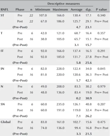
Changes in RNFL thickness from baseline to postope-ratively and averages of the absolute values of the RNFL thickness obtained by sectoral analyses are shown in table 1. No statistically significant difference (p≥0.05) was seen between pre- and post-treatment measurements in relation to each RNFL variable, i.e., on an avera-ge, the pre-treatment measurements are the same as post-treatment measurements. Further, despite the po-sitive differences between the pre- and post-treatment measurements (pre-post>0), these differences were not statistically significant. As shown in figures 2 and 3, the averages of each of the RNFL variables for pre-treatment are higher than those for post-treatment, but this was not statistically significant (p≥0.05).
Figure 1. Example of RNFL thickness measurements using circular scans of 1.8 mm radius.
measurements, with the greatest degree of correlation obtained between N values (r=0.77 → R2=59.3%). A significant positive correlation indicates that cases with high scores on the pre-treatment variable tend to score high on the post-treatment variable. In contrast, those with low scores on the pre-treatment variable also tend to have low scores on the post-treatment variable (in this case, the correlation was positive).
DISCUSSION
Visual field defects after pars plana vitrectomy have been a frequent topic of discussion in recent years. Pos-sible mechanisms include RNFL “dehydration” during fluid-air exchange(4-5), elevated IOP during vitrectomy, or
Table 1. Descriptive and comparative measures between pre-and post-treat ment RNFL measurements
Descriptive measures
RNFL Phase n Minimum Maximum Average d.p. P value
ST Pre 22 107.0 166.0 130.4 17.1 0.340
Post 22 067.0 186.0 125.7 29.1 Pre= Post
(Pre-Post) 004.7 23.1
T Pre 06 042.0 121.0 068.7 16.4 0.357
Post 16 038.0 105.0 065.7 15.1 Pre= Post
(Pre-Post) 3.1 15.7
IT Pre 06 092.0 166.0 137.4 16.5 0.291
Post 16 092.0 185.0 131.7 27.8 Pre= Post
(Pre-Post) 005.8 25.6
IN Pre 06 082.0 228.0 122.4 34.0 0.845
Post 16 081.0 220.0 120.6 36.3 Pre= Post
(Pre-Post) 001.7 42.1
N Pre 06 049.0 208.0 083.5 30.2 0.979
Post 16 048.0 136.0 083.4 19.0 Pre= Post
(Pre-Post) 000.2 30.9
SN Pre 06 060.0 235.0 126.1 40.8 0.207
Post 16 060.0 191.0 119.0 32.4 Pre= Post
(Pre-Post) 007.1 26.2
Global Pre 06 083.0 161.0 102.7 15.6 0.475
Post 16 074.0 136.0 099.4 16.8 Pre= Post
(Pre-Post) 003.3 21.5
Database: 22 eyes.
The p value refers to the significance probability on Student t test for paired samples. ST= superior temporal; T= temporal; IT= inferior temporal; IN= inferior nasal; N= nasal; SN= superior nasal.
Database: 22 eyes.
Note: With the inclusion of global RNFL.
Figure 3. Average of comparisons between pre- and post-treatment RNFL measurements.
Database: 22 eyes.
Note: Without the inclusion of global RNFL.
Figure 2. Average of comparisons between pre-and post-treatment RNFL measurements
As shown in the Results section, there was no statis-tically significant difference between pre- and posto-perative RNFL thickness around the optic nerve in any of the studied regions; this difference was also not significant when comparing sex and age.
It is important to note that the patients in this study were not tested for visual field defects pre- or postope-ratively. These results only demonstrate that ILM peeling has no direct impact on RNFL thickness, and the visual field defects shown in the literature might not be related to RNFL thickness changes.
The strong correlation between the extent of nasal and superior nasal thickness and pre-and postoperative global
Table 2. Correlation analysis between pre- and post-treatment RNFL measurements
Correlation of interest r P value
STpre X STpost 0.61 <0.002
Tpre X Tpost 0.50 <0.014
ITpre X ITpost 0.43 <0.043
INpré X INpost 0.29 <0.187
Npre X Npost 0.28 <0.204
SNpre X SNpost 0.77 <0.001
Globalpre X Globalpost 0.12 <0.576
Database: 22 eyes.
thicknesses must be considered. As described above, the analysis assumes that knowing the extent of both pre- and postoperative nasal thickness, it is possible to deter-mine an approximate value for the global measurement (and vice versa). However, further studies with a greater number of eyes are necessary to justify this finding.
In some eyes, a possible cause for increased postope-rative thickness is edema of the inner retinal layers in the first postoperative week, which leads to greater RNFL thickness measurements using OCT.
In conclusion, there appears to be no significant re-duction in RNFL thickness measurements after macular hole surgery, regardless of age or sex, in patients not diag-nosed with glaucoma or undergoing prior retinal surgery.
REFERENCES
1. Brooks Jr HL. Macular hole surgery with and without internal li-miting membrane peeling. Ophthalmology. 2000;107(10):1939-49. 2. Christensen UC, Krøyer K, Sander B, Larsen M, Henning V, Villumsen
J, et al. Value of internal limiting membrane peeling in surgery for idiopathic macular hole stage 2 and 3: a randomised clinical trial. Br J Ophthalmol. 2009;93(8):1005-15.
3. Lois N, Burr J, Norrie J, Vale L, Cook J, McDonald A, Boachie C, Ternent L, McPherson G. Full-thickness Macular Hole and Internal Limiting Membrane Peeling Study (FILMS) Group. Internal limiting membrane peeling versus no peeling for idiopathic full-thickness macular hole: a pragmatic randomized controlled trial. Invest Oph-thalmol Vis Sci. 2011;52(3):1586-92.
4. Welch JC. Dehydration injury as a possible cause of visual field defect after pars plana vitrectomy for macular hole. Am J Ophthalmol. 1997;124(5):698-9.
5. Hirata A, Yonemura N, Hasumura T, Murata Y, Negi A. Effect of infusion air pressure on visual field defects after macular hole surgery. Am J Ophthalmol. 2000;130(5):611-6.
6. Hutton WL, Fuller DG, Snyder WB, Fellman RL, Swanson WH. Visual field defects after macular hole surgery. A new finding. Ophthalmology. 1996;103(12):2152-8; discussion 2158-9. 7. Ejstrup R, la Cour M, Heegaard S, Kiilgaard JF. Toxicity profiles of
subretinal indocyanine green, Brilliant Blue G, and triamcinolone acetonide: a comparative study. Graefes Arch Clin Exp Ophthalmol. 2012;250(5):669-77.
8. Toba Y, Machida S, Kurosaka D. Comparisons of retinal nerve fiber layer thickness after indocyanine green, brilliant blue G, or triamci-nolone acetonide-assisted macular hole surgery. J Ophthalmol. 2014;2014:187308. doi: 10.1155/2014/187308.
9. Schuman JS. Spectral domain optical coherence tomography for glaucoma (An AOS thesis). Trans Am Ophthalmol Soc. 2008;106: 426-58.
10. Arora S, Goel N, Arora T, Sharma P, Raina UK, Thakar M, Ghosh B. Comparative evaluation of retinal nerve fiber layer thickness after conventional brilliant blue assisted internal limiting membrane peeling versus brilliant blue selective staining using whole blood in macular hole surgery. Ophthalmic Surg Lasers Imaging Retina. 2016;47(5):436-42.
11. Balducci N, Morara M, Veronese C, Torrazza C, Pichi F, Ciardella AP. Retinal nerve fiber layer thickness modification after internal limiting membrane peeling. Retina. 2014;34(4):655-63.