Daniel Alejandro Palma de Freitas
PREVALENCE OF ANTIBIOTIC RESISTANCE GENES IN PERIODONTAL INFECTIONS: TETRACYCLINE, METRONIDAZOLE, ERYTHROMYCIN
Universidade Fernando Pessoa
Faculdade de Ciências da Saúde
Daniel Alejandro Palma de Freitas
PREVALENCE OF ANTIBIOTIC RESISTANCE GENES IN PERIODONTAL INFECTIONS: TETRACYCLINE, METRONIDAZOLE, ERYTHROMYCIN
Universidade Fernando Pessoa
Faculdade de Ciências da Saúde
PREVALENCE OF ANTIBIOTIC RESISTANCE GENES IN PERIODONTAL INFECTIONS: TETRACYCLINE, METRONIDAZOLE, ERYTHROMYCIN
Trabalho apresentado à Universidade Fernando Pessoa
Como parte dos requisitos para a obtenção do grau de
Mestre em Medicina Dentária
RESUMO
Porphyromonas gingivalis e Prevotella intermedia são conhecidos patogénios na periodontite em adultos. É normalmente necessária antibioterapia no tratamento de periodontite sendo estes muitas vezes prescritos empiricamente. Para a prescrição de um tratamento com um antibiótico específico, é crucial a identificação de genes de resistência. O objetivo deste estudo foi a identificação de genes de resistência a antibióticos em estirpes bacterianas previamente isoladas de infeções periodônticas. A técnica de PCR foi usada na identificação dos genes de resistência a antibióticos TetM, TetQ, MefA, ErmB e Nim nas estirpes isoladas. Observou-se que 8% dos isolados tinham um dos genes de resistência a tetraciclina testados. Também foi observado que 2% dos isolados tinham um dos genes de resistência a eritromicina. Nenhum dos isolados demonstrou a presença do gene de resistência a metronidazol. Não foram detetados genes de resistência a tetraciclina nas estirpes de P. gingivalis.
Palavras-chave: Resistência a antibióticos; Porphyromonas sp.; Prevotella sp.; genes de resistência a antibióticos
Porphyromonas gingivalis and Prevotella intermedia are thought to be pathogens in adult periodontitis. Antibiotherapy is usually needed in the treatment of periodontitis being often prescribed empirically. To allow prescription of a specific antibiotic treatment, identification of resistance genes is crucial. The aim of this study was the identification of antibiotic resistance genes in strains previously isolated from periodontal infections. PCR was used for the identification of TetM, TetQ, MefA, ErmB and Nim resistance genes in isolated strains. It was seen that 8% of isolates had one of the tested tetracycline resistance genes. It was also observed that 2% of isolates had one of the analysed erythromycin resistance genes. None of the isolates showed the presence of the metronidazole resistance gene. No tetracycline resistance gene was observed in P. gingivalis strains.
Keywords: Antibiotic resistance; Porphyromonas sp.; Prevotella sp.; antibiotic resistance genes
INDEX
RESUMO ... v
ABSTRACT ... vi
I. INTRODUCTION ... 1
1.1. Periodontitis ... 2
1.1.1. Bacterial acquisition of antibiotic resistance ... 3
1.2. Tetracycline mode of action ... 4
1.2.1. Tetracycline resistance genes and resistance mechanisms ... 5
1.3. Metronidazole mode of action ... 6
1.3.1. Metronidazole resistance genes and resistance mechanisms ... 7
1.4. Erythromycin mode of action ... 8
1.4.1. Erythromycin resistance genes and resistance mechanisms ... 9
1.5. Antibiotics in Dentistry ... 9
II. MATERIALS AND METHODS ... 12
2.1. Patients and Microbiological procedures ... 12
2.2. PCR analysis ... 12
III. RESULTS AND DISCUSSION ... 13
IV. CONCLUSION ... 15
I. INTRODUCTION
The oral cavity constitutes a special environment in which more than 700 commensal or resident bacterial species may store and exchange their genetic material (Dupin et al., 2015).
Two of the most common human oral diseases (caries and inflammatory periodontitis) result from the accumulation of bacterial biofilms (plaques) on tooth surfaces. Oral health is the result of a balance between the resident flora and defence systems of the host. When this balance is disturbed, commensal and transient bacteria, viruses and fungi may be responsible for various local infections (Dupin et al., 2015).
Concerning periodontal disease, black-pigmented, Gram negative oral anaerobes such as Porphyromonas gingivalis and Prevotella intermedia are thought to be pathogens in the development of adult periodontitis (Andrés et al., 1998; Gatignol et al., 2003). These bacteria are frequently isolated from patients under periodontitis treatment. Odontogenic local infections require surgical treatment and, if required, a probabilistic antibiotherapy is needed that is effective on most recognized oral pathogens (Weeda and Backer, 2016).
In most cases, antibiotic prescription is empirical and based on the clinical condition of the patient. As a result, treatment is often inappropriate and leads to the development of bacterial resistance and even multiple resistances (Salinas et al., 2006). The intensive or inadequate use of antibiotics in medicine and dentistry favors the selection of bacteria that have acquired resistance to other antibiotics. Antibiotic resistance genes gradually spread among other pathogenic bacterial species by horizontal gene transfer in resident or transient bacterial populations. So, antibiotic resistance has become a serious problem in nowadays medical and dental practice, and data from the literature suggest that antibiotic resistance in the periodontal microbiota has increased. For this reason, antibiotic resistances have been under extensive microbiological, biochemical and genetic investigations (Weeda and Backer, 2016).
Reports from different countries show an increasing prevalence of patients with oral and subgingival antibiotic resistant bacteria. The use of antibiotics (defined as daily doses/100,000 inhabitants) was significantly higher in Mediterranean countries than in the rest of Europe (Van Winkelhoff and Winkel, 2005). However, in Portugal there is no data concerning antibiotic resistance of oral flora (Handal et al., 2005).
According to European experts, only in Europe, "superbugs" or bacteria cause more than 33,000 deaths per year, all due to antibiotic resistance (Cassini et al., 2019). This study observed higher number of deaths resulting from antibiotic resistance bacteria than the number of deaths from tuberculosis and HIV/AIDs combined (Cassini et al., 2019). In 2015, Italy was the country with the highest number of deaths (10,762), followed by France (5,543). In contrast, Netherlands, Sweden, Denmark, Slovenia,
Prevalence of antibiotic resistance genes in periodontal infections: tetracycline, metronidazole, erythromycin
Lithuania and Finland have their highest pick at 200 deaths. Countries with lowest number of deaths are Norway with 69, Luxembourg with 19, Estonia with 15, and last but certainly not least Iceland which has achieved a remarkable number of 1 (Cassini et al., 2019).
Taking the above numbers into consideration, antibiotic prescription in Europe, is not done in the same way in all countries. Surely in Italy, doctors concern about giving away antibiotic prescription is not as high as in Iceland and other Nordic countries, where bacterial resistance levels are very low, being given only when it is actually needed. Another key factor, even when an antibiotic is responsibly used, prescription should be as specific as possible. For that, it should always be certified what bacterial strain is responsible for a certain infection (Cassini et al., 2019).
The determination of in vitro antimicrobial susceptibility can be important in certain situations, for example, to monitor patterns of susceptibility and resistance in the population and to help in the selection of the appropriate antibiotic in dentistry treatments (Jacinto et al., 2006). Although antibiotic sensitivity can be determined from standard cultural microbiological analysis, this generally takes several days due to the slow growth of fastidious anaerobic bacteria. Since infection can spread rapidly and cause severe complications such as sepsis and obstruction of the airway, such a delay can prove problematic and undesirable. The introduction of PCR-based techniques has resulted in the development of tests that can detect specific pathogens and genes directly and rapidly from clinical samples. Indeed, conventional PCR has already become an important tool in clinical diagnostic and research laboratories (Iwahara et al., 2006).
The main goal of this study was to identify the presence of antibiotic resistance genes in strains isolated from periodontal pockets of Portuguese adults suffering from periodontal infections. Analyzed genes were TetM and TetQ genes that confer resistance to tetracycline, nim gene responsible for metronidazole resistance and ermB and mefA genes involved in erythromycin resistance. These correspond to the most frequently prescribed antibiotics for periodontitis treatment.
1.1. Periodontitis
Periodontitis is a complex inflammatory and infectious disease in the oral cavity, affecting all oral tissues. Besides soft tissues, this pathology also affects hard tissue and teeth. It is caused by unbalanced numbers of specific microorganisms in the oral cavity (Green, 2019).
In the USA, between 50 and 90% of the population has gingivitis, a reversable condition that, if left untreated, leads to periodontitis, an irreversible disorder (Green, 2019).
The contamination of the supporting tissue will spread and destroy progressively all structures, including periodontal ligament, leading to the formation of pockets where bacteria can accumulate, which then will lead to gingiva recession, followed by bone and tooth lost (Nazir, 2017).
The development of periodontitis results from the combination of bacterial infection and host response. Hence, it varies among people, since one person might bare 10 million bacteria and another 20 million, both without having any sign of periodontitis (Saini et al., 2009).
Statistically, periodontal diseases are widespread, breaking race and age barriers, (individuals can develop this disorder at any age) to the extent that it is considered a public health concern, affecting around 20 to 50% of the population worldwide (Saini et al., 2009).
Moreover, environmental and genetic factors may promote or protect (to a certain point) the occurrence of periodontal disease (Saini et al., 2009). It is, therefore, considered is a multifactorial disorder whose etiology involves environmental factors such as poor oral hygiene, smoking, age, some pharmaceutical products and stress (Nazir, 2017). Moreover, systemic conditions such as diabetes, cardiovascular disease, and adverse pregnancy outcomes (hormonal changes in women´s body), have also a strong association with periodontitis (Nazir, 2017).
Periodontitis begins with the accumulation of a biofilm on the tooth surface and surroundings, followed by the accumulation of subgingival microbiota, forming an adherent layer of plaque. This plaque becomes solid, and underneath is the perfect shelter for growth of anaerobic bacteria (Pacios et al., 2015).
These subgingival bacteria can interact with host tissues without penetrating them, inducing an immune inflammatory response. Periodontal infection stimulates innate and adaptative immune responses (Pacios et al., 2015), leading to the production of cytokines such as the tumor necrosis factor that induce osteoclastogenesis (Chang et al., 2009). This inflammation is a pathologic process that will also lead to the uncoupling of bone formation and its resorption (Graves et al., 2011).The inflammation process starts with the activation of cells of the blood stream (T cells and macrophages) by the presence of an antigen of the pathogenic bacteria. The cells respond by the production and release of proinflammatory cytokines, that will promote vasodilatation, allowing a faster neutralization or inducing apoptosis of pathogenic cells. Moreover, during the inflammation process, osteoclasts are also stimulated by cytokines, leading to bone degradation (Pacios et al., 2015).
Bacteria present in periodontitis keep on multiplying, triggering more destruction of soft and bone tissues. Without a stable bone, periodontal structure crashes, the ligament is detached, and elastic fibers are lost, leading to teeth movement, subgingival cavities and finally tooth loss (Saini et al., 2009). 1.1.1. Bacterial acquisition of antibiotic resistance
Prevotella species (Prevotella intermedia and Prevotella nigrescens) are black pigmented strictly anaerobic bacteria, that can be found in human digestive tract and in dental plaque, being able to trigger gingivitis and periodontal disease. These bacterial species are close taxonomic relatives of the oral pathogen Porphyromonas gingivalis. These bacteria are able to transfer genetic material between each
Prevalence of antibiotic resistance genes in periodontal infections: tetracycline, metronidazole, erythromycin
other and to other species, since they possess a multitude of genetic elements such as transposons that can easily be transferred by bacterial conjugation (Tribble et al., 2011). One of such transposons is a large conjugative transposon (more than 60 kb), commonly associated with tetracycline resistance (Tribble et al., 2011).
Although some studies show that antibiotic resistance is often found in these oral anaerobic species, the level of participation of each transposable element in the acquisition of antibiotic resistance is still unknown (Tribble et al., 2011). In one hand, there are studies that state the presence of 4% of tetracycline resistant P. intermedia isolates (Tribble et al., 2011). On the other hand, others have shown 26% of resistance in similar species (Tribble et al., 2011).
Moreover, some researchers suggested the hypothesis for the acquisition of antibiotic resistance based on the selective pressure in the form of antibiotic therapy being responsible for the rise in the antibiotic resistance in oral environment (Tribble et al., 2011). On the other hand, other scientists believe that it is the duration of the treatment that being either shorter or longer than it should, is triggering resistance development (Wilson et al., 2019).
Since all those theories have been proven to be reliable, it is more likely that acquisition of antibiotic resistance might result from the combination of several factors, like bacteria´s transfer mechanisms combined with the selective pressure in the form of antibiotic, and the short duration of the antibiotic treatment. Moreover, the posology for periodontitis should not be the same as for the treatment of an apical tooth infection (Tribble et al., 2011).
Today´s concept regarding periodontitis treatment, is that it should always include a mechanical phase followed by antibiotic therapy. It is known that if mechanical treatment is applied without antibiotic therapy, periodontitis bacteria will persist (Kapoor et al., 2012). On the other way around, if an antibiotic monotherapy is prescribed but no mechanical treatment is applied, a rise in the expression of resistance genes and their transfer through bacterial conjugation is observed, transforming a medium periodontitis into an aggressive type of periodontitis, with the development of multiple periodontal abscesses and overall worse treatment outcome when compared with the combined therapy (Heitz‐ Mayfield, 2009).
1.2. Tetracycline mode of action
Tetracyclines were discovered in the 1940s, showing a great efficiency against a wide variety of microorganisms, among them parasites and bacteria, such as Gram-positive and negative, chlamydia, protozoan parasites, mycoplasma and rickettsia (Chopra and Roberts, 2001).
Furthermore, this group of antibiotics is useful to treat patients allergic to penicillin, against bacteria such as N. gonorrhoeae, T. pallidum, C. tetani, L. monocytogenes and P. multocida (Sousa, 2006).
This class of antibiotics is relatively affordable, leading to their use in an extensive range of situations such as prophylaxis in humans (prevention of malaria caused by mefloquine-resistant Plasmodium falciparum) and in animals (subtherapeutic levels in animal feed as growth promoters) (Chopra and Roberts, 2001).
This involves crossing of bacterial membranes by passive diffusion in order to interact with their targets (Shutter and Akhondi, 2019). This antibiotic inhibits bacterial growth, by interfering with the synthesis of bacterial proteins and making impossible the anchorage of aminoacyl-tRNA in the bacterial ribosomal acceptor (A) site. It is a bacteriostatic antibiotic, inhibiting the link between the codon and the anticodon at the 30S subunit of the ribosome (Sousa, 2006).
Tetracycline can also act by destroying the cellular membrane (Shutter and Akhondi, 2019). 1.2.1. Tetracycline resistance genes and resistance mechanisms
Nowadays, three types of tetracycline resistance mechanisms are known: ribosome protection, tetracycline efflux pumps and tetracycline modification (Shutter and Akhondi, 2019).
The first one is the most common in all bacteria, from Gram-positive and Gram-negative bacteria to mycoplasmas (Shutter and Akhondi, 2019). This mechanism involves an EF-G-like protein that confers ribosome protection (Gzyl and Wieden, 2017). The tetracycline resistance works by binding of a specific cytoplasmic protein to the ribosome, in the same site where tetracycline binds. This protein is a GTPase, with sequence and structural similarities with the elongation factor EF-G, that protects the bacterial ribosome from binding the antibiotic tetracycline (Gzyl and Wieden, 2017). So far, 38 different tetracycline resistance genes have already been identified (Roberts, 2005). The tetM, tetO, tetQ and tetS genes encode proteins that act by the ribosome protection mechanism described above, encoding a protein of 642 amino acids called GTPase (Li et al., 2013).
As an example, the tetO gene product acts on the 30S ribosomal subunit binding site and is able to free the ribosome from the tetracycline, leading to its hydrolysis. In this way, the ribosome returns to the elongation cycle (Connell et al., 2003).
On the other hand, the tetM encoded protein, has a similar sequence to EF-G, that is responsible for the translocation of the ribosome during the elongation phase of protein synthesis. In this way, this protein prevents tetracycline action (Connell et al., 2003).
The second most frequent resistance mechanism is the efflux pumps type that has been found mostly in Gram-positive and y-purple bacteria such as Enterobacteriaceae (Grossman, 2016). In this type of tetracycline resistance, membrane proteins, that can be encoded by several resistance genes, actively pump tetracycline from the cell by exchanging protons with a tetracycline cation and consequently lead to a reduction on tetracycline concentration inside the cell (Li, 2017). This tetracycline resistance is conferred by the presence of tetracycline resistance genes that code for proteins that alter membrane
Prevalence of antibiotic resistance genes in periodontal infections: tetracycline, metronidazole, erythromycin
permeability to the antibiotic. This is the case of the TetA, TetB, TetC, TetD and TetE genes (Chopra and Roberts, 2001). These genes only confer resistance to tetracycline but the TetB gene is able to pump not only tetracycline but also minocycline, giving resistance to these two antibiotics (Tuckman et al., 2007).
Efflux pumps proteins are encoded by genes present on the bacteria chromosome. Most of these proteins have low antibiotics specificity and can therefore confer resistance to a wide group of antibiotics (Sousa, 2016).
The last mechanism of resistance, the tetracycline modification mechanism involves the modification of the 16S rRNA present at the tetracycline-binding site of the small ribosomal subunit. Tetracycline prevents the positioning of the aminoacyl-tRNA within the A site of the 30S ribosomal subunit, acting as a bacteriostatic agent. Bacterial species that are resistant to this antibiotic have a mutation in the 16S rRNA that prevents the elongation of protein synthesis, leading to peptide chain termination (Chukwudi, 2016).
1.3. Metronidazole mode of action
Metronidazole was discovered in 1959, then in 1962 a patient was cured from both acute ulcerative gingivitis and from Trichomonas vaginalis through the use of this antibiotic (Lofmark et al., 2010). Nevertheless, only in 1970s, metronidazole´s success was widespread among world´s population, regarding its effect in Gram-negative anaerobes infections like Bacteroides or Gram-positive anaerobes such as Clostridium (Lofmark et al., 2010).
Metronidazole inhibits anaerobic and microaerophilic organisms as they both work in a similar way in terms of their enzyme catalysing fermentation, called pyruvate ferredoxin oxidoreductase (Sousa, 2006).
Nowadays, this drug is often prescribed for the treatment of Vincent´s angina which is an acute necrotizing ulcerative gingivitis, Helicobacter pylori infections such as gastritis and gastroenteritis caused by Campylobacter spp. (Sousa, 2006).
There are those who argue about the toxicity of metronidazole, but it has been proven that even though the reduction of the nitro group in 5-nitroimidazoles, such as metronidazole, causes some short-lived cytotoxic intermediates, still this antibiotic is considered to be nontoxic, as those intermediates end up being nontoxic products. The interaction between those intermediates and deoxyribonucleic acid causes that mild toxicity (Lofmark et al., 2010).
It is well known that the efficacy to reduce nitroimidazoles depends on metabolic pathways of low redox potential that are linked to ferredoxin and flavodoxin like electron transport components (Campbell et al., 2019). These pathways are common in anaerobic protozoa and bacteria that are found in periodontitis and vaginal infections and are not present in aerobic cells of the host (Hrdý et al., 2005).
This selective toxic mechanism of metronidazole depends on the reduction of nitro groups which affects the rate of entry of the drug into the cell, conducted by ferredoxin-linked mechanism (or similar) reactions in the cell (Hrdý et al, 2005). The reduced agent leads to breakage of DNA, that is dependent on the A+T content of DNA. Furthermore, these drugs prevent DNA repair, which leads to permanent damage (Weir and Le, 2019).
Anaerobic bacteria, due to their low redox potential, reduce this drug to its active metabolite by an electron transfer step (Wouden et al., 2001).
However, in micro-aerophilic bacteria (including H. pylori) the metabolism of metronidazole is different from anaerobic bacteria. The obtained metabolites produced by the electron transfer step are re-oxidized - "futile cycling". During this process, toxic oxygen radicals are produced, that must be eliminated by an active scavenger system (Wouden et al., 2001).
Some studies showed that in H. pylori the activation of metronidazole is done by a two-electron transfer step catalysed by an oxygen-insensitive nitroreductase encoded by the rdxA gene. This prevents the functioning of the futile cycle, allowing metronidazole activation (Wouden et al., 2001).
1.3.1. Metronidazole resistance genes and resistance mechanisms
Since nitroimidazoles such as metronidazole, are vastly used in the treatment of anaerobic infections, a huge concern rises among experts related to multidrug-resistant anaerobic bacteria (Alauzet et al., 2019).
Nowadays, many and complex bacterial resistance mechanisms towards metronidazole are known. According to literature, nitro-imidazole-reductase enzymes are responsible for the inactivation of this drug. These enzymes are encoded by the nim genes (Alauzet et al., 2019). So far, 11 nim genes (nim A to nim K) have been identified and depending on the resistance gene expression, anaerobic bacteria harbouring these genes may develop low or high level of resistance to these kind of drugs (Alauzet et al., 2019).
Resistance of metronidazole in H. pylori may happen, when the gene rdxA is mutated by the occurrence of point mutations and/or insertions, that lead to the appearance of a premature stop codon. Consequently, the encoded oxygen-insensitive nitroreductase is not produced or a shorter version of this enzyme is synthesized that has no enzymatic activity. As a result, metronidazole is not activated (Alauzet et al., 2019).
The unsuccess of nitroimidazoles can also result from 2 other mechanisms of inactivation of these drugs, such as aminothiol radical scavengers and radioprotectors that are normally present in the cell or due to the presence of other organisms able to inactivate those drugs in the same environment (Olender et al., 2018).
Prevalence of antibiotic resistance genes in periodontal infections: tetracycline, metronidazole, erythromycin
The radical scavengers, also called antioxidants, react with oxidising agents such as radicals and prevent their oxidative action (Choppin et al., 2013).
The radioprotectors are compounds that reduce radiation damage in tissues. These compounds are usually antioxidants that, for being effective, must be present in the cell before or at the moment of the exposure to radiation (Raviraj et al., 2014).
1.4. Erythromycin mode of action
Erythromycin was isolated from the bacteria Saccharopolyspora erythraea in 1949, and belongs to the macrolides antibiotic´s group (Sauberan and Bradley, 2018).
Since then, erythromycin has been used not only in the treatment of a wide range of infections such as respiratory (pneumonia, legionnaires disease) and skin infections, intestinal amebiasis, syphilis and pelvic inflammatory disease, but also of non-infectious conditions as a prophylactic measure such as neonatal conjunctivitis and chlamydia and prophylaxis of rheumatic fever (Farzam and Quick, 2019). Moreover, in combination with other drugs in creams, erythromycin is used to treat skin conditions like acne and can be useful in the prevention of group B streptococcal infection in the newborn (Farzam and Quick, 2019).
Erythromycin has a vast efficacy on different types of microorganisms, not only Gram-positive and Gram-negative bacteria, but also other microorganisms such as Treponema pallidum, Ureaplasma urealyticum, Chlamydia trachomatis, Entamoeba histolytica and Mycoplasma pneumoniae. Among the affected Gram-negative bacteria are Neisseria gonorrhoeae, Haemophilus influenzae, Bordetella pertussis and Legionella pneumophila. The Gram-positive strains include Corynebacterium minutissimum, Corynebacterium diphtheria, Streptococcus pneumoniae, Streptococcus pyogenes, Staphylococcus aureus and Listeria monocytogenes (Farzam and Quick, 2019).
Erythromycin is a bacteriostatic antibiotic, whose mode of action is based on protein synthesis arrest, by binding to the 23S rRNA molecule present in the 50S subunit of bacterial ribosomes. In this way, this drug prevents the release of the peptide chain from the ribosome, blocking the growth of the polypeptide chain (Farzam and Quick, 2019).
It is worth pointing out that erythromycin does not affect protein synthesis in human´s tissues since eukaryotic cells do not have 50S ribosomal subunits (do not have the 23S rRNA molecule). Instead, ribosomes of eukaryotic organisms are composed of a 40S and a 60S subunits (Farzam and Quick, 2019).
Erythromycin can be administered orally, intravenously, or in the topic and ophthalmic form. This drug is metabolised mainly by the liver through the action of a cytochrome P450 enzyme (CYP3A4) that is responsible for its demethylation, allowing its renal excretion (Farzam and Quick, 2019). Therefore, caution is advised to hepatic patients.
1.4.1. Erythromycin resistance genes and resistance mechanisms
Resistance to erythromyccin can result from changes in the 23S rRNA that prevent the attachment of the drug to this molecule and consequently protein synthesis can proceed (Farzam and Quick, 2019). This mechanism of erythromycin resistance identified in Streptococcus is determined by the presence of a methylase enzyme (Desjardins et al., 2014). This enzyme induces conformational changes in bacterial ribosomes that reduce the capacity of antibiotics such as macrolides, to bind to the target molecule in the 50S ribosomal subunit (Pokkunuri and Champney, 2007).
The methylase enzyme is encoded by an erm gene (Desjardins et al., 2014). The expression of antibiotic resistance conferred by this group of genes can be constitutive or inducible and most of this resistance is due to the ermB gene (Desjardins et al., 2014). Recently, a new erm gene (ermTR) has been identified in Streptococcus pyogenes (Desjardins et al., 2014).
Another erythromycin resistance mechanism results from drug efflux (often referred as the M phenotype), being encoded by the mefA gene, among others, which creates low-level of resistance to erythromycin (Desjardins et al., 2014).
Erythromycin resistance can also be conferred by the presence of the linB gene. This gene encodes a lincosamide nucleotidyltransferase, which confers resistance to licosamide and macrolides. The enzyme nucleotidyltransferase has a role in DNA repair. It is a component of the excision repair mechanism, where a glicosylase enzyme recognizes an incorrectly matched or mutated nucleotide that has to be removed. After removal, the left gap is filled in by the nucleotidyltransferase enzyme, that inserts the correct nucleotide using the complementary sequence as a template (Liu et al., 2007).
1.5. Antibiotics in Dentistry
Acute symptoms of dental infection or abscess can be managed temporally in hospitals or general practice, but the original cause of the infection should be managed by a dentist providing the correct diagnosis and treatment. In cases of infected areas needing treatment such as root canal treatment or tooth extraction, previous antibiotic treatment is required. Moreover, antibiotic therapy is always required whenever there are signs of severe systemic infection, such as fever (Ytreland, 2016).
Despite this essay focus on Tetracycline, Metronidazole and Erythromycin resistance, these chemicals are not the first-line antibiotics used against oral infections. Instead, β-lactam antibiotics are the most prescribed ones in cases of oral diseases, and they are the theme of the thesis of another student. Antibiotic therapy in Dentistry, including dose and combination of antibiotic agents, varies depending on many factors such as pathology, microorganisms present, and the degree of affectedness. For instance, metronidazole or clindamycin are prescribed in cases of infections by anaerobes such as necrotizing ulcerative acute periodontitis (Leite, 2014).
Prevalence of antibiotic resistance genes in periodontal infections: tetracycline, metronidazole, erythromycin
In some cases, a combination of this antibiotic with amoxicillin is desired depending on the microorganism's type. In cellulitis, there is a high risk of sepsis, so oral antibiotics can be prescribed by the dentist with a careful assessment of the severity. In some of these cases, even hospital care is required (Leite, 2014).
For this reason, bacterial samples should be taken for antibiotic sensitivity testing. In countries like Norway, dentists follow this strategy and therefore tend to more accurately prescribe narrow spectrum antibiotics (Ytreland, 2016).
According with Ikram (2012), metronidazole or amoxicillin can be prescribed in cases of dental infections/abscesses, however this author is not clear on which one to use first. Moreover, it is referred that for severe dental infections it is more appropriate to use the combination of metronidazole and amoxicillin (Ikram, 2012). The recommended prescribed dose can vary from 400 mg, 3 times a day for five days (Ikram, 2012) to 500-750 mg for 8 days (Roda, 2007). Special attention to the prescribed dose should be taken in cases of patients under warfarin treatment, since metronidazole potentiates the effect of warfarin. Metronidazole inhibits warfarin metabolism, leading to the rise of its serum levels, increasing its anticoagulant effects. So, INR should be monitored in these patients to adjust the dose of warfarin if required (Ikram, 2012). In patients suffering from chronic renal insufficiency, metronidazole intake should be done every 12 hours, instead of 8 hours intervals (Infarmed, 2017). In chronic hepatic insufficiency or failure, the serum levels of metronidazole should be monitored (Infarmed, 2017). Metronidazole is contraindicated during the first trimester of pregnancy (Infarmed, 2017).
Tetracycline can be prescribed in some cases of periodontal disease (aggressive, chronic and necrotizing ulcerative acute periodontitis), refractory periodontitis and some gingivitis such as necrotizing ulcerative acute gingivitis. Other cases of its use are juvenile periodontitis when associated with Actinobacillus actinomycetemcomitans, dental abscess and connective tissue abscess (Leite, 2014).
When prescribing tetracycline, dentists should be aware that the level of this drug is higher in gingiva and gingival crevicular fluid than in the serum (Kumar, 2015). This drug possesses antibacterial activity, inhibits matrix metalloproteinases such as collagenases, anti-collagenase activity (enzyme that participates in the destruction of connective tissue) and anti-inflammatory activity. Therefore, promotes wound-healing and reduces bone loss (Kumar, 2015). Doxycycline has a stronger effect on metalloproteinases inhibition, so, more connective tissue is preserved (Kumar, 2015). On the other hand, minocycline stimulates osteoblasts, enhancing periodontal healing (Kumar, 2015).
There are forms of topic tetracyclines, that can be applied directly into the gingiva, allowing the slow release of the drug. Since it is a local use, it does not have any systemic undesirable side effect (Leite, 2014).
The recommended dose for tetracycline is 250mg, every 6 hours for 7 days (Leite, 2014), and for doxycycline 100mg/day (Kumar, 2015). Local treatment should be performed with 20 mg of doxycycline twice a day for three months (Leite, 2014).
The use of tetracyclines is contraindicated during pregnancy and breastfeeding. It should not be used in children under 12 years of age (Leite, 2014), since it might affect enamel formation, leading to brown or yellow lines in teeth (Leite, 2014). This antibiotic has some hepatotoxic effects, so caution is required in patients with liver conditions (Leite, 2014).
Erythromycin is used as a second-line antibiotic in cases of dental abscesses, as an alternative to penicillins (Wray et al., 2011). The recommended dose for adults and children over 12 years of age or with more than 40 kg of weight is 1-2 g/day, for a period of 5 to 14 days (Infarmed, 2019). In cases of severe infections, the dose can be increased to 1 g every 6 hours (total of 4 g/day) (Infarmed, 2019). This antibiotic should not be taking during pregnancy or breastfeeding unless it is strictly necessary (Infarmed, 2019).
Future research should aim not only to create new narrow-spectrum antibiotic with selective killing or neutralizing targets, but also, to achieve the right drug combination for each case, assessing the nature and spot of the infection, microorganism(s) responsible for it and patient as a whole (Dingsdag and Hunter, 2017).
According with Dingsdag and Hunter (2017), broad-spectrum antibiotics kill off-target microorganisms, alter the physiology of some of them, and promote overgrowth of certain strains. It may cause disequilibrium among several types of strains, that may also lead to the overgrowth of endogenous pathogens such as C. difficile (Dingsdag and Hunter, 2017).
In order to circumvent drug resistance, scientists have been trying to develop new nitroimidazole drugs strategies (Dingsdag and Hunter, 2017). For instance, metronidazole/ triazoleconjugates but also a novel narrow-spectrum more selective antibiotic and selective killing mechanism called deuteroporphyrin–nitroimidazole adducts have been designed. These adducts are taken up by porphyrin receptors of P. gingivalis, reducing the number of bacteria receiving the drug and thus reducing the rates of resistance (Dingsdag and Hunter, 2017). This is not a real solution for periodontal disease therapy, since there are more bacteria than just P. gingivalis but it is a step forward as a therapy to control pathogenic microorganisms and also lower resistance (Dingsdag and Hunter, 2017).
Prevalence of antibiotic resistance genes in periodontal infections: tetracycline, metronidazole, erythromycin
II. MATERIALS AND METHODS
2.1. Patients and Microbiological procedures
This study was based on 50 patients treated at the Dentistry Clinic of the Health Sciences Faculty, Fernando Pessoa University, Porto, Portugal. Adult patients with 35 to 75 years old, diagnosed with refractory marginal periodontitis and that did not receive antimicrobial therapy in the previous 30 days were included in the study. Sampling was performed with aseptic surgical techniques. Samples were collected according with Herrera et al. (2000) using four sequential sterile paper points inserted into the subgingival sulcus after cleaning the supragingival plaque and held in place for 60 s. A sterilized prereduced transport fluid (RTF) was used to transport samples to the laboratory where they were immediately processed. Isolation of black pigmented bacteria was performed using culture procedures: samples were vortexed for 60 s and diluted in PBS solution using a 10-fold serial dilution to 10-3. A
volume of 100 µL of each dilution was spread on non-selective 5% horse blood agar plates enriched with hemin (5mg/L) and vitamin K (1mg/L). Incubation was performed at 37ºC in an anaerobic atmosphere by Macs Mics (Whitley Jar Gassing System). Plates were red at days 7 and 14. Preliminary characterization of microbial species was based on colony pigmentation and morphology, Gram staining and catalase production. Bacterial identification had been previously performed using the API Rapid ID 32 A system (bioMérieux) and confirmed by PCR analysis (Pina et al., 2011).
2.2. PCR analysis
DNA was extracted as described by Ashimoto et al. (1996).
For the identification of the tetracycline resistance genes, DNA (5 µL) was amplified in a reaction mixture containing 10 µL of 5x PCR buffer, 3 µL of MgCl2 25mM, 1 µL of dNTP mixture 10 mM, 2,5
µL of each primer 10 µM and 0,25 µL Taq polymerase in a total volume of 50 µL (Koukos et al., 2015). Primers used for identification of the TetM gene were GACACGCCAGGACATATGG-3' and 5'-TGCTTTCCTCTTGTTCGAG-3' (Koukos et al., 2015). PCR was performed according with Koukos et al. (2015) as followed: 5 minutes at 94ºC; 37 cycles of 30 seconds at 94ºC, 1 minute at 55ºC and 90 seconds at 72ºC; final extension for 10 minutes at 72ºC.
Primers used for identification of the TetQ gene were GGCTTCTACGACATCTATTA-3' and 5'-CATCAACATTTATCTCTCTG-3' (Koukos et al., 2015). PCR was performed according with Koukos et al. (2015) as followed: 5 minutes at 94ºC; 37 cycles of 30 seconds at 94ºC, 1 minute at 50ºC and 160 seconds at 72ºC; final extension for 10 minutes at 72ºC.
For identification of the nim gene, DNA (5 µL) was amplified in a reaction mixture containing 10 µL of 5x PCR buffer, 3 µL of MgCl2 25mM, 1 µL of dNTP mixture 10 mM, 5 µL of each primer 10 µM
and 0,25 µL Taq polymerase in a total volume of 50 µL (Koukos et al., 2015). Primers used for identification of the nim gene were ATGTTCAGAGAAATGCGGCGTAAGCG-3' and
5’-GCTTCCTTGCCTGTCATGTGCTC-3’ (Koukos et al., 2015). PCR was performed according with Koukos et al. (2015) as followed: 5 minutes at 94ºC; 35 cycles of 30 seconds at 94ºC, 1 minute at 62ºC and 1 minute at 72ºC; final extension for 10 minutes at 72ºC.
For identification of the ermB and mefA genes, DNA (1 µL) was amplified in a reaction mixture containing 4 µL of 5x PCR buffer, 0,4 µL of MgCl2 25mM, 0,8 µL of dNTP mixture 10 mM, 1 µL of
each primer 10 µM and 0,25 µL Taq polymerase in a total volume of 20 µL (Najafi Mosleh et al., 2014). Primers used for identification of the ermB gene were 5’-CGTACCTTGGATATTCACCG-3' and 5’-GTAAACAGTTGACGATATTC-3’ (Najafi Mosleh et al., 2014). Primers used for identification of the mefA gene were CCCAGCTTAGGTATACGTAC-3' and 5’-CTGTATGGAGCTACCTGTCTGG-3’ (Najafi Mosleh et al., 2014). PCR was performed according with Ubukata et al. (2003) as followed: 5 minutes at 94ºC; 30 cycles of 20 seconds at 94ºC, 20 seconds at 52ºC and 15 seconds at 72ºC; final extension for 10 minutes at 72ºC.
PCR products were analyzed by electrophoresis on a 1% agarose gel.
III. RESULTS AND DISCUSSION
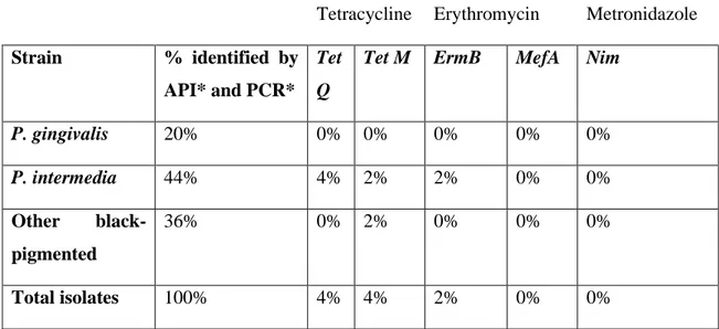
Isolated black pigmented bacterial strains identified by conventional and molecular techniques in a previous study (Pina et al., 2011), were screened for the presence of antibiotic resistance genes (Table 1).
Table 1. Results obtained in the identification of isolated strains using API and PCR (Pina et al., 2011) and in the identification of antibiotic resistance genes.
Antibiotic Resistance Genes
Tetracycline Erythromycin Metronidazole Strain % identified by
API* and PCR*
Tet Q
Tet M ErmB MefA Nim
P. gingivalis 20% 0% 0% 0% 0% 0% P. intermedia 44% 4% 2% 2% 0% 0% Other black-pigmented 36% 0% 2% 0% 0% 0% Total isolates 100% 4% 4% 2% 0% 0% *Pina et al. (2011).
Prevalence of antibiotic resistance genes in periodontal infections: tetracycline, metronidazole, erythromycin
Previously isolated strains were identified as Prevotella intermedia that represented 44% and as Porphyromonas gingivalis that were 20% of total isolates. The remaining 36% strains belonged to other black-pigmented species (Table 1) (Pina et al., 2011).
Concerning all tested antibiotic resistance genes, a total of 42% of antibiotic resistance genes in strains isolated from periodontal infections was observed. These results agree with most studies of other countries that show high levels of antibiotic resistance among anaerobes (Maestre et al., 2007; Dubreuil et al., 2003; Handal et al., 2005; Koukos et al., 2015; Collins et al., 2016).
Concerning tetracycline resistance genes, it was observed that 8% of isolated strains had one of the analysed tetracycline resistance genes. The TetQ gene was detected in 4% of total isolates. The TetQ harbouring strains were all identified as P. intermedia. The TetM gene was also detected in 4% of total isolates, corresponding 1% of these to P. intermedia strains. The remaining 3% belonged to other black-pigmented unidentified strains. None of these tetracycline resistance genes was present in P. gingivalis strains (Table 1).
Ioannidis et al. (2009) also observed high levels of tetQ (70-80%) and tetM (76-82%) genes in samples collected from periodontal infections.
Moreover, unlike our data, another study (Koukos et al., 2015) on implants showed that the most abundant genes were the tetracycline resistance genes. Collins et al. (2016) also detected seven tetracycline resistance genes in bacterial isolates from chronic periodontitis, including the tetQ gene present in 72% of tested patients.
Isolated strains were also tested for the presence of the erythromycin resistance genes ermB and mefA. Only 2% of total isolates showed the presence of the ermB gene and none harboured the mefA gene (Table 1).
The nim gene, responsible for metronidazole resistance, was not detected in any of the isolates. Although, in accordance with this work, some studies did not detect the nim gene in collected samples (Ioannidis et al., 2009; Koukos et al., 2016), Xie et al. (2014) reported the presence of this gene in strains isolated from periodontal abscesses.
β-Lactam resistance has been associated with resistance to tetracycline (Tet genes) and to erythromycin (erm genes) (Falagas et al., 2000). A recent study of oral anaerobes from patients with periodontitis identified a high prevalence (97%) of CfxA β-lactamase production by aminopenicillin-resistant Prevotella in subgingival plaque (Handal et al., 2005).
Samples used in this study were also screened for the presence of β-lactam resistance genes (TEM and cfxA genes) in the work of the master thesis of Laura Gonçalves. In the same way, it was observed that strains with tetracycline resistance genes (TetQ or TetM) also harboured the TEM gene. Moreover, the ermB gene was detected in strains that also had the TEM and the TetM genes. These results may indicate
a combined transfer of antibiotic resistance genes. However, due to the sampling size, these results should be confirmed with a bigger collection of sample isolates from periodontal infections.
Divergence in bacterial frequencies observed in this study and in other reports might be explained by geographical differences and divergences in sampling (Fosse et al., 1999; Dubreuil et al., 2003) as well as sample size. This study should be enlarged to the analysis of a bigger number of patients carrying periodontal infections.
IV. CONCLUSION
As expected, oral bacterial species also carry in addition other resistance genes, such as tetracycline and erythromycin antibiotic resistance genes, probably due to the diversity of mechanisms of transfer of genetic material.
Due to the increasing prevalence of antibiotic resistance among bacterial strains isolated from periodontal infections, the empirical prescription of these antibiotics as a therapy strategy for periodontitis should be avoided.
This study contributes to the knowledge on subgingival microbiota and its resistance genes present in periodontal infections. Knowing the prevalence of resistance genes can have impact on their clinical prescription and might raise awareness to the appropriate use of antibiotics.
Prevalence of antibiotic resistance genes in periodontal infections: tetracycline, metronidazole, erythromycin
V. REFERENCES
Alauzet, C., Lozniewski, A., Marchandin, H. (2019). Metronidazole resistance and nim genes in anaerobes: a review. Anaerobe, 55, pp. 40-53.
Andrés, M. T. et al. (1998). Antimicrobial susceptibilities of Porphyromonas gingivalis, Prevotella intermedia and Prevotella nigrescens spp. isolated in Spain. Antimicrobial Agents and Chemotherapy, 42(11), pp. 3022-3023.
Ashimoto, A. et al. (1996). Polymerase chain reaction detection of 8 putative periodontal pathogens in subgingival plaque of gingivitis and advanced periodontitis lesions. Oral Microbiology and Immunology, 11, pp. 266-273.
Binta, B., Patel, M. (2016). Detection of cfxA2, cfxA3 and cfxA6 genes in beta-lactamase producing oral anaerobes. Journal of Applied Oral Sciences, 24(2), pp. 142-147.
Campbell, I. J. et al. (2019). Evolutionary relationships between low potential ferredoxin and flavodoxin electron carriers. Frontiers in Energy Research, 7, pp.79.
Cassini, A. et al. (2018). Attributable deaths and disability-adjusted life-years caused by infections with antibiotic-resistant bacteria in the EU and the European Economic Area in 2015: a population-level modelling analysis. The Lancet, 19(1), pp. 56-66.
Cassini, A. et al. (2019). Attributable deaths and disability-adjusted life-years caused by infections with antibiotic-resistant bacteria in the EU and the European Economic Area in 2015: a population-level modelling analysis. The Lancet, 19(1), pp. 56-66.
Chang, J. et al. (2009). Inhibition of osteoblastic bone formation by nuclear factor-kappa B. Nature Medicine, 15, pp. 682-689.
Choppin, G. et al. (2013). Radiation Biology and Radiation Protection. In: Radiochemistry and Nuclear Chemistry, 4th ed. Academic Press, pp. 445-491.
Chukwudi C. U. (2016). rRNA binding sites and the molecular mechanism of action of the tetracyclines. Antimicrobial Agents and Chemotherapy, 60(8), pp.4433-4441.
Collins, J. R. et al. (2016). Periodontal pathogens and tetracycline resistance genes in subgingival biofilm of periodontally healthy and diseased Dominican adults. Clinical Oral Investigations, 20, pp. 349-356.
Connell, S. et al. (2003). Ribosomal protection proteins and their mechanism of tetracycline resistance. Antimicrobial Agents and Chemotherapy. 47(12), pp.3675-3681.
Desjardins, M. et al. (2004). Prevalence and mechanisms of erythromycin resistance in group A and group B Streptococcus: implications for reporting susceptibility results. Journal of Clinical Microbiology, 42(12), pp. 5620-5623.
Dingsdag, S. A., Hunter, N. (2017). Metronidazole: an update on metabolism, structure, cytotoxicity and resistance mechanisms. Journal of Antimicrobial Chemotherapy, 73, pp. 265-279.
Dubreuil, L. et al. (2003). β-lactamase production in Prevotella and in vitro susceptibilities to selected β-lactam antibiotics. International Journal of Antimicrobial Agents, 21, pp. 267-273.
Dupin, C. et al. (2015). Oral Gram-negative anaerobic bacilli as a reservoir of β-lactam resistance genes facilitating infections with multiresistant bacteria. International Journal of Antimicrobial Agents, 45, pp. 99-105.
Falagas, M. E., Siakavellas, E. (2000). Bacteroides, Prevotella and Porphyromonas species: a review of antibiotic resistance and therapeutic options. International Journal of Antimicrobial Agents, 15, pp. 1-9.
Farzam, K., Quick, J. (2019). Erythromycin. StatPearls [Online]. Available at: https://www.ncbi.nlm.nih.gov/books/NBK430685/ (Accessed: 15 October 2019).
Fosse, T. et al. (1999). Prevalence of β-lactamase-producing strains among 149 anaerobic gram-negative rods isolated from periodontal pockets. Oral Microbiology Immunology, 14, pp. 352-357.
Gatignol, J. P. et al. (2003). Comparison of laboratory methods for detecting β-lactamase positive strains in the species Prevotella intermedia sensu lato isolated from periodontal pockets. European Journal of Clinical Microbiology and Infectious Diseases, 22, pp. 389-391.
Graves, D. T., Li, J., Cochran, D. L. (2011). Inflammation and uncoupling as mechanisms of periodontal bone loss. Journal of Dental Research, 90(2), pp. 143-153.
Prevalence of antibiotic resistance genes in periodontal infections: tetracycline, metronidazole, erythromycin
Green, J. (2019). 8 Common oral infections, Colgate [Online]. Available at:
https://www.colgate.com/en-us/oral-health/conditions/mouth-sores-and-infections/eight-common-oral-infections-0615 (Accessed: 7 October 2019).
Grossman, T. H. (2016). Tetracycline antibiotics and resistance. Cold Spring Harbor Perspectives in Medicine, 6(4), pp. a025387.
Gzyl, K. E., Wieden, H. J. (2017). Tetracycline does not directly inhibit the function of bacterial elongation factor Tu. PLoS One, 12(5), pp. e0178523.
Handal, T. et al. (2005). Detection and characterization of beta-lactamase genes in subgingival bacteria from patients with refractory periodontitis. FEMS Microbiology Letters, 242(2), pp. 319-324.
Heitz‐Mayfield, L. (2009). Systemic antibiotics in periodontal therapy. Australian Dental Journal, 54(1), pp. 96-101.
Herrera, D. et al. (2000). β-lactamase producing bacteria in the subgingival microflora of adult patients with periodontitis. A comparison between Spain and The Netherlands. Journal of Clinical Periodontology, 27, pp. 520-525.
Hrdý, I. et al. (2005). Alternative pathway of metronidazole activation in Trichomonas vaginalis hydrogenosomes. Antimicrobial Agents and Chemotherapy, 49(12), pp. 5033-5036.
Ikram, R. (2012). Appropriate use of metronidazole. Best Practice Journal, 43, pp. 2-5.
Infarmed, (2017). Metronidazol, Folheto informativo, Infarmed [Online]. Available at: http://app7.infarmed.pt/infomed/download_ficheiro.php?med_id=5540&tipo_doc=fi
(Accessed: 15-01-2020 )
Infarmed (2019). Eritromicina, Folheto informativo, Infarmed [Online]. Available at:
http://app7.infarmed.pt/infomed/download_ficheiro.php?med_id=3070&tipo_doc=fi (Accessed: 15-01-2020)
Ioannidis, I. et al. (2009). Prevalence of tetM, tetQ, nim and blaTEM genes in the oral cavities of Greek subjects: a pilot study. Journal of Clinical Periodontology, 36, pp. 569-574.
Iwahara, K. et al. (2006). Detection of cfxA and cfxA2, the β-lactamase genes of Prevotella spp., in clinical samples from dentoalveolar infection by real-time PCR. Journal of Clinical Microbiology, 44(1), pp. 172-176.
Jacinto, R. C. et al. (2006). Incidence and antimicrobial susceptibilities of Porphyromonas gingivalis isolated from mixed endodontic infections. International Endodontics Journal, 39, pp. 62-70.
Kapoor, A. et al. (2012). Systemic antibiotic therapy in periodontics. Dental Research Journal, 9(5), pp. 505-515.
Koukos, G. et al. (2015). Prevalence of antibiotic resistance genes in subjects with successful and failing dental implants. A pilot study. The Open Dentistry Journal, 8, pp. 257-263.
Koukos, G. et al. (2016). Prevalence of β-lactam (blaTEM) and metronidazole (nim) resistance genes
in the oral cavity of Greek subjects. The Open Dentistry Journal, 10, pp. 89-98.
Kumar, L. S. (2015). Pharmaceuticals: Tetracyclines and periodontal disease. British Dental Journal, 218, pp. 213.
Leite, J. A. (2014). Antibioterapia em Medicina Dentária. Veritati - Repositório Institucional da Universidade Católica Portuguesa, pp. 5-81.
Li, W. et al. (2013). Mechanism of tetracycline resistance by ribosomal protection protein Tet(O). Nature Communications, 4, pp. 1477.
Li, L. et al. (2017). Insight into synergetic mechanisms of tetracycline and the selective serotonin reuptake inhibitor, sertraline, in a tetracycline-resistant strain of Escherichia coli. The Journal of Antibiotics, 70, pp.944-953.
Liu, Y. et al. (2007). Coordination of steps in single-nucleotide base excision repair mediated by apurinic/apyrimidinic endonuclease 1 and DNA polymerase. Journal of Biological Chemistry, 282(18), pp. 13532-13541.
Lofmark, S. et al. (2010). Metronidazole is still the drug of choice for treatment of anaerobic infections. Clinical Infectious Diseases, 50(Suppl 1), pp. S16-S23.
Prevalence of antibiotic resistance genes in periodontal infections: tetracycline, metronidazole, erythromycin
Maestre, J. R. et al. (2007). Odontogenic bacteria in periodontal disease and resistance patterns to common antibiotics used as treatment and prophylaxis in odontology in Spain. Revista Española de Quimioterapia, 20(1), pp. 61-67.
Najafi Mosleh, M. et al. (2014). Antimicrobial susceptibility and analysis of macrolide resistance genes in Streptococcus pneumoniae isolated in Hamadan. Iranian Journal of Basic Medical Sciences, 17, pp. 595-599.
Nazir, M. A. (2017). Prevalence of periodontal disease, its association with systemic diseases and prevention. International Journal of Health Sciences, 11(2), pp. 72-80.
Olender, D. et al. (2018). Multidirectional efficacy of biologically active nitro compounds included in medicines. Pharmaceuticals, 11(2), pp. 54.
Pacios, S. et al. (2015). Osteoblast lineage cells play an essential role in periodontal bone loss through activation of nuclear factor-kappa B. Scientific Reports, 5, pp. 16694.
Pina, P. et al. (2011). Antimicrobial susceptibilities of Porphyromonas and Prevotella species isolated from periodontitis infections in the north of Portugal. In: IV International Conference on Environmental, Industrial and applied Microbiology (BIOMICROWORLD2011). Málaga, Spain. Microbes in Applied Research: Current Advances and Challenges. World Scientific Publishing Co. Pte. Ltd., 492-494.
Pokkunuri, I., Champney, W. S. (2007). Characteristics of a 50S ribosomal subunit precursor particle as a substrate for ermE methyltransferase activity and erythromycin binding in Staphylococcus aureus. RNA Biology, 4(3), pp. 147-153.
Raviraj, J. et al. (2014). Radiosensitizers, radioprotectors, and radiation mitigators. Indian Journal of Dental Research, 25(1), pp. 83-90.
Roberts, M. C. (2005). Update on acquired tetracycline resistance genes. FEMS Microbiology Letters, 245(2), pp.195-203.
Roda, R. P. (2007). Antibiotic use in dental practice. Medicina Oral Patologia Oral Cirurgia Bucal, 12, pp. 86-92.
Saini, R. et al. (2009). Periodontitis, a true infection. Journal of Global Infectious Diseases. 1(2), pp. 149-150.
Salinas, M. B. et al. (2006). Antibiotic susceptibility of the bacteria causing odontogenic infections. Medicina Oral Patologia Oral Cirurgia Bucal, 11, pp. E70-E75.
Sauberan, J. B., Bradley, J. S. (2018). Antimicrobial agents. Principles and Practice of Pediatric Infectious Diseases, 5, pp. 1499-1531.
Shutter, M. C., Akhondi, H. (2019). Tetracycline, StatPearls. [Online]. Available at: https://www.ncbi.nlm.nih.gov/books/NBK549905/. Accessed: 10-01-2020.
Sousa, J. C. (2006). Manual de Antibióticos Antimicrobianos. Edições Universidade Fernando Pessoa. Porto, Portugal.
Sousa, J. C. et al. (2016). Antibióticos. Edições Universidade Fernando Pessoa. Porto, Portugal.
Tribble, G. et al. (2010). Genetic analysis of mobile tetQ elements in oral Prevotella species. Anaerobe, 16(6), pp. 604-609.
Tuckman, M. et al. (2007). Occurrence of tetracycline resistance genes among Escherichia coli isolates from the phase 3 clinical trials for tigecycline. Antimicrobial Agents and Chemotherapy, 51(9), pp. 3205-3211.
Ubukata, K., Iwata, S., Sunakawa, K. (2003). In vitro activity of new ketolide, telithromycin, and eight other macrolide antibiotics against Streptococcus pneumoniae having mefA and ermB genes that mediate macrolide resistance. Journal of Infection and Chemotherapy, 9, pp. 221-226.
Van Winkelhoff, A. J., Winkel, E. G. (2005). Microbiological diagnostics in periodontics: biological significance and clinical validity. Periodontology 2000, 39, pp. 40-52.
Weeda, L. Jr., Backer, M. J. (2016). Antibiotic therapy in the treatment of odontogenic infections - an evolving landscape. The Journal of the Tennessee Dental Association, 96(2), pp.13-17.
Weir, C. B., Le, J. K. (2019). Metronidazole. StatPearls [Online]. Available at: https://www.ncbi.nlm.nih.gov/books/NBK539728/. (Accessed:10-01-2020).
Wilson, H. et al. (2019). Optimal antimicrobial duration for common bacterial infections. Australian Prescriber, 42(1), pp. 5–9.
Prevalence of antibiotic resistance genes in periodontal infections: tetracycline, metronidazole, erythromycin
Wouden, E. J. et al. (2001). Mechanism and clinical significance of metronidazole resistance in Helicobacter pylori. Scandinavian Journal of Gastroenterology, 36(234), pp. 10-14.
Wray, D. et al. (2011). Drug prescribing for dentistry - Dental clinical guidance. Scottish Dental Clinical Effectiveness Programme, 3ª ed.
Xie, Y. et al. (2014). Antimicrobial resistance and prevalence of resistance genes of obligate anaerobes isolated from periodontal abscesses. Journal of Periodontology, 85(2), pp. 327-334.
Ytreland, K. J. (2016). Antibiotic use and antibiotic resistance in dental practice, The Artic University of Norway, pp.10.