2018/2019
Ana Filipa Santos Martins
Acute Inflammatory myelitis: a 10-year
clinical review
Revisão de Mielites Inflamatórias em 10
anos num Serviço Hospitalar de Neurologia
Mestrado Integrado em Medicina
Área: Neurologia
Tipologia: Dissertação
Trabalho efetuado sob a Orientação de:
Professora Doutora Joana da Cruz Guimarães Ferreira de Almeida
E sob a Coorientação de:
Doutor Luís Carlos Pereira Braz
Trabalho organizado de acordo com as normas da revista:
Multiple Sclerosis and Related Disorders
Ana Filipa Santos Martins
Acute Inflammatory Myelitis: a 10-year
clinical review
Revisão de Mielites Inflamatórias em 10
anos num Serviço Hospitalar de Neurologia
À Professora Doutora Joana Guimarães, pela inspiração, pelo apoio e pelo bom
exemplo que transmitiu: quer como professora, quer como médica.
Ao Doutor Luís Braz, por todas as horas em que se disponibilizou para me
ajudar, na estruturação, planificação e crescimento de um trabalho, que lhe era
tão querido.
Aos amigos e “estrelinhas da sorte”!, que ouviram todas as lamúrias, que me
seguraram nos momentos de descrença, que deram aquele abraço
estatisticamente significativo e que estiveram comigo até ao fim, neste
derradeiro e stressante ano.
À minha família, os que sempre acreditaram.
Um obrigada do fundo do coração.
Filipa
1
Acute inflammatory myelitis – a 10-year clinical review
Ana Filipa Martins*, Luís Braz*, Leonor Almeida, Mafalda Seabra, Maria José Sá, Joana
Guimarães
* Equally contributing authors
Authors:
Ana Filipa Martins - address: Alameda Professor Hernâni Monteiro, 4200-319 Porto, Portugal
– affiliatons: Faculty of Medicine, University of Porto (FMUP), Porto, Portugal
Luís Braz - address: Alameda Professor Hernâni Monteiro, 4200-319 Porto, Portugal –
affiliations: Department of Neurology, Centro Hospitalar Universitário São João (CHUSJ), Porto, Portugal; Neurology and Neurosurgery Unit, Department of Clinical Neurosciences and Mental Health, Faculty of Medicine, University of Porto (FMUP), Porto, Portugal
Leonor Almeida - address: Alameda Professor Hernâni Monteiro, 4200-319 Porto, Portugal –
affiliatons: Faculty of Medicine, University of Porto (FMUP), Porto, Portugal
Mafalda Seabra - address: Alameda Professor Hernâni Monteiro, 4200-319 Porto, Portugal –
affiliations: Department of Neurology, Centro Hospitalar Universitário São João (CHUSJ), Porto, Portugal; Neurology and Neurosurgery Unit, Department of Clinical Neurosciences and Mental Health, Faculty of Medicine, University of Porto (FMUP), Porto, Portugal
Maria José Sá - address: Alameda Professor Hernâni Monteiro, 4200-319 Porto, Portugal –
affiliations: Department of Neurology, Centro Hospitalar Universitário São João (CHUSJ), Porto, Portugal; address: Praça 9 de abril – 349, 4249-004, Porto, Portugal - affiliations: Faculty of Health Sciences, University Fernando Pessoa, Porto, Portugal
Joana Guimarães - address: Alameda Professor Hernâni Monteiro, 4200-319 Porto, Portugal –
affiliations: Department of Neurology, Centro Hospitalar Universitário São João (CHUSJ), Porto, Portugal; Neurology and Neurosurgery Unit, Department of Clinical Neurosciences and Mental Health, Faculty of Medicine, University of Porto (FMUP), Porto, Portugal
Corresponding author:
Ana Filipa Martins - ORCID: 0000-0002-6220-3124; e-mail address:
[email protected], [email protected]; address: Alameda Professor Hernâni
2
Abstract
Background: Myelitis is an inflammatory condition that affects spinal cord (SC) and can be a
clinical presentation of many diseases affecting the Central Nervous System (CNS). Though Multiple Sclerosis (MS) is the most recognized, other clinical entities may also be a cause of myelitis, so we aim to characterize each entity and its impact in the future of patients.
Aim: To characterize clinical and paraclinical findings and follow-up data of patients admitted to
a Portuguese university hospital ward, presenting an acute first inflammatory myelitis episode.
Methods: The study was designed as a retrospective analysis of all adult patients with a first
episode of myelitis, admitted to the ward of Neurology Department of Centro Hospitalar Universitário São João (CHUSJ), EPE, Portugal, from 1st January of 2007 to 31st December of
2016. Statistical analysis comprised descriptive statistics as well as ANOVA, Mann-Whitney U, Kruskal-Wallis, chi-square, Fisher’s exact and Bonferroni correction tests, using SPSS Software V.25 and p values <0,05 were considered of statistical significance. Odds Ratio (OR) was the measure of association used.
Results: Of 244 acute SC syndromes identified, 71 were included as a first myelitis event. MS,
including Clinically isolated syndrome (CIS), was the most frequent diagnosis established (66,2%) Were found statistically significant differences concerning autonomous walking (p<0,001), sphincter dysfunction (p=0,002), pain (p=0,011) among MS/CIS vs other diagnostic entities. Related to MSSS results were found statistically significant differences when comparing MS/CIS vs Neuromyelitis optica spectrum disorders (NMOSD) patients (p=0.011). In what concerns follow-up, 19 (26,8%) patients had a full recovery and 34 (49,3%) had a relapse of its pathology, showing statistically significant differences among etiologies (p=0,013). The association of having MSSS > 2,5 at last appointment and presence of motor symptoms (OR=5,24 [1,74-15,87]) and walking impairment (OR=2,88 [1,73-4,80]) at inaugural episode were evaluated, as well as MSSS > 2,5 and evidence of myelitis relapses (OR=1,80 [1,01-3,21]).
Conclusion: Traducing different pathological processes, clinical and paraclinical signs evaluated
have differences between MS/CIS group and other etiologies. Considering the low rate of full recovery, these disorders represent an important cause of impairment and, therefore, we should recognize and act promptly to reduce their burden.
Keywords: inflammatory myelopathy, myelitis, demyelinating disease, multiple sclerosis,
3
Highlights:
Myelopathies can be caused by multiple etiologies, including inflammatory.
MS is the most prevalent and studied cause of inflammatory myelitis.
Not all causes of myelitis have the same course of disease and prognosis.
4
Abbreviations
SC – Spinal Cord
CNS – Central Nervous System MS – Multiple Sclerosis
CIS – Clinically isolated syndrome
NMOSD – Neuromyelitis optica spectrum disorders IATM – Idiopathic acute transverse myelitis
PI – Post-infectious
ADEM – Acute disseminated encephalomyelitis SLE – Systemic Lupus Erythematosus
MRI – Magnetic resonance imaging
LETM – Longitudinal extensive transverse myelitis CSF – Cerebrospinal fluid
IgG – Immunoglobulin G OCB – Oligoclonal IgG bands AQP4 – Water channel aquaporin-4
MOG – Myelin oligodendrocyte glycoprotein VEP – Visual evoked potentials
EDSS – Expanded Disability Status Scale MSSS – Multiple Sclerosis Severity Score
5
1. Introduction:
Acute transverse myelitis is an inflammatory SC syndrome (TMCWG, 2002), meaning it presents with motor, sensory and/or autonomic impairment, reflecting SC dysfunction (Beh et al., 2013). Since this clinical presentation is common to all myelopathies, diagnostic workup should be supported by a detailed history and a complete physical examination and helped by diagnostic imaging and laboratory exams (Beh et al., 2013; Cho and Bhattacharyya, 2018; Greenberg and Frohman, 2015).
According to clinical context, the clinician might perform additional tests, and ruling out treatable causes should be a priority (Cho and Bhattacharyya, 2018; Tobin et al., 2014).
Gathered all information, diagnosis must fit in one of these categories (Zalewski et al., 2018): inflammatory (including demyelinating, infectious and systemic inflammatory diseases), compressive, neoplastic, vascular, toxic or metabolic cause of myelopathy.
Non-infectious inflammatory myelopathies are a common but heterogeneous group of disorders affecting SC (Greenberg and Frohman, 2015). An immune-mediated process is responsible for CNS injuries, which might present with a neurological deficits spectrum, such as myelitis when SC is the region involved (Kaplin et al., 2005).
An accurate diagnose is of great importance, providing to the patient an attempt intervention to prevent further CNS injury and recurrence. Furthermore, it may reduce the long-term burden associated with this event, as well as prevent side effects because of a more selective therapy choice (Greenberg and Frohman, 2015; Yeh and Hintzen, 2018). Therefore, it is important to recognize at presentation those predictors of worse prognosis in order to defeat with stronger therapeutic tools (Greenberg et al., 2019).
Conditions vary on their course, including their tendency to relapse or risk of disability progression, and have specific disease immunological and imaging biomarkers. Descriptive and comparative studies of their characteristics provide to clinicians important clues to their management as well as some security to patients (Debette et al., 2009).
2. Aims:
The aim of our study is to characterize clinical and paraclinical findings of patients admitted to a Portuguese university hospital ward presenting with an acute first non-infectious myelitis episode, and their follow-up. After defining the final diagnosis, we aim to compare characteristics of MS-related myelitis group, including CIS, to those of myelitis of other etiologies. We also intend to recognize features at admission which will allow appropriate distinction and prediction of inflammatory myelitis’ neurologic evolution.
6
3. Material and Methods:
We performed a 10-year’s retrospective and descriptive analysis of data gathered prospectively from medical records. We selected adult patients (≥18 years old) admitted for study and treatment to Neurology department ward at Centro Hospitalar Universitário São João (CHUSJ), Portugal, presenting with clinical or imaging SC syndrome compatible findings from 1st January 2007 to
31st December 2016. From these cases, the ones who presented a first episode of acute
noninfectious myelitis were included and defined a database.
3.1. Clinical Data:
Clinical and paraclinical data were collected from medical records: gender, age, date of admission, time of symptoms’ onset to nadir, neurological exam findings, evidence of previous neurological symptom/disease, family history of neurological disease, supplementary diagnosis tests’ results, current diagnosis according to the most recent diagnostic criteria and dysfunction staged by EDSS (Kurtzke, 1983) at admission, discharge and last appointment.
3.2. Definition of cases:
3.2.1. Clinical Presentation:
The patients selected to our database presented sensory (paresthesia, dysesthesia, hypoesthesia, sensory level, Lhermitte’s sign), motor (monoparesis, hemiparesis, paraparesis, tetraparesis, impaired walking) and/or autonomic (urinary retention, bowel or bladder incontinence, incomplete evacuation or constipation) symptoms. From symptoms onset to nadir, the time considered compatible with an inflammatory etiology ranged from 4 hours to 21 days, as settled by Transverse Myelitis Consortium Working Group (Schmalstieg and Weinshenker, 2010; TMCWG, 2002).
3.2.2. Etiologies:
Eight etiological subgroups were defined: 1) CIS and 2) MS, defined according to 2017 McDonald criteria (Thompson et al., 2018); 3) IATM, as an exclusion diagnosis and according to Transverse Myelitis Consortium Working Group (TMCWG, 2002) 4) PI myelitis, defined by evidence of recent infection responsible for an autoimmune reaction (Cho and Bhattacharyya, 2018; Kaplin et al., 2005; Schmalstieg and Weinshenker, 2010); 5) NMOSD (Tan et al., 2016); 6) ADEM (Krupp et al., 2007); and 7) other systemic autoimmune diseases with neurological involvement.
7
3.3. Supplementary diagnosis tests:
3.3.1. Neuroimaging:
Brain and SC MRI were performed in order to identify and measure any inflammatory sign along CNS and their results were reviewed by a neurologist. Brain MRI results were analyzed as Normal or evidence of brain affection, being that these last cases were summarized according to Barkhof criteria (Barkhof et al., 1997): if 3 or more criteria present classified as suggestive of MS. SC MRI, including sagittal and axial planes, were performed and analyzed in order to assess inflammatory signs, such as signal in T2-weighed scans, enhancement by gadolinium contrast and cord swelling. We also registered the number of lesions, their longitudinal extension - according to these subgroups: ≤ 2 segments and > 3 segments, this one there forward defined LETM - and their sagittal (cervical, thoracic, lumbar, sacral, conus medullaris or hollomedullar) and transversal (centromedullary, peripheral, hollocordic and mixed) localization.
3.3.2. CSF and serum analysis:
CSF analysis is a useful diagnostic aid in clinical neurology traducing CNS inflammation, that can be defined by CSF cytology and leucocyte count, considering pleocytosis ≥10 total cells/mm3; IgG index in CSF/serum (> 0,5) and presence of OCB. Serum antibodies are useful
tools in this diagnosis workup: anti-AQP4 antibodies and anti-MOG antibodies were detected in serum samples using indirect immunofluorescence techniques, according to a commercial kit (NMOSD Screen 1, EUROIMMUN, Lübeck, Germany).
3.3.3. Additional tests:
VEP is an important tool to assess optic nerve involvement and their results were subdivided into Normal and Increased P100 wave Latencies. In some cases, according to clinical context, additional tests might be requested to exclude secondary etiologies.
3.4. Follow-up:
To characterize each patient follow-up, we decided to take into account the time from diagnosis to last clinical appointment, their most recent EDSS classification and the number of relapses, when applied.
All relapses were identified according to description of new symptoms or signs presented for at least 24 hours, not associated with fever or other medical condition that might unmask subclinical lesions (Inglese, 2006).
8
3.5. Exclusion criteria:
Our intention was to describe acute myelitis in adult population, so patients below 18 years were excluded. Findings compatible with compressive, vascular, neoplastic/paraneoplastic, metabolic, infectious and irradiation etiologies were also excluded from the final database (Cho and Bhattacharyya, 2018; Jacob and Weinshenker, 2008; Schmalstieg and Weinshenker, 2010; TMCWG, 2002).
3.6. Statistical analysis:
Data was saved and analyzed using IBM SPSS Statistics V.25. Categorical variables were expressed as percentages. Continuous variables were presented as means with standard deviation, considering their normal distribution by assessing kurtosis values between -1 and 1 and Kolmogorov-Smirnov values > 0,05; or as median and minimum-maximum range, when the permission above described not applied. The different diagnostic groups were compared using ANOVA, Mann-Whitney U, Kruskal-Wallis, chi-square, Fisher’s exact test, Bonferroni correction test and p values <0,05 were considered of statistical significance. Odds Ratio (OR) was calculated and used as a measure of association.
3.7. Ethical aspects:
The study was approved by Ethics Committee for Health of Centro Hospitalar Universitário São João (CHUSJ), E.P.E, Porto, Portugal.
4. Results
4.1. Population findings and diagnostic etiologies:
A retrospective analysis of inpatient database identified 1327 patients manifesting myelopathy compatible symptoms. Among these, 244 had an acute onset and we selected 71 (29,1%) for presenting a first acute noninfectious inflammatory event (Figure A.1 - appendices).
In this myelitis selected group, all individuals were caucasian, 45 (63.4%) were female and 26 (36,6%) were male and were diagnosed with different clinical entities: 7 (9,9%) were diagnosed with CIS, 40 (56,3%) were diagnosed with MS, 9 (12,7%) were diagnosed with NMOSD (4 were NMO seronegative and 4 were NMO seropositive, of 8 patients tested), 8 (11,3%) were diagnosed with IATM, 3 (4,2%) were diagnosed with PI, 2 (2,8%) were diagnosed with ADEM, 2 were
9 diagnosed with a systemic autoimmune disease with neurological involvement - 1 of them (1,4%) was diagnosed with SLE and the other (1,4%) was diagnosed with Behçet.
Their age at admission ranged from 18 to 80, with a median age of 32 years old. We found statistically significant differences among groups concerning age at presentation (p=0,006). The onset of symptoms occurred with a median of 7 days, ranging from 2 to 21 days, and there were no statistically significant differences among all etiologies (p=0,440).
4.2. Diagnosis comparisons:
Information concerning clinical presentation, follow-up and demographic data of each diagnostic entity are summarized in Table B.1 - appendices.
All symptoms and neurological signs were compared, and a statistically significant difference was assessed to autonomous walking in MS/CIS patients versus other groups of diagnosis (p<0,001), as well as for sphincter’s related symptoms (p=0,002) and pain (p=0,011) – other comparisons showed no statistically significant differences.
MS/CIS patients showed some specificities, absented on other etiologies, such as positive medical family history in 7 (14,9%) of these patients and 5 (10,6%) MS/CIS patients presented Lhermitte’s sign.
When it comes to complementary diagnostic tests, for cranial MRI results there is a statistically significant difference for imaging findings compatible with Barkhof criteria when MS/CIS patients were compared with other diagnosis categories (p<0,001). SC MRI results were also compared: when it comes to longitudinal extension of lesions’ subclasses there is also a statistically significant difference between MS/CIS versus others (p<0,001); however, there is a trend nearly significant difference between NMOSD versus IATM (p=0,05); transversal localization was subcategorized in peripheral lesions and other localizations and no statistically significant difference was observed when comparing MS/CIS versus other diagnostics (p=0,374). Immunological test results were also compared: there is a significant statistic difference concerning OCB positivity when compared all diagnosis (p=0,006); AQP4-antibody positivity showed also statistically significant differences between NMOSD and other diagnoses (p=0,007). We evaluated EDSS at admission and a statistically significant difference was found between MS/CIS versus NMOSD (p=0,001).
Considering follow-up and prognosis variables was found a statically significant difference between MS/CIS versus other diagnoses (p=0,004) concerning evidence of relapse, but it did not apply to myelitis relapses. Finally, all MSSS results were also compared and a statistically significant difference was found between MS/CIS versus NMOSD (p=0,011).
MS/CIS, IATM, NMOSD were the bigger groups of our cohort of diagnosis, with 47, 8 and 9 patients, respectively. Considering only these 64 patients, the median age at admission was 31
10 [18-80] years old and there was a statistically significant difference between MS/CIS and NMOSD patients (p=0,001). Overall, a female predominance of 42 (65,6%) women versus 22 (34,4%) men cases in all groups persists.
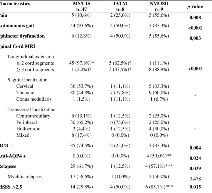
Table 1 – Comparison of MS/CIS, IATM and NMOSD patients.
Characteristics MS/CIS n=47 IATM n=8 NMOSD n=9 p value Pain 5 (10,6%) 2 (25,0%) 5 (55,6%) 0,008 Autonomous gait 44 (93,6%) 4 (50,0%) 3 (33,3%) <0,001 Sphincter dysfunction 6 (12,8%) 4 (50,0%) 5 (55,6%) 0,003
Spinal Cord MRI
Longitudinal extension ≤ 2 cord segments 45 (97,8%)* 5 (62,5%)* 1 (11,1%) <0,001 ≥ 3 cord segments 1 (2,2%)* 3 (37,5%)* 8 (88,9%) Sagittal localization Cervical 36 (53,7%) 1 (11,1%) 5 (33,3%) - Thoracic 30 (44,8%) 7 (77,8%) 9 (60,0%) Conus medullaris 1 (1,5%) 1 (11,1%) 1 (6,7%) Transversal localization Centromedullary 6 (13,1%) 1 (12,5%) 2 (25,0%) - Peripheral 30 (65,2%) 6 (75,0%) 2 (25,0%) Hollocordic 2 (4,4%) 1 (12,5%) 4 (50,0%) Mixed 8 (17,4%) 0 (0,0%) 0 (0,0%) OCB + 35 (74,5%) 2 (25,0%) 3 (33,3%) 0,004 Anti-AQP4 + 0 (0,0%) 0 (0,0%) 4 (50,0%)** 0.024 Relapses 29 (61,7%) 1 (12.5%) 4 (57,1%)*** 0.039 Myelitis relapses 17 (58,6%) 1 (100%) 2 (50,0%) 0,478 MSSS >2,5 14 (29,8%) 4 (50,0%) 6 (85,7%)*** 0,015
Description: The three biggest diagnostic groups were selected and compared. Categorical variables were analyzed using chi-square or Fisher’s
exacttest. Sagittal and transversal localization were defined considering anatomic references and, if present in more than one anatomic segment scored in more than one category. For sagittal and transversal localizations no comparison tests were performed, only descriptive statistics.
Abbreviations: MS: Multiple Sclerosis; CIS: Clinically Isolated Syndrome; IATM: Idiopathic acute transverse myelitis; NMOSD:
Neuromyelitis optica spectrum disorders; MRI – Magnetic Resonance Imaging; OCB + : positive oligoclonal bands in cerebrospinal fluid; Anti-AQP4 +: NMO – antibody positivity; MSSS – Multiple Sclerosis Severity Score.
*: one patient from MS/CIS group and IATM group did not show any lesion on spinal cord MRI; **: one patient missed this test; ***: two patients were lost (one died during ward admission and the other was a foreign patient); - : not possible to infer any differences statistically significant considering not all categories have the same representativity.
Table 1 refers to these three groups comparison, concerning clinical presentation, supplementary tests’ results and follow-up data.
11
4.3. Supplementary tests
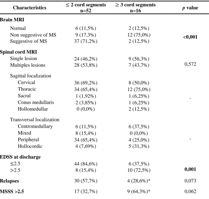
Table 2 – Comparison among longitudinal extension of cord lesions’ categories.
Characteristics ≤ 2 cord segments n=52 ≥ 3 cord segments n=16 p value Brain MRI Normal 6 (11,5%) 2 (12,5%) <0,001 Non suggestive of MS 9 (17,3%) 12 (75,0%) Suggestive of MS 37 (71,2%) 2 (12,5%)
Spinal cord MRI
Single lesion 24 (46,2%) 9 (56,3%) 0,572 Multiples lesions 28 (53,8%) 7 (43,7%) Sagittal localization Cervical 36 (69,2%) 8 (50,0%) - Thoracic 34 (65,4%) 12 (75,0%) Sacral 1 (1,92%) 1 (6,25%) Conus medullaris 2 (3,85%) 1 (6,25%) Hollomedullar 0 (0,0%) 2 (12,5%) Transversal localization Centromedullary 6 (11,5%) 6 (37,5%) - Mixed 8 (15,4%) 0 (0,0%) Peripheral 34 (65,4%) 4 (25,0%) Hollocordic 4 (7,69%) 5 (31,3%) EDSS at discharge ≤2.5 44 (84,6%) 6 (37,5%) 0,001 >2.5 8 (15,4%) 10 (72,5%) Relapses 30 (57,7%) 4 (28,6%)* 0,073 MSSS >2.5 17 (32,7%) 9 (64,3%)* 0,062
Description: Patients were gathered considering the longitudinal extension of their lesions. Sagittal and transversal localization were defined
considering anatomic references and, if present in more than one anatomic segment scored in more than one category. Categorical variables were compared using chi-square and Fisher’s exacttest.
Abbreviations: MRI: Magnetic Resonance Imaging; EDSS – Expanded Disability Status Scale; MSSS: Multiple Sclerosis Severity Score; *:
two patients were lost follow-up; -: not possible to infer any differences statistically significant considering not all categories have the same representativity.
Table 2 summarizes some clinical and follow-up findings when compared longitudinal extension categories of lesions detected on SC MRI. We observed LETM was associated with higher EDSS at discharge (p=0,001), higher MSSS but lower relapsing rates, even though these two last observations did not show statistically significant differences.
12 In what concerns immunological tests, anti-AQP4 were positive in 4 of 24 (16,7%) tests requested. Anti-MOG were requested only two times and were both negative – one of the patients was diagnosed with ADEM and the other with NMOSD.
35 patients presented multiple lesions on SC MRI, with a median of 1 lesion detected and a maximum of 9 lesions, and were found statistically significant differences among etiologies (p=0,047). SC MRI did not reveal any lesion in 3 patients, which were diagnosed with different diseases: ADEM, Behçet and MS. Brain MRI was not performed on Behçet’s patient.
4.4. Prognosis
69 patients completed follow-up and their follow-up mean time was 52,13 ± 34,87 months. However, 2 out of 71 patients were lost follow-up: one of them died during myelitis event and the second was a foreigner, whose process was transferred to his home country.
34 patients (49,3%) had a relapse of its inflammatory myelopathy etiology and 20 (58,8%) of them had myelitis recurrence. Were found differences statistically significant concerning relapses’ number among different etiologies (p=0,013) but it did not apply when compared the number of myelitis relapses (p=0,621). Were found differences statistically significant concerning EDSS at ward’s discharge (p<0,001), with highest results attributable to NMOSD and IATM. MSSS results showed, also, highest scores for NMOSD and lowest for SLE and Behçet patients. 19 patients (26,8%) had a full recovery and was found a statistically significant difference when compared the presence of motor symptoms in those who had achieved full recovery and those who had not (p=0,028). Furthermore, we assessed the presence of motor symptoms was associated with a lower rate of full recovery (OR = 0,398 [0,18 – 0,89]).
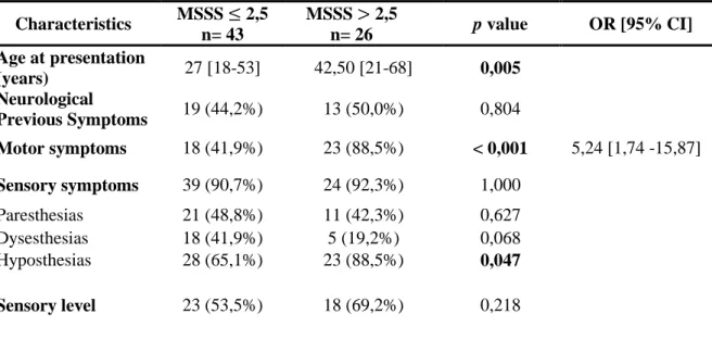
Table 3 – Clinical and follow-up variables associated with long-term outcome.
Characteristics MSSS ≤ 2,5 n= 43 MSSS > 2,5 n= 26 p value OR [95% CI] Age at presentation (years) 27 [18-53] 42,50 [21-68] 0,005 Neurological Previous Symptoms 19 (44,2%) 13 (50,0%) 0,804 Motor symptoms 18 (41,9%) 23 (88,5%) < 0,001 5,24 [1,74 -15,87] Sensory symptoms 39 (90,7%) 24 (92,3%) 1,000 Paresthesias 21 (48,8%) 11 (42,3%) 0,627 Dysesthesias 18 (41,9%) 5 (19,2%) 0,068 Hyposthesias 28 (65,1%) 23 (88,5%) 0,047 Sensory level 23 (53,5%) 18 (69,2%) 0,218
13 Deep sensory symptoms 14 (32,6%) 12 (46,2%) 0,31 Bilateral symptoms 23 (53,5%) 17 (65,4%) 0,451 Pyramidal tract symptoms 15 (65,4%) 15 (25,0%) 0,082 Walking impairment 3 (7,0 %) 11 (42,3%) 0,001 2,88 [1,73 – 4,80] Autonomic symptoms 10 (23,3%) 10 (38,5%) 0,273 Pain 6 (14,0%) 8 (30,8%) 0,125 EDSS at onset 2,50 [0-7] 4,00 [2-8,5] 0,009 EDSS at discharge 1,50 ±1,31 3,19±1,71 <0,001 Relapses 18 (41,9%) 16 (61,5%) 0,140 Myelitis relapses 9 (20,9%) 11 (42,3%) 0,099 1,80 [1,01 – 3,21]
Description: Patients were gathered considering their MSSS category: MSSS ≤ 2,5 or MSSS > 2,5. We evaluated all continuous variables in order to assess their normal distribution or not, and EDSS at discharge was the one who showed a normal distribution considering both criteria. Meanwhile, age at admission only obeyed to kurtosis assumption. All continuous variables that did not show a normal distribution were described using median and minimum-maximum range and were analyzed using non-parametric tests to compare median results. All continuous variables that did show a normal distribution were described using mean and standard deviation and were compared using ANOVA tests and Levene’s test to assure equal variances. Categorical variables were compared using chi-square or Fisher’s exacttest.
Risk of having MSSS > 2,5 when certain finding at admission/registered in follow-up data was presented was calculated using Odds Ratio (OR) and respective 95% confidence interval (95% CI) when proved association.
Abbreviations: EDSS – Expanded Disability Status Scale; MSSS: Multiple Sclerosis Severity Score; *: two patients were lost follow-up.
26 (37,7%) presented MSSS above 2,5 at the last medical appointment recorded and Table 3 summarizes symptoms presented at admission, follow-up data and their prognosis on long-term, represented by MSSS classes.
5. Discussion
Diagnosis categories were established according to most recent criteria and, when appropriated, reconsidered according to most updated data at the time.
MS is the most well recognized demyelinating disease and, therefore, the most studied and in 2017 new McDonald MS diagnostic criteria were published (Carroll, 2018).
Our MS/CIS patients presented a female predominance and median age lower than other etiologies, reflecting a predominant affection of adults in their 3rd – 4th decade of life. These
findings are compatible with demographic characterization in the literature (Raffel et al., 2016). Sensory symptoms were more prevalent than motor ones, with a high prevalence of cases with autonomous gait preserved. Pain is relatively uncommon in these patients as well as autonomic impairment, as observed. Lhermitte’s sign reflects a demyelinating lesion of SC posterior columns and is frequently associated with MS, which is consistent with our study - only MS/CIS patients presented Lhermitte’s sign.
14 As expectable, brain MRI had typical findings considered in Barhkof criteria and 26.3% presented increased latencies, reflecting optical nerve involvement as a possible lesion in MS.
OCB positivity is also an important marker, contemplated in new McDonald MS diagnostic criteria, and the majority of patients presented it in CSF tests, as well as an increased IgG index (McNicholas et al., 2018). Pleocytosis, another marker of CNS inflammation, was not present in the majority of MS/CIS, which is consistent with the literature (Gastaldi et al., 2017; Wingerchuk, 2018).
As observed in our study, lesions are usually small in extension (less than 3 vertebral segments) and have a peripheral transversal localization (Jacob and Weinshenker, 2008).
Even though only 17,5% reached complete recovery, MS presented lower EDSS and MSSS than other relapsing etiologies.
NMOSD, out of all etiologies studied, it was the one with a higher median age at presentation and MSSS, as well as a lower recovery rate.
The increased latencies on VEP are compatible with well recognized optic neuritis that names this disorders group.
On MRI, as expected, majority presented LETM and even though none had a normal cranial MRI, vast majority did not obey Barkhof criteria, reflecting a different affection of CNS than MS/CIS (Jurynczyk et al., 2015). Anti-AQP4 is a classic immunologic marker of NMOSD and was only positive in 4 of 8 patients tested. However, it is known some patients presenting typical phenotype of NMOSD are anti-AQP4 seronegative, and it has been suggested that these patients should be tested for anti-MOG (de Seze, 2017; Jurynczyk et al., 2017; Zamvil and Slavin, 2015). Anti-MOG is neither a stable nor a specific immunological marker for NMOSD and is described in many other neurologic disorders, such as ADEM (de Seze, 2017). It has also been described anti-MOG NMOSD phenotype differs from anti-AQP4 NMOSD, concerning clinical and prognostic features (Kitley et al., 2014), and double positivity is not usually seen, suggesting distinct pathological mechanisms (Dos Passos et al., 2018). In our study, only two patients were tested for anti-MOG antibody, none of them were positive and only one was diagnosed with anti-AQP4 seronegative NMOSD. We believe this low rate of anti-MOG assessment happened because of the retrospective design and timeline of our study since anti-MOG seropositive NMOSD is a recent and emergent entity (Mader et al., 2011).
ADEM is also a well-known inflammatory demyelinating disorder of CNS in pediatric age rather rarer in adult population. It traditionally presents as a monophasic condition, compatible with our no-relapses findings, and brain MRI usually shows multiple white matter lesions that do not obey Barkhof criteria, also compatible with our results (Wingerchuk and Weinshenker, 2013). It is a differential diagnosis for LETM and typically presents with bilateral symptoms, all these characteristics described in the literature are also consistent with our findings. Immunology tests,
15 such as OCB and anti-AQP4, were all negative as expected (Wingerchuk and Weinshenker, 2013), as well as anti-MOG in the patient tested.
Recent developments in neuroimaging, as well as the discovery of specific neuroinflammatory biomarkers, have been responsible to identify and come to a diagnostic conclusion about patients who, otherwise, would have an idiopathic condition diagnosed (Yeh and Hintzen, 2018). IATM criteria were published before the last years’ advances in diagnostic tools and not reviewed after that (Yeh and Hintzen, 2018; Zalewski et al., 2018). Besides that, IATM was one of the three bigger diagnostic groups.
SLE and Behçet’s Disease (Lukjanowicz and Brzosko, 2009; Piquet and Clardy, 2018; Yu et al., 2014) are both systemic diseases with neurological affection but not two classic diagnoses for myelitis. In our study, we had a very small sample of systemic diseases as a cause of myelitis, which makes it harder to come up with some conclusions. Furthermore, these two patients did not complete all diagnosis workup: they were not tested for anti-AQP4, which might be important since some SLE patients have coexisting NMOSD (Kim et al., 2017), OCB status was not assessed and did not complete MRI study.
ADEM, NMOSD, PI and SLE were the etiologies with higher rates of LETM, an imaging entity typically associated with higher disability grades (Wingerchuk and Weinshenker, 2013), as observed in our study. In 3 cases, no lesion was identified on SC MRI, which was presumed to be a consequence of MRI image obtained in a too short period of time.
Long-term prognosis was analyzed in order to come up with some conclusions about prognostic predictors at admission that might guide clinicians to prevent further impairment. As observed, the presence of motor symptoms predicts a worse prognosis (OR = 5,24 [1,74 – 15,87]), as well as the absence of autonomous walking (OR=2,88 [1,73 – 4,80]), but no association was found in what concerns autonomic dysfunction. We found statistically significant differences concerning EDSS at admission and at discharge when comparing MSSS above vs equal or below 2,5, but it did not apply to relapses. Therefore, we may conclude that impairment at admission and at discharge are truly important long-term conditioners to functional capacity, so an effective approach is of essential importance to reduce this burden (Greenberg et al., 2019). Time delay to get a diagnosis or number of wrong diagnosis were not assessed but it is intuitive that this might contribute to a worse outcome for these patients, especially those with a relapsing course of disease.
We believe not considering therapeutic approach is an important limitation of our study because many of these demyelinating diseases already have modifying prognosis therapy, which might have an impact in prognosis outcomes, as evaluated through relapses and MSSS variables. Also, the retrospective design of this study made it impossible to assure a uniform approach, in what concerns all diagnostic workup and data collected from medical records. Another limitation that impaired our ability to achieve some conclusions was the small group representation of
non-16 MS/CIS diagnosis, which might be overcome if performed a prospective national database study so that we can have a greater representation of non-MS/CIS cases.
We used MSSS in order to obtain information about disability progression over time, avoiding time as a confounder, in all these patients. However, we are aware of this scale was not validated for other diseases besides MS, which have different disease mechanisms as well as a different course.
Despite that, we believe our study has accomplished its main aims and even though this is a small sample, it resembles findings validated in the most recent literature.
6. Conclusion
Noninfectious inflammatory myelopathies have heterogeneous courses but present overlapping features (Wingerchuk, 2018).
No single feature is enough to define a disorder with absolute certainty and diagnostic workup of SC inflammatory processes should be no strict but rather broaden, reflecting clinical context to look after a proper diagnosis, and time saver to exclude other reversible differential diagnoses (Greenberg and Frohman, 2015; Stangel et al., 2013). While SC MRI is the gold standard imaging technique (Greenberg and Frohman, 2015), brain MRI and CSF analysis are essential tools that complement all information about CNS affection (Cho and Bhattacharyya, 2018). As in everything in medicine, diagnosis is a probabilistic game where all information gathered help us go through clinical thinking.
A proper diagnosis is critical to assess effective and targeted therapy, so we can reduce the burden of adverse effects and estimate patients’ prognosis, considering relapsing and impairment features (Greenberg and Frohman, 2015; Wingerchuk, 2018).
7. Conflict of interests: None.
8. Financial disclosure: The authors have nothing to disclose.
9. Acknowledgments: Professor Joselina Barbosa and Doctor Hélio Alves, for helping with
statistical analysis.
10. References
Barkhof, F., Filippi, M., Miller, D.H., Scheltens, P., Campi, A., Polman, C.H., Comi, G., Ader, H.J., Losseff, N., Valk, J., 1997. Comparison of MRI criteria at first presentation to predict
17 conversion to clinically definite multiple sclerosis. Brain : a journal of neurology 120 ( Pt 11), 2059-2069.
Beh, S.C., Greenberg, B.M., Frohman, T., Frohman, E.M., 2013. Transverse myelitis. Neurologic clinics 31(1), 79-138.
Carroll, W.M., 2018. 2017 McDonald MS diagnostic criteria: Evidence-based revisions. Multiple sclerosis (Houndmills, Basingstoke, England) 24(2), 92-95.
Cho, T.A., Bhattacharyya, S., 2018. Approach to Myelopathy. Continuum (Minneapolis, Minn.) 24(2, Spinal Cord Disorders), 386-406.
Davatchi, F., Assaad-Khalil, S., Calamia, K.T., Crook, J.E., Sadeghi-Abdollahi, B., Schirmer, M., Tzellos, T., Zouboulis, C.C., Akhlagi, M., Al-Dalaan, A., Alekberova, Z.S., Ali, A.A., Altenburg, A., Arromdee, E., Baltaci, M., Bastos, M., Benamour, S., Ben Ghorbel, I., Boyvat, A., Carvalho, L., Chen, W., Ben-Chetrit, E., Chams-Davatchi, C., Correia, J.A., Crespo, J., Dias, C., Dong, Y., Paixão-Duarte, F., Elmuntaser, K., Elonakov, A.V., Graña Gil, J., Haghdoost, A.-A., Hayani, R.M., Houman, H., Isayeva, A.R., Jamshidi, A.R., Kaklamanis, P., Kumar, A., Kyrgidis, A., Madanat, W., Nadji, A., Namba, K., Ohno, S., Olivieri, I., Vaz Patto, J., Pipitone, N., de Queiroz, M.V., Ramos, F., Resende, C., Rosa, C.M., Salvarani, C., Serra, M.J., Shahram, F., Shams, H., Sharquie, K.E., Sliti-Khanfir, M., Tribolet de Abreu, T., Vasconcelos, C., Vedes, J., Wechsler, B., Cheng, Y.K., Zhang, Z., Ziaei, N., 2014. The International Criteria for Behçet's Disease (ICBD): a collaborative study of 27 countries on the sensitivity and specificity of the new criteria. Journal of the European Academy of Dermatology and Venereology 28(3), 338-347.
de Seze, J., 2017. MOG-antibody neuromyelitis optica spectrum disorder: is it a separate disease? Brain : a journal of neurology 140(12), 3072-3075.
Debette, S., de Seze, J., Pruvo, J.P., Zephir, H., Pasquier, F., Leys, D., Vermersch, P., 2009. Long-term outcome of acute and subacute myelopathies. Journal of neurology 256(6), 980-988. Dos Passos, G.R., Oliveira, L.M., da Costa, B.K., Apostolos-Pereira, S.L., Callegaro, D., Fujihara, K., Sato, D.K., 2018. MOG-IgG-Associated Optic Neuritis, Encephalitis, and Myelitis: Lessons Learned From Neuromyelitis Optica Spectrum Disorder. Frontiers in neurology 9, 217-217. Gastaldi, M., Zardini, E., Franciotta, D., 2017. An update on the use of cerebrospinal fluid analysis as a diagnostic tool in multiple sclerosis. Expert review of molecular diagnostics 17(1), 31-46. Greenberg, B.M., Frohman, E.M., 2015. Immune-mediated myelopathies. Continuum (Minneapolis, Minn.) 21(1 Spinal Cord Disorders), 121-131.
Greenberg, B.M., Krishnan, C., Harder, L., 2019. New onset transverse myelitis diagnostic accuracy and patient experiences. Multiple sclerosis and related disorders 30, 42-44.
18 Inglese, M., 2006. Multiple sclerosis: new insights and trends. AJNR. American journal of neuroradiology 27(5), 954-957.
Jacob, A., Weinshenker, B.G., 2008. An approach to the diagnosis of acute transverse myelitis. Seminars in neurology 28(1), 105-120.
Jurynczyk, M., Craner, M., Palace, J., 2015. Overlapping CNS inflammatory diseases: differentiating features of NMO and MS. Journal of neurology, neurosurgery, and psychiatry 86(1), 20-25.
Jurynczyk, M., Messina, S., Woodhall, M.R., Raza, N., Everett, R., Roca-Fernandez, A., Tackley, G., Hamid, S., Sheard, A., Reynolds, G., Chandratre, S., Hemingway, C., Jacob, A., Vincent, A., Leite, M.I., Waters, P., Palace, J., 2017. Clinical presentation and prognosis in MOG-antibody disease: a UK study. Brain : a journal of neurology 140(12), 3128-3138.
Kaplin, A.I., Krishnan, C., Deshpande, D.M., Pardo, C.A., Kerr, D.A., 2005. Diagnosis and management of acute myelopathies. The neurologist 11(1), 2-18.
Kim, S.M., Kim, S.J., Lee, H.J., Kuroda, H., Palace, J., Fujihara, K., 2017. Differential diagnosis of neuromyelitis optica spectrum disorders. Therapeutic advances in neurological disorders 10(7), 265-289.
Kitley, J., Waters, P., Vincent, A., Palace, J., 2014. Features of neuromyelitis optica spectrum disorders and aquaporin-4 with myelin-oligodendrocyte glycoprotein antibodies-reply. JAMA neurology 71(7), 924.
Krupp, L.B., Banwell, B., Tenembaum, S., 2007. Consensus definitions proposed for pediatric multiple sclerosis and related disorders. Neurology 68(16 suppl 2), S7-S12.
Kurtzke, J.F., 1983. Rating neurologic impairment in multiple sclerosis: an expanded disability status scale (EDSS). Neurology 33(11), 1444-1452.
Lukjanowicz, M., Brzosko, M., 2009. Myelitis in the course of systemic lupus erythematosus: review. Polskie Archiwum Medycyny Wewnetrznej 119(1-2), 67-72.
Mader, S., Gredler, V., Schanda, K., Rostasy, K., Dujmovic, I., Pfaller, K., Lutterotti, A., Jarius, S., Di Pauli, F., Kuenz, B., Ehling, R., Hegen, H., Deisenhammer, F., Aboul-Enein, F., Storch, M.K., Koson, P., Drulovic, J., Kristoferitsch, W., Berger, T., Reindl, M., 2011. Complement activating antibodies to myelin oligodendrocyte glycoprotein in neuromyelitis optica and related disorders. Journal of Neuroinflammation 8(1), 184.
McNicholas, N., Hutchinson, M., McGuigan, C., Chataway, J., 2018. 2017 McDonald diagnostic criteria: A review of the evidence. Multiple sclerosis and related disorders 24, 48-54.
19 Piquet, A.L., Clardy, S.L., 2018. Infection, Immunodeficiency, and Inflammatory Diseases in Autoimmune Neurology. Seminars in neurology 38(3), 379-391.
Raffel, J., Wakerley, B., Nicholas, R., 2016. Multiple sclerosis. Medicine 44(9), 537-541. Roxburgh, R.H., Seaman, S.R., Masterman, T., Hensiek, A.E., Sawcer, S.J., Vukusic, S., Achiti, I., Confavreux, C., Coustans, M., le Page, E., Edan, G., McDonnell, G.V., Hawkins, S., Trojano, M., Liguori, M., Cocco, E., Marrosu, M.G., Tesser, F., Leone, M.A., Weber, A., Zipp, F., Miterski, B., Epplen, J.T., Oturai, A., Sorensen, P.S., Celius, E.G., Lara, N.T., Montalban, X., Villoslada, P., Silva, A.M., Marta, M., Leite, I., Dubois, B., Rubio, J., Butzkueven, H., Kilpatrick, T., Mycko, M.P., Selmaj, K.W., Rio, M.E., Sa, M., Salemi, G., Savettieri, G., Hillert, J., Compston, D.A., 2005. Multiple Sclerosis Severity Score: using disability and disease duration to rate disease severity. Neurology 64(7), 1144-1151.
Schmalstieg, W.F., Weinshenker, B.G., 2010. Approach to acute or subacute myelopathy. Neurology 75(18 Suppl 1), S2-8.
Stangel, M., Fredrikson, S., Meinl, E., Petzold, A., Stüve, O., Tumani, H., 2013. The utility of cerebrospinal fluid analysis in patients with multiple sclerosis. Nature Reviews Neurology 9, 267. Tan, C.T., Mao, Z., Qiu, W., Hu, X., Wingerchuk, D.M., Weinshenker, B.G., 2016. International consensus diagnostic criteria for neuromyelitis optica spectrum disorders. Neurology 86(5), 491-492.
Thompson, A.J., Banwell, B.L., Barkhof, F., Carroll, W.M., Coetzee, T., Comi, G., Correale, J., Fazekas, F., Filippi, M., Freedman, M.S., Fujihara, K., Galetta, S.L., Hartung, H.P., Kappos, L., Lublin, F.D., Marrie, R.A., Miller, A.E., Miller, D.H., Montalban, X., Mowry, E.M., Sorensen, P.S., Tintore, M., Traboulsee, A.L., Trojano, M., Uitdehaag, B.M.J., Vukusic, S., Waubant, E., Weinshenker, B.G., Reingold, S.C., Cohen, J.A., 2018. Diagnosis of multiple sclerosis: 2017 revisions of the McDonald criteria. The Lancet. Neurology 17(2), 162-173.
TMCWG, 2002. Proposed diagnostic criteria and nosology of acute transverse myelitis. Neurology 59(4), 499-505.
Tobin, W.O., Weinshenker, B.G., Lucchinetti, C.F., 2014. Longitudinally extensive transverse myelitis. Current opinion in neurology 27(3), 279-289.
Wingerchuk, D.M., 2018. Immune-Mediated Myelopathies. Continuum (Minneapolis, Minn.) 24(2, Spinal Cord Disorders), 497-522.
Wingerchuk, D.M., Weinshenker, B.G., 2013. Acute disseminated encephalomyelitis, transverse myelitis, and neuromyelitis optica. Continuum (Minneapolis, Minn.) 19(4 Multiple Sclerosis), 944-967.
20 Yeh, E.A., Hintzen, R.Q., 2018. Specific myelopathy diagnoses using advancing diagnostics: Idiopathic no more. Neurology 90(2), 51-52.
Yu, C., Gershwin, M.E., Chang, C., 2014. Diagnostic criteria for systemic lupus erythematosus: a critical review. Journal of autoimmunity 48-49, 10-13.
Zalewski, N.L., Flanagan, E.P., Keegan, B.M., 2018. Evaluation of idiopathic transverse myelitis revealing specific myelopathy diagnoses. Neurology 90(2), e96-e102.
Zamvil, S.S., Slavin, A.J., 2015. Does MOG Ig-positive AQP4-seronegative opticospinal inflammatory disease justify a diagnosis of NMO spectrum disorder? Neurology(R) neuroimmunology & neuroinflammation 2(1), e62.
Anexos
I. Normas de Publicação da Revista
Multiple Sclerosis and Related Disorders
II. Parecer e autorização da Comissão de Ética do Centro
Hospitalar Universitário São João, E.P.E, Porto, PORTUGAL,
para a realização da investigação
AUTHOR INFORMATION PACK 8 Mar 2019 www.elsevier.com/locate/msard 1
MULTIPLE SCLEROSIS AND RELATED
DISORDERS
AUTHOR INFORMATION PACK
TABLE OF CONTENTS
.XXX
.• Description
• Audience
• Impact Factor
• Abstracting and Indexing
• Editorial Board
• Guide for Authors
p.1
p.1
p.1
p.2
p.2
p.4
ISSN: 2211-0348DESCRIPTION
.Multiple Sclerosis is an area of ever expanding research and escalating publications. Multiple Sclerosis and Related Disorders is a wide ranging international journal supported by key researchers from all neuroscience domains that focus on MS and associated disease of the central nervous system. The primary aim of this new journal is the rapid publication of high quality original research in the field. Important secondary aims will be timely updates and editorials on important scientific and clinical care advances, controversies in the field, and invited opinion articles from current thought leaders on topical issues. One section of the journal will focus on teaching, written to enhance the practice of community and academic neurologists involved in the care of MS patients. Summaries of key articles written for a lay audience will be provided as an on-line resource.
A team of four chief editors is supported by leading section editors who will commission and appraise original and review articles concerning: clinical neurology, neuroimaging, neuropathology, neuroepidemiology, therapeutics, genetics / transcriptomics, experimental models, neuroimmunology, biomarkers, neuropsychology, neurorehabilitation, measurement scales, teaching, neuroethics and lay communication.
The journal will publish the following types of articles: Reviews; Original Research Articles; Editorials; Comment; Clinical Trial papers; Letter to the Editors; Case Reports; Book reviews; News. The
submission of an on-line summary of selected papers of relevance for lay audience, Teaching Lessons
and supporting images and datasets is also encouraged.
AUDIENCE
.
All branches of neuroscience: clinical neurologists, neurophysiologists, geneticists, psychologist, molecular biologists, MRI and allied imaging specialists, immunologists, major pharmaceutical companies, ethical and legal specialists, MS specialist nurses, drug trial nurses.
IMPACT FACTOR
.
AUTHOR INFORMATION PACK 8 Mar 2019 www.elsevier.com/locate/msard 2
ABSTRACTING AND INDEXING
.
EMBASE Scopus
Google Scholar
Current Contents / Clinical Medicine Science Citation Index Expanded Medline/Index Medicus
PubMed
EDITORIAL BOARD
.
Editors in Chief
C.H. Hawkes, Centre for Neuroscience and Trauma, Barts and the London School of Medicine and Dentistry, 4
Newark Street, E1 2AT, London, UK
M. Levy, Dept. of Neurobiology, Harvard Medical School, 220 Longwood Ave., , Boston, Massachusetts, MA
02115, USA
E. Waubant, UCSF School of Medicine, Dept. of Neurology, 675 Nelson Rising Lane, San Francisco, CA 94158,
California, USA
Executive Editor
G. Giovannoni, Centre for Neuroscience and Trauma, Barts and the London School of Medicine and Dentistry,
4 Newark Street, E1 2AT, London, UK
Section Editors Experimental Models
S. Amor, Vrije Universiteit Medisch Centrum (VUMC), Amsterdam, Netherlands
Epidemiology
N. Koch-Henriksen, Aarhus University Hospital in Aalborg, Aalborg, Denmark
Neuro-ophthalmology
L.J. Balcer, University of Pennsylvania School of Medicine, Philadelphia, Pennsylvania, USA
Infection
J.R. Berger, University of Pennsylvania, Philadelphia, Pennsylvania, USA
Neuroethics
J.L. Bernat, Dartmouth Hitchcock Medical Center, Lebanon, New Hampshire, USA
Pediatric MS
R. Dale, The University of Sydney, Sydney, New South Wales, Australia
Biomarkers
F. Deisenhammer, Medizinische Universität Innsbruck, Innsbruck, Austria
Genetics / transcriptomics
R.Q. Hintzen, Erasmus MC: Universitair Medisch Centrum Rotterdam, Rotterdam, Netherlands
Measurement / quality of life
J.C. Hobart, Peninsula Medical School, Plymouth, UK
Teaching
B.M. Keegan, Mayo Clinic, Rochester, Minnesota, USA
Rehabilitation
J. Kesselring, Universität Zürich, Zürich, Switzerland
Clinical Neurology
J.-I. Kira, Kyushu University, Fukuoka, Japan
Psychology and Fatigue
AUTHOR INFORMATION PACK 8 Mar 2019 www.elsevier.com/locate/msard 3 Stem Cell Research
G. Martino, San Raffaele Institute, Milano, Italy
Therapy
A. Miller, Mount Sinai School of Medicine, New York, New York, USA
Immunology
F.T. Sellebjerg, Copenhagen University Hospital, Copenhagen, Denmark
Imaging
J. Wolinsky, University of Texas Health Science Center at Houston (UTHealth), Houston, Texas, USA
Neuropathology
C. Stadelmann, Georg-August-Universität Göttingen, Göttingen, Germany
Lay Summaries
A. Thomson, Barts and the London Queen Mary's School of Medicine and Dentistry, London, England, UK
Statistics
M.P. Sormani, Università degli Studi di Genova, Genoa, Italy
Former Co Editor-in-Chief
B. Banwell, The Children's Hospital of Philadelphia F. Lublin, Icahn School of Medicine at Mount Sinai
Editorial Board
B. Banwell, Philadelphia, USA
V. Brinar, Sveučilišta u Zagrebu, Zagreb, Croatia
H. Butzkueven, Royal Melbourne Hospital, Melbourne, Victoria, Australia
J. Correale, Institute for Neurological Research Dr. Raul Carrea, Buenos Aires, Argentina P. Coyle, Stony Brook University, New York, USA
O. Fernandez, Hospital Regional Universitario Carlos Haya, Málaga, Spain H.-P. Hartung, Heinrich-Heine-Universität Düsseldorf, Düsseldorf, Germany E. Havrdova, Charles University, Prague 2, Czech Republic
L. Kappos, Universitätsspital Basel, Basel, Switzerland P. Li, Queen Elizabeth Hospital, Kowloon, Hong Kong
J. Losy, Poznań University of Medical Sciences, Poznan, Poland C. Lubetzki, Sorbonne Université, Paris cedex 13
C.F. Lucchinetti, Mayo Clinic, Rochester, Minnesota, USA M.A. Macías, Universidad de Guadalajara, Guadalajara, Mexico
A.H. Maghzi, Cedars-Sinai Medical Center, Los Angeles, California, USA A. Miller, Carmel Medical Center, Haifa, Israel
N. Prayoonwiwat, Mahidol University, Bangkok, Thailand T. Saida, Institute of Neurotherapeutics, Kyoto, Japan
B.S. Singhal, Bombay Hospital Institute of Medical Sciences, Mumbai, India S. Sveinbjornsdottir, Landspitali University Hospital, Reykjavik, Iceland J. Toro, Fundación Santa Fé de Bogotá, Bogotá, Colombia
C.-P. Tsai, National Yang-Ming University, Taipei, Taiwan
I. van der Mei, University of Tasmania, Hobart, Tasmania, Australia L. Vécsei, University of Szeged, Szeged, Hungary
E. Willoughby, Auckland City Hospital, Auckland, New Zealand B. Yamout, American University of Beirut (AUB), Beirut, Lebanon M. Zakaria, Ain Shams University, Cairo, Egypt
AUTHOR INFORMATION PACK 8 Mar 2019 www.elsevier.com/locate/msard 4
GUIDE FOR AUTHORS
.
Your Paper Your Way
We now differentiate between the requirements for new and revised submissions. You may choose to submit your manuscript as a single Word or PDF file to be used in the refereeing process. Only when your paper is at the revision stage, will you be requested to put your paper in to a 'correct format' for acceptance and provide the items required for the publication of your article.
To find out more, please visit the Preparation section below. INTRODUCTION
Types of article
Original Research Articles
Full length research papers will not normally be more than 3500 words in length from the Introduction through the Discussion section and will preferably be shorter. Submission of a paper to Multiple Sclerosis and Related Disorders will be held to imply that it represents original research not previously published (except in the form of an abstract or preliminary report), that it is not being considered for publication elsewhere, and that if accepted by Multiple Sclerosis and Related Disorders it will not be published elsewhere in the same form in any language without the consent of the Publisher. Major papers of topical content will be given priority in publication.
Book Reviews
These are normally submitted by the Book Review Editors, but they welcome suggestions of books for review.
Case Reports
Case reports should detail the clinical, laboratory and neuroimaging features of informative patients. Informative patients should provide insights that inform on genetic contributions to disease, rare clinical manifestations, novel laboratory or imaging features, or highlight important concepts in the differential of MS and related disorders. Case reports should be approximately 1200 words, and should have no more than five key references
Comment
Comments should focus on specific issues relevant to MS and related disorders, or should discuss recent publications. Comments should be less than 800 words and should reference the article(s) upon which the commentary is based.
Clinical Trial papers
Manuscripts detailing the results of clinical trials in MS and related disorders are encouraged. The trial methodology should account for all screened participants, and analyses should observe an intention-to-treat model where appropriate. All sources of funding for the study must be disclosed, and the involvement of the study sponsor must be detailed. Clinical trial manuscripts should be a maximum of 3500 words.
Editorials
The Editors welcome suggestions for editorials which give personal and topical views on subjects within the Journal's area of interest. They should not normally exceed 1500 words in total, including references.
Letters to the Editors
These normally refer to articles previously published in the Journal. The Editors are also willing to consider letters on subjects of direct relevance to the Journal's interest. Letters should not exceed 1000 words in total and, where appropriate, must begin with the reference to the published article about which the author is commenting. Research letters should be submitted as 'letter to the Editors' Review Articles
Review papers are normally 4000-5000 words in total. Authors are advised to consult one of the Editors with an outline before submitting a review.
Contact details for submission
Authors may send queries concerning the submission process, manuscript status, or journal procedures to the Editorial Office at:
MSARD, Editorial Office, ELSEVIER. E-mail: [email protected]
AUTHOR INFORMATION PACK 8 Mar 2019 www.elsevier.com/locate/msard 5
Submission checklist
You can use this list to carry out a final check of your submission before you send it to the journal for review. Please check the relevant section in this Guide for Authors for more details.
Ensure that the following items are present:
One author has been designated as the corresponding author with contact details: • E-mail address
• Full postal address
All necessary files have been uploaded: Manuscript:
• Include keywords
• All figures (include relevant captions)
• All tables (including titles, description, footnotes)
• Ensure all figure and table citations in the text match the files provided • Indicate clearly if color should be used for any figures in print
Graphical Abstracts / Highlights files (where applicable) Supplemental files (where applicable)
Further considerations
• Manuscript has been 'spell checked' and 'grammar checked'
• All references mentioned in the Reference List are cited in the text, and vice versa
• Permission has been obtained for use of copyrighted material from other sources (including the Internet)
• A competing interests statement is provided, even if the authors have no competing interests to declare
• Journal policies detailed in this guide have been reviewed
• Referee suggestions and contact details provided, based on journal requirements For further information, visit our Support Center.
BEFORE YOU BEGIN Ethics in publishing
Please see our information pages on Ethics in publishing and Ethical guidelines for journal publication.
Studies in humans and animals
If the work involves the use of human subjects, the author should ensure that the work described has been carried out in accordance with The Code of Ethics of the World Medical Association (Declaration of Helsinki) for experiments involving humans. The manuscript should be in line with the Recommendations for the Conduct, Reporting, Editing and Publication of Scholarly Work in Medical
Journals and aim for the inclusion of representative human populations (sex, age and ethnicity) as
per those recommendations. The terms sex and gender should be used correctly.
Authors should include a statement in the manuscript that informed consent was obtained for experimentation with human subjects. The privacy rights of human subjects must always be observed. All animal experiments should comply with the ARRIVE guidelines and should be carried out in accordance with the U.K. Animals (Scientific Procedures) Act, 1986 and associated guidelines, EU
Directive 2010/63/EU for animal experiments, or the National Institutes of Health guide for the care
and use of Laboratory animals (NIH Publications No. 8023, revised 1978) and the authors should clearly indicate in the manuscript that such guidelines have been followed. The sex of animals must be indicated, and where appropriate, the influence (or association) of sex on the results of the study. Checklist for reporting and reviewing studies of experimental animal models of multiple sclerosis and related disorders
The guide, reported here, is intended to act as a checklist to aid both authors and referees of manuscripts, just as the Consolidated Standards of Reporting Trials (CONSORT) guidelines are a compulsory part of reporting clinical trials.
AUTHOR INFORMATION PACK 8 Mar 2019 www.elsevier.com/locate/msard 6
Declaration of interest
All authors must disclose any financial and personal relationships with other people or organizations that could inappropriately influence (bias) their work. Examples of potential competing interests include employment, consultancies, stock ownership, honoraria, paid expert testimony, patent applications/registrations, and grants or other funding. Authors must disclose any interests in two places: 1. A summary declaration of interest statement in the title page file (if double-blind) or the manuscript file (if single-blind). If there are no interests to declare then please state this: 'Declarations of interest: none'. This summary statement will be ultimately published if the article is accepted. 2. Detailed disclosures as part of a separate Declaration of Interest form, which forms part of the journal's official records. It is important for potential interests to be declared in both places and that the information matches. More information.
Submission declaration and verification
Submission of an article implies that the work described has not been published previously (except in the form of an abstract, a published lecture or academic thesis, see 'Multiple, redundant or concurrent
publication' for more information), that it is not under consideration for publication elsewhere, that
its publication is approved by all authors and tacitly or explicitly by the responsible authorities where the work was carried out, and that, if accepted, it will not be published elsewhere in the same form, in English or in any other language, including electronically without the written consent of the copyright-holder. To verify originality, your article may be checked by the originality detection service Crossref
Similarity Check.
Preprints
Please note that preprints can be shared anywhere at any time, in line with Elsevier's sharing policy. Sharing your preprints e.g. on a preprint server will not count as prior publication (see 'Multiple,
redundant or concurrent publication' for more information).
Use of inclusive language
Inclusive language acknowledges diversity, conveys respect to all people, is sensitive to differences, and promotes equal opportunities. Articles should make no assumptions about the beliefs or commitments of any reader, should contain nothing which might imply that one individual is superior to another on the grounds of race, sex, culture or any other characteristic, and should use inclusive language throughout. Authors should ensure that writing is free from bias, for instance by using 'he or she', 'his/her' instead of 'he' or 'his', and by making use of job titles that are free of stereotyping (e.g. 'chairperson' instead of 'chairman' and 'flight attendant' instead of 'stewardess').
Changes to authorship
Authors are expected to consider carefully the list and order of authors before submitting their manuscript and provide the definitive list of authors at the time of the original submission. Any addition, deletion or rearrangement of author names in the authorship list should be made only before the manuscript has been accepted and only if approved by the journal Editor. To request such a change, the Editor must receive the following from the corresponding author: (a) the reason for the change in author list and (b) written confirmation (e-mail, letter) from all authors that they agree with the addition, removal or rearrangement. In the case of addition or removal of authors, this includes confirmation from the author being added or removed.
Only in exceptional circumstances will the Editor consider the addition, deletion or rearrangement of authors after the manuscript has been accepted. While the Editor considers the request, publication of the manuscript will be suspended. If the manuscript has already been published in an online issue, any requests approved by the Editor will result in a corrigendum.
Clinical trial results
In line with the position of the International Committee of Medical Journal Editors, the journal will not consider results posted in the same clinical trials registry in which primary registration resides to be prior publication if the results posted are presented in the form of a brief structured (less than 500 words) abstract or table. However, divulging results in other circumstances (e.g., investors' meetings) is discouraged and may jeopardise consideration of the manuscript. Authors should fully disclose all posting in registries of results of the same or closely related work.