O R I G I N A L PA P E R
GracËa Porto ? Helena Alves ? Pedro Rodrigues
Jose M Cabeda ? Cristina Portal ? AnunciacËaÄo Ruivo
Benvindo JusticËa ? Roger Wolff ? Maria De Sousa
Major histocompatibility complex class I associations in iron overload:
evidence for a new link between the HFE H63D mutation, HLA-A29,
and non-classical forms of hemochromatosis
Received: 11 June 1997 / Revised: 29 October 1997
AbstractmThe present study is an analysis of the
frequen-cies of HFE mutations in patients with different forms of
iron overload compared with the frequencies found in
healthy subjects from the same region. The frequencies of
HLA-A and -B antigens and HLA haplotypes were also
analyzed in the same subjects. The study population
in-cluded: 71 healthy individuals; 39 genetically and clinically
well-characterized patients with genetic hemochromatosis
(HH); and 25 patients with non-classical forms of iron
overload (NCH), excluding secondary hemochromatosis.
All subjects were HLA-typed and HFE-genotyped by the
oligonucleotide ligation assay (OLA). The gene frequencies
found for the C282Y and H63D mutations of HFE were
respectively: 0.03 and 0.23 in healthy individuals, 0.86 and
0.04 in HH patients, and 0.08 and 0.48 in NCH patients. An
expected significant association between HH and HLA-A3
was observed, which was found to be in linkage
disequili-brium with the C282Y mutation. A new association was
seen, however, between HLA-A29 and NCH, in linkage
disequilibrium with the H63D mutation. Again as expected,
the HLA-B antigen B7 was associated with HH in linkage
disequilibrium with HLA-A3. In addition, the HLA-B
antigen B44 was found to be associated with NCH but
not in linkage disequilibrium with either A29 or the H63D
mutation. In conclusion, a new association of the HFE
H63D mutation with forms of hemochromatosis other than
HH and a new association between the HLA phenotype
A29 and the HFE H63D mutation were found in the same
patients. These findings reinforce evidence for the
involve-ment of the major histocompatibility class I in iron
metab-olism, supporting the notion of a physiological role for the
immunological system in the regulation of iron load.
Key wordsmIron ? Hemochromatosis ? MHC ? HLA ? HFE
Introduction
The genetic basis of hemochromatosis (HH) has been
known for more than 20 years, following the demonstration
of the association between the major histocompatibility
complex (MHC) class I antigen HLA-A3 and the disease
(Edwards et al. 1977; Simon et al. 1976). These and
subsequent studies of the strong linkage disequilibrium
between HH and various markers in the MHC region all
led to the expectation that a putative HH gene would lie
within approximately 1 cM of the HLA-A locus (Gasparini
et al. 1993; Jazwinska et al. 1993; Raha-Chowdhury et al.
1995; Yaouanq et al. 1994). While the HLA association has
had profound implications both for the clinical diagnosis
and cloning strategies of the HH gene, the possible
biolo-gical significance of that association is only now becoming
evident. Two major recent developments strongly support
the implication of the MHC class I in the pathogenesis of
hemochromatosis. The first was the demonstration that b
2-microglobulin (b
2M)-deficient mice, which lack MHC
class I molecules, display a progressive hepatic iron
over-load similar to that observed in human HH (De Sousa et al.
1994; Rothenberg and Voland 1996; Santos et al. 1996).
The second was the identification by Feder and co-workers
(1996) of the candidate gene for HH, termed HFE. This is a
new MHC class I-like gene, where homozygosity for a
single base pair mutation (C282Y), resulting in a cystein to
tyrosine substitution at position 282, was found in 83% of
HH patients. This finding was promptly confirmed by other
groups who described the presence of the C282Y mutation
in varying, but always very high, numbers of
hemochro-G. Porto ? J.M. Cabeda ? C. Portal ? A. RuivoSanto AntoÂnio General Hospital, Largo do Prof. Abel Salazar, no.1, P-4050 Porto, Portugal
G. Porto ( ) ?P. Rodrigues ? J.M. Cabeda ? B. JusticËa ? M. De Sousa Abel Salazar Institute for the Biomedical Sciences, Largo do Prof. Abel Salazar, no. 2, P-4050 Porto, Portugal
H. Alves
North Histocompatibility Center, Lusotransplante, Rua Dr. Roberto Frias, P-4200 Porto, Portugal R. Wolff
Mercator Genetics Inc., 4040 Campbell Avenue, Menlo Park, CA 94025, USA
matosis patients in several parts of the world (Barton et al.
1997; Beutler et al. 1996; Borot et al. 1997; Carella et al.
1997; Jazwinska et al. 1996; Jouanolle et al. 1996). A
second mutation was also found in the HFE gene (H63D)
resulting in a histidine to aspartic acid substitution at
position 63. This variant was found to be highly frequent
in normal chromosomes (17%) (Feder et al. 1996) but its
contribution to HH is less clear.
From the clinical point of view, identification of the
HFE mutations is of great relevance. It not only allows a
better screening of HH but it provides the opportunity to
characterize more precisely other iron overload syndromes
where the HH gene implication has always been
contro-versial. These include forms of hemochromatosis
asso-ciated with other hematological disorders, chronic alcoholic
or viral hepatitis, or the new syndrome of iron overload
with normal transferrin saturation recently described by
Moirand and co-workers (1997). The lack of association of
the HLA-A3 phenotype in those forms of iron overload has
been classically used as an argument supporting the
non-HLA linkage of the disease. To our knowledge, however,
associations with other HLA phenotypes or with HFE have
not yet been demonstrated. The same is true for the African
dietary iron overload, where a still unknown genetic factor
has been identified (Gordeuk et al. 1992).
In this study, the frequencies of HFE mutations in a
group of patients with several forms of non-transfusional
iron overload are compared with the frequencies found in
HH patients and healthy subjects from the same
geogra-phical area. Looking for possible interactions between HLA
phenotypes and HFE mutations, we estimated the
frequen-cies of various HLA-A and -B antigens and HLA haplotypes
in the same groups of subjects.
Materials and methods
Patients
A total of 64 patients with iron overload were included in this study. Of those patients, 39 (25 unrelated probands and 14 family members) were genetically and clinically well-characterized HH patients according to previously described criteria (Porto et al. 1997), and 25 (20 unrelated probands and 5 family members) were diagnosed as hemochromatosis patients based on biochemical and histological grounds but who did not fulfill the criteria to be classified as having genetic hemocromatosis, either because they had other associated clinical conditions (hemato-logical disorders, chronic alcoholic or viral liver disease, disturbance of lipid metabolism), and/or showed lower levels of iron stores (55 g) estimated by quantitative phlebotomies. These patients were classified as having non-classical hemochromatosis (NCH). The most relevant clinical data in the group of NCH patients are summarized in Table 1. These patients constituted a clinically heterogeneous group but had the common characteristic of excessive iron deposits in parenchymal cells demonstrated by liver biopsy and/or an hepatic iron index greater than 2, making difficult the differential diagnosis with early forms of HH. Two main characteristics, besides the presence of associated clinical conditions, may distinguish the NCH group from HH probands: 1. The level of total body iron stores is usually in the range of 2±4 g (see Table 1mClinical characterization of the 20 NCH patients
Patient
# Sex Age(Years)Trans-ferrin satura-tion (%) Serum ferritin (ng/ml) Hepatic iron index (Perls grade) Iron re-moved (g) Report to
family Clinical conditionsassociated HLA A and Bphenotype HFE genotype
1 Male 48 56 2446 46 (IV) n.a. Proband Hereditary xerocytosis A 29,2; B 12,35 C282Y(±/±)H63D(+/+) 2 Male 39 92 1500 46 (IV) 9.8 Sibling Hereditary xerocytosis A 29,2; B 14,35 C282Y(±/±)H63D(+/±) 3 Male 37 78 2240 (III) 8.6 Proband Haemolytic anaemia A 2,9; B 27,51 C282Y(±/±)H63D(+/±) 4 Male 25 85 810 2.2 2.4 Proband Haemophilia A A 29,X; B 38,44 C282Y(±/±)H63D(+/+) 5 Male 36 81 811 4.0 1.8 Nephew Haemophilia A A 29,X; B 38,27 C282Y(±/±)H63D(+/+) 6 Male 60 100 3000 2.8 n.a. Proband Chronic hepatitis C A 29,2; B 7,51 C282Y(±/±)H63D(+/±) 7 Male 64 92 752 (III) 2.0 Proband Chronic hepatitis C A 24,28; B 51,44 C282Y(±/±)H63D(+/±) 8 Male 44 78 605 3.4 1.5 Proband Chronic hepatitis C A 2,26; B 27,44 C282Y(±/±)H63D(±/±) 9 Male 59 60 714 (III) 2.6 Proband Chronic hepatitis B A 29,2; B 51,40 C282Y(±/±)H63D(+/+) 10 Male 68 87 509 2.6 3.2 Proband Chronic hepatitis B A 26,33; B 7,14 C282Y(±/±)H63D(+/+) 11 Male 30 62 620 (III) 2.9 Proband Chronic hepatitis B A 3,24; B 35,X C282Y(±/±)H63D(±/±) 12 Male 44 57 527 (III) 3.7 Proband Alcohol abuse+Chr.
hepatitis B A 10,11; B 14,12 C282Y(±/±)H63D(±/±) 13 Male 57 65 525 1.6 4.2 Proband Alcohol abuse A 29,1; B 8,44 C282Y(±/±)H63D(+/±) 14 Male 49 51 412 (III) 3.0 Proband Alcohol abuse A 12,X; B 35,12 C282Y(±/±)H63D(±/±) 15 Female 55 60 1254 1,5 (III) 5.3 Proband Obesity+steatohepatitis A 29,2; B 35,12 C282Y(±/±)H63D(+/+)
16 Male 58 53 907 2.2 4.0 Proband Obesity n.a. C282Y(±/±)H63D(+/±)
17 Male 28 50 652 2.9 3.2 Son n.a. C282Y(±/±)H63D(+/±)
18 Male 31 68 362 4.8 2.3 Proband A 29,28; B 37,X C282Y(+/±)H63D(+/±)
19 Male 28 27 144 (II) n.a. Proband A 3,30; B 7,14 C282Y(+/±)H63D(+/±)
20 Male 25 22 281 (II) n.a. Sibling A 3,11; B 7,37 C282Y(±/±)H63D(+/±)
21 Male 44 47 438 2.0 1.9 Proband A 2,11; B 44,X C282Y(+/±)H63D(±/±)
22 Male 34 30 563 2.6 2.1 Proband A 1,X; B 8,X C282Y(±/±)H63D(+/+)
23 Male 46 59 773 1.4 (II) n.a. Proband A 24,28; B 44,X C282Y(±/±)H63D(±/±)
24 Male 44 41 1000 (III) 3.5 Proband n.a. C282Y(±/±)H63D(+/±)
Table 1), in marked contrast with HH where a level of iron stores greater than 5 g was used as a diagnostic criterion (the range in the present group of HH probands was 5±13 g). 2. The transferrin saturation, which is a homogeneous biochemical marker of iron over-load in HH patients (ranging from 60% to 110% in the present group of HH patients), showed some heterogeneity in the NCH group. Seven patients in the present series had normal transferrin saturation (550%) with unexplained hepatic iron overload and hyperferritinemia (see Table 1). These were clinically related to those described by Moirand and co-workers (1997) with the new syndrome of iron overload with normal transferrin saturation. In this study we did not include patients with secondary forms of iron overload such as blood transfusion.
A group of 71 unrelated apparently healthy subjects from the same geographical area in the north of Portugal was used as a control population. None of the controls had evidence of iron overload in terms of biochemical parameters. All participants in this study gave their informed consent.
C282Y and H63D mutation detection
HFE genotyping was performed by OLA on polymerase chain reaction (PCR)-amplified genomic DNA samples using amplification primers for the C282Y or H63D mutations and the normal allele as previously described (Feder et al. 1996).
HLA-A and -B typing
HLA typing was performed in freshly collected venous blood samples by the standard complement-dependent micro-lymphocytotoxicity assay using a battery of sera which enabled the definition of the following HLA antigens: A-locus: 1, 2, 3, 9 (23,24,25), 10 (26,34), 11, 28, 29, 32 and 33; B-locus: 5 (51), 7, 8, 12 (44,45), 13, 14, 15 (62), 16 (38), 17 (57), 18, 21 (49,50), 22, 27, 35, 37, 40, 70.
Family studies and haplotype analysis
Family studies for the screening of hemochromatosis were done systematically in all first-degree relatives of all patients and included HLA typing. For most of those relatives, frozen material stored at the time of the study was available, permitting the present detection of the HFE mutations. Segregation analysis was then performed, allowing in most cases the assignment of the HLA haplotypes as well as the definition of the HLA haplotypes associated with the HFE mutations.
Statistics
In all statistical analyses, patients were divided into two groups according to the clinical presentation: HH patients and those with NCH. Allele frequencies were estimated for the HFE mutations and HLA antigens in the two groups of patients and compared with the same frequencies in the control population. For those estimations, only unrelated probands were included. Whenever a single HLA antigen was defined in one subject and the family study could not check the segregation, its frequency was not taken into account in calculating the value for the blank allele. If, on the other hand, family studies showed segregation in two different haplotypes, it was taken as the specificity of the blank. To analyze the relative strength in disease association of the different HFE or HLA alleles, the etiological fraction d was calculated as described according to the formula d= (FAD-FAP)/(1-FAP), where FAD is the allele frequency in the diseased population and FAP the allele frequency in the control population (Bengtsson and Thomson 1981; Thomson et al. 1983). The same analysis was done for haplotype frequencies where FHD is the frequency of the haplotype in the diseased population. Since no family studies were done in controls and, therefore, no haplotypes were defined, we estimated the range of d values [d] for haplotype associations by comparing the FHD with the range of frequencies in the control population [FHP]. This range may vary from zero, where the pairs of alleles in question are assumed to be always in different chromosomes, to the maximum frequency esti-mated assuming that the alleles in question are always in the same chromosome. Comparisons between frequencies in different groups were done by the chi-square test with Yates' correction for small samples.
Results
HFE and HLA allele frequencies
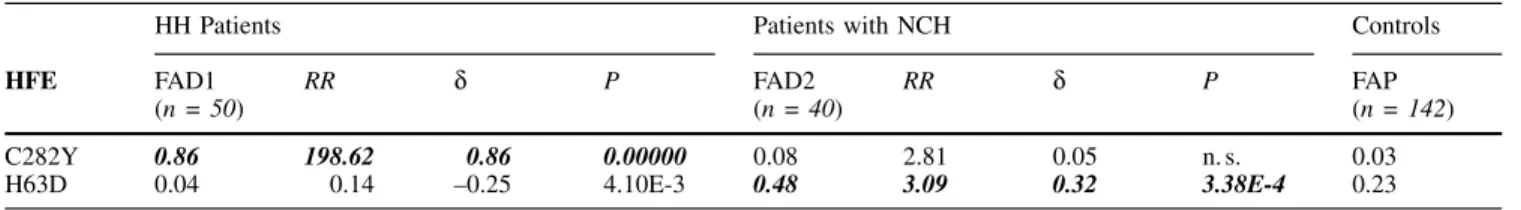
Of the 25 unrelated HH patients (21 males and 4 females),
all carried at least one of the described HFE mutations: 21
were found homozygous for the C282Y mutation, two were
heterozygous, one was a double heterozygote for the
C282Y and the H63D mutations, and one was a
hetero-zygote for the H63D mutation only. This corresponds to
gene frequencies of 0.86 for the C282Y and 0.04 for the
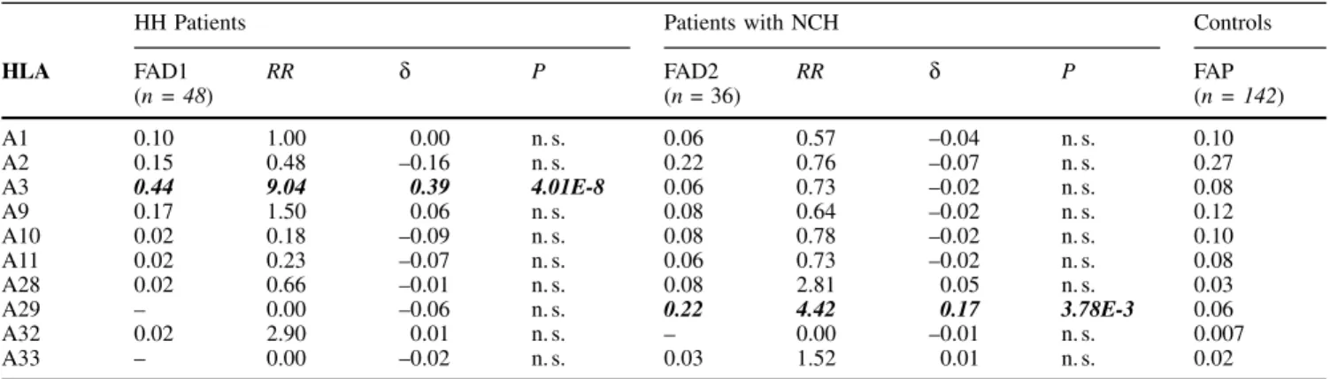
Table 2mAllele frequencies of HLA-A antigens in the two groups of patientswith iron overload (FAD1 and FAD2) in comparison with the control group (FAP)HH Patients Patients with NCH Controls
HLA FAD1 (n = 48) RR d P FAD2(n = 36) RR d P FAP(n = 142) A1 0.10 1.00 0.00 n.s. 0.06 0.57 ±0.04 n.s. 0.10 A2 0.15 0.48 ±0.16 n.s. 0.22 0.76 ±0.07 n.s. 0.27 A3 0.44 9.04 0.39 4.01E-8 0.06 0.73 ±0.02 n.s. 0.08 A9 0.17 1.50 0.06 n.s. 0.08 0.64 ±0.02 n.s. 0.12 A10 0.02 0.18 ±0.09 n.s. 0.08 0.78 ±0.02 n.s. 0.10 A11 0.02 0.23 ±0.07 n.s. 0.06 0.73 ±0.02 n.s. 0.08 A28 0.02 0.66 ±0.01 n.s. 0.08 2.81 0.05 n.s. 0.03 A29 ± 0.00 ±0.06 n.s. 0.22 4.42 0.17 3.78E-3 0.06 A32 0.02 2.90 0.01 n.s. ± 0.00 ±0.01 n.s. 0.007 A33 ± 0.00 ±0.02 n.s. 0.03 1.52 0.01 n.s. 0.02 RR = Relative risk d = Etiologic fraction
n = Total number of alleles in each group P = Level of significance of the associations n.s. = Not significant
H63D mutations, frequencies not different from those
previously reported in other HH populations. Of the 71
healthy individuals, four were found heterozygous for the
C282Y mutation, five were homozygous, and 22
hetero-zygous for the H63D mutation; all other subjects carried
neither mutation. This corresponds to gene frequencies of
0.03 and 0.23 for the C282Y and H63D mutations,
respec-tively, again similar to the previously reported results in a
healthy USA population (Feder et al. 1996). Of the 20
unrelated NCH patients (19 males and 1 female): one was
heterozygous for the C282Y mutation, two were compound
heterozygotes, six were homozygous and five heterozygous
for the H63D mutation, and six carried neither mutation.
This corresponds to a gene frequency of 0.08 for the C282Y
mutation (significantly lower than in classical HH) and an
unexpectedly high frequency of 0.48 for the H63D
muta-tion.
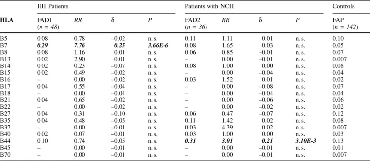
The allele frequencies of the HLA antigens found in the
two groups of patients are listed in Tables 2 and 3 in
comparison with the same frequencies in the control
popu-lation. As expected, significant associations were found for
HLA antigens A3 and B7 in HH patients, the association of
A3 being stronger than B7 but none of them stronger than
the association with the C282Y mutation (see d values in
Tables 2±4). The frequency of the C282Y mutation in HH
patients with the HLA A3 and/or B7 phenotypes was 1.00,
confirming the strong linkage disequilibrium between the
three loci. In patients with NCH, significant associations
were found for HLA antigens A29 and B44. Again, none of
them was stronger than the association with the H63D
mutation (see d values in Tables 2±4). In order to test
whether a linkage disequilibrium also existed among H63D,
A29, and B44, the H63D mutation frequencies were
com-pared in patients and controls with the HLA phenotypes
A29 or B44. Of a total of 17 subjects with the HLA
phenotype A29 (7 NCH patients and 10 controls), 13 (7
patients and 6 controls) had the H63D mutation. This
frequency (76%) was significantly higher (P50.002) than
those found in NCH patients or controls lacking the HLA
A29 phenotype (36% and 34%, respectively). In contrast,
the frequencies of appearance of the H63D mutations in
patients or controls with the phenotype B44 were not
significantly different from those in subjects without the
B44 phenotype. Although a linkage disequilibrium between
A29 and B44 has been described in the Portuguese
popula-tion as well as in other European Caucasoid populapopula-tions
(Silva Carvalho 1983), in the present groups of subjects we
could not confirm the existence of such linkage. The fact
that the present control population comes from a
geogra-phically restricted area in the north of Portugal could be
influencing this result. To further test the existence of
linkage disequilibria between the HFE mutations and
HLA alleles, we proceeded with the analysis of haplotype
associations in both groups of patients.
Haplotype analysis
The segregation analysis done with family studies
per-mitted the definition of 76 HLA haplotypes: 42 in HH
Table 3mAllele frequencies of HLA-B antigens in the two groups of patients with iron overload (FAD1 and FAD2) in comparison with the control group (FAP)HH Patients Patients with NCH Controls
HLA FAD1 (n = 48) RR d P FAD2(n = 36) RR d P FAP(n = 142) B5 0.08 0.78 ±0.02 n.s. 0.11 1.11 0.01 n.s. 0.10 B7 0.29 7.76 0.25 3.66E-6 0.08 1.65 0.03 n.s. 0.05 B8 0.08 1.16 0.01 n.s. 0.06 0.85 ±0.01 n.s. 0.07 B13 0.02 2.90 0.01 n.s. ± 0.00 ±0.01 n.s. 0.007 B14 0.02 0.23 ±0.07 n.s. 0.08 1.00 0.00 n.s. 0.08 B15 0.02 0.49 ±0.02 n.s. ± 0.00 ±0.04 n.s. 0.04 B16 ± 0.00 ±0.02 n.s. 0.03 1.52 0.01 n.s. 0.02 B17 0.04 0.55 ±0.04 n.s. ± 0.00 ±0.08 n.s. 0.07 B18 ± 0.00 ±0.04 n.s. ± 0.00 ±0.04 n.s. 0.04 B21 0.04 0.65 ±0.02 n.s. ± 0.00 ±0.06 n.s. 0.06 B22 ± 0.00 ±0.02 n.s. ± 0.00 ±0.02 n.s. 0.02 B27 0.04 0.31 ±0.10 n.s. 0.06 0.47 ±0.07 n.s. 0.12 B35 0.04 0.48 ±0.05 n.s. 0.11 1.42 0.02 n.s. 0.08 B37 ± 0.00 ±0.01 n.s. 0.03 4.39 0.02 n.s. 0.007 B40 0.02 0.07 ±0.01 n.s. 0.03 1.00 0.00 n.s. 0.03 B44 0.10 0.74 ±0.05 n.s. 0.31 3.01 0.21 3.10E-3 0.13 B45 ± 0.00 ±0.01 n.s. ± 0.00 ±0.01 n.s. 0.01 B70 ± 0.00 ±0.01 n.s. ± 0.00 ±0.01 n.s. 0.007 RR = Relative risk d = Etiologic fraction
n = Total number of alleles in each group P = Level of significance of the associations n.s. = Not significant
patients (36 from the probands and 6 additional from other
affected family members) and 34 in NCH patients (31 from
the probands and 3 additional from other affected family
members). All except one of the 42 haplotypes defined in
HH patients segregated with the C282Y mutation. Among
these, A3/B7 was the most common with a haplotype
frequency of 0.19. Of the 34 haplotypes defined in NCH
patients, 17 segregated with the H63D mutation. Among
these, A29/B44 was the most common with a haplotype
frequency of 0.12. The d values for the different
combina-tions of the most frequent alleles in the two groups are
shown in Table 5. The simple comparison of those d values
shows that in both forms of hemochromatosis the strongest
associations occur with the HFE mutations alone (C282Y
and H63D, respectively, in HH and NCH). Significant
associations were observed between the C282Y or H63D
mutations and the HLA antigens A3 or A29, respectively
(see Table 5), reflecting the strong linkage disequilibrium
between the HFE and HLA-A loci. The linkage between
HLA-A and -B loci was also observed for A3 and B7 in HH
patients but it was not significant for A29 and B44 in NCH
patients. This result contrasts with the significant allelic
association of B44 alone in the same patients (see Table 3).
Discussion
It is becoming increasingly evident that the clinical
spec-trum of iron overload disorders is changing. Early forms of
unexpressed HH are much more common, making the
differential diagnosis with other less severe forms of iron
overload difficult. The recent description by Moirand and
co-workers (1997) of a new syndrome of iron overload with
lower iron stores is of clinical relevance and raises an
important question about the mechanisms which may lead
to parenchymal iron loading in the presence of normal
plasma transferrin saturation. As well as that particular
syndrome, other clinical situations exist where the
patho-genesis of iron overload remains unclear. They include
forms of hemochromatosis associated with other
hemato-logical disorders or chronic liver disease, in which
paren-chymal cell iron accumulation is found in the liver biopsy.
The present findings of an association between the HFE
H63D mutation with those iron overload conditions may
represent one step forward in the search for a common
pathogenic basis in all forms of iron overload, possibly in
close association with MHC class I molecules.
Table 4mAllele frequencies of HFE mutations in the two groups of patients with iron overload (FAD1 and FAD2) in comparison with the control group (FAP)
HH Patients Patients with NCH Controls
HFE FAD1 (n = 50) RR d P FAD2(n = 40) RR d P FAP(n = 142) C282Y 0.86 198.62 0.86 0.00000 0.08 2.81 0.05 n.s. 0.03 H63D 0.04 0.14 ±0.25 4.10E-3 0.48 3.09 0.32 3.38E-4 0.23 RR = Relative risk d = Etiologic fraction
n = Total number of alleles in each group P = Level of significance of the associations n.s. = Not significant
Table 5mFrequencies of the relevant haplotype combinations in HH or NCH patients (FHD) in comparison with their frequency ranges in normal controls [FHP]. For comparison are also indicated the frequencies and d values of the mutations H63D and C282Y in the respective groups
FHD
(n = 34) [FHP](n = 142) [d] P
H63D 0.48 0.23 0.32 3.38E-4
NCH Patients H63D/A29 0.21 0±0.04 0.18±0.21 1.28E-4
H63D/B44 0.18 0.02±0.07 0.12±0.16 n.s.
A29/B44 0.12 0±0.03 0.09±0.12 n.s.
H63D/A29/B44 ª ª ª ª
(n = 42) (n = 142)
C282Y 0.86 0.03 0.86 0.00000
HH Patients C282Y/A3 0.45 0.007 0.446±0.450 3.33E-16
C282Y/B7 0.26 0±0.007 0.19±0.26 4.19E-9
A3/B7 0.19 0.000 0.19 3.77E-15
C282Y/A3/B7 ª ª ª ª
[d] = Etiologic fraction range
P = Significance level of the associations n.s. = Not significant
The strong association observed between the HLA-A3
and -B7 phenotypes and the C282Y mutation was not
surprising, given the well established linkage
disequili-brium between those antigens and HH. A new association
of HLA-A29 with the H63D mutation was found,
howe-ver,mfurther supporting a strong linkage disequilibrium
between the HFE and HLA-A loci. The biological
signifi-cance of that association with non-classical forms of iron
overload is still not clear. Two alternative explanations
could be put forward: 1. The H63D/A29 association could
be a marker of a still unidentified cellular iron loading gene
that would predispose to a different (usually less severe)
form of iron overload, most commonly manifested when
other clinical conditions were associated, such as alcoholic
or viral disease. Under this assumption, the H63D/A29
haplotype could correspond to an ancestral haplotype
mark-ing that putative gene, similarly to the haplotype C282Y/A3/
B7 in classical HH. 2. An alternative hypothesis could be
that the MHC complex itself would influence iron transport
either directly or indirectly through the presentation of
particular viral peptides or other endogenous peptides.
Within this perspective, some particular MHC
configura-tions could constitute additional susceptibility markers of
iron overload besides the HFE or any other putative iron
loading gene, thus providing an explanation for the
ob-served strong association of B44 with NCH independently
of the H63D/A29 association.
If the MHC is either directly or indirectly involved in
iron metabolism, one could further speculate that, in human
evolution, some particular HLA antigens or haplotype
combinations could be positively selected to protect against
iron deficiency in populations with poor iron availability, as
proposed earlier by Jancovic and co-workers (1989), for the
haplotype A3/B7 linked to the hemochromatosis gene.
Whether the H63D/A29 association merely represents a
bystander marker or has some functional role in iron
over-load is not determined in the present analysis but requires
further investigation. It may be anticipated that the
inclu-sion of the characterization of T lymphocytes in these
patients may help to elucidate this association. Peripheral
CD8
+T lymphocytes are known to be activated and their
numbers to be set in the context of MHC class I molecules
(Freitas and Rocha 1997; Tanchot et al. 1997). Abnormally
low numbers of those cells have been found in HLA-A3
+HH patients with the most severe forms of iron overload
(Porto et al. 1997), and the same patients also showed
abnormalities in the TCR repertoire of CD8
+, but not CD4
+T cells (Cabeda et al. 1995).
In conclusion, the present data, showing an association
of the H63D mutation with forms of hemochromatosis other
than HH and a new association of the HLA phenotype A29
with the H63D mutation in the same patients, reinforces
evidence for MHC class I involvement in iron metabolism
and supports the notion that the immunological system has
a physiological role in the regulation of iron load (De Sousa
1989; De Sousa et al. 1992).
AcknowledgmentsmFor their invaluable technical assistance, we gratefully acknowledge all the laboratory staff of the Clinical Chem-istry Laboratory in Santo AntoÂnio General Hospital and the North Histocompatibility Centre, and especially Rosa Lacerda from the Abel Salazar Institute for the Biomedical Sciences. We also acknowledge all clinicians who referred to us their hemochromatosis patients. This work was funded by JNICT grant PECS/C/SAU/59/95.
References
Barton, J.C., Shih, W.H., Sawada-Hirai, R., Acton, R.T., Harmon, L., Rivers, C., and Rothenberg, B.E. Genetic and clinical description of hemochromatosis probands and heterozygotes: evidence that multiple genes linked to the major histocompatibility complex are responsible for hemochromatosis. Blood Cells Mol Dis 30: 135± 145, 1997
Bengtsson, B.O. and Thomson, G. Measuring the strength of associa-tions between HLA antigens and diseases. Tissue Antigens, 18: 356±363, 1981
Beutler, E., Gelbart.T., West, C., Lee, P., Adams, M., Blackstone, R., Pockros, P., Kosty, M., Venditti, C.P., Pradyumna, D.P., Seese, N.K., Chorney, K.A., Ten Elshof, A.E., Gerhard, G.S., and Chor-ney, M. Mutation analysis in hereditary hemochromatosis. Blood Cells Mol Dis 22: 187±194, 1996
Borot, N., Roth, M.P., Malfroy, L., Demangel, C., Vinel, J.P., Pascal, J.P., and Copin, H. Mutations in the MHC class I-like candidate gene for hemochromatosis in French patients. Immunogenetics 45: 320±324, 1997
Cabeda, J.M., Porto, G., Lacerda, R., Jeddi-Tehranni, M., Silva, B.M., Gigliotti, D., Hultcratz, R., Andersson, R., Wigzell, H., and De Sousa, M. Anomalies of the CD8+ population in genetic hemo-chromatosis: characterization of the T-cell-receptor repertoire (ab-stract). FASEB J 9: 3051, 1995
Carella, M., D'Ambrosio, L., Totaro, A., Grifa, A., Valentino, M.A., Piperno., A., Girelli, D., Roetto, A., Franco, B., Gasparini, P., and Camaschella, C. Mutation analysis of the HFE gene in Italian hemochromatosis patients. Am J Hum Genet 60: 828±832, 1997 De Sousa, M. The immunology of iron overload. In M. De Sousa and J.
Brock (eds): Iron in Immunity, Cancer and Inflamation, pp. 247± 258, Wiley, Chichester, 1989
De Sousa, M. T lymphocytes and iron overload: new correlation of possible significance to the biology of the immunological system. Mem Inst Oswaldo Cruz 87 [Suppl V]: 23±29, 1992
De Sousa, M., ReimaÄo, R., Lacerda, R., Hugo, P., Kaufmann, S.H.E., and Porto, G. Iron overload in b2-microglobulin-deficient mice.
Immunol Lett 39: 105±111, 1994
Edwards, C.Q., Carroll, M., Bray, P., and Cartwright, G.E. Hereditary hemochromatosis: diagnosis in siblings and children. N Engl J Med 297: 7±13, 1977
Feder, J.N., Gnirke, A., Thomas, W., Tsuchihashi, Z., Ruddy, D.A., Basava, A., Dormishian, F., Domingo, R., Ellis, M.C., Fullan, A., Hinton, L.M., Jones, N.L., Kimmel, B.E., Kronmal, G.S., Lauer, P., Lee, V.K., Loeb, D.B., Mapa, F.A., McClelland, E., Meyer, N.C., Mintier, G.A., Moeller, N., Moore, T., Morikang, E., Prass, C.E., Quintana, L., Starnes, S.M., Schatzman, R.C., Brunke, K.J., Drayna, D.T., Risch, N.J., Bacon, B.R., and Wolff, R.K. A new MHC class I-like gene is mutated in patients with hereditary hemochromatosis. Nat Genet 13: 399±408, 1996
Freitas, A. and Rocha, B. Lymphocyte survival: a red queen hypoth-esis. Science 277: 1950, 1997
Gasparini, P., Borgato, L., Piperno, A., Girelli, D., Olivieri, O., and Gottardi, E. Linkage Analysis of 6p21 polymorphic markers and the hereditary hemochromatosis: localization of the gene centro-meric to HLA-F. Hum Mol Genet 2: 571±576, 1993
Gordeuk, V., Mukiibi, J., Hasstedt, S.J., Samowitz, W., Edwards, C.Q., West, G., Ndambire, S., Emmanual, J., Nkanza, N., Chapanduka, Z., Randall, M., Boone, P., Romano, P., Martell, R.W., Yamashita, T., Effler, P., and Brittenham, G. Iron overload in Africa. Interac-tion between a gene and dietary iron content. N Engl J Med 326: 95±100, 1992
Jancovic, G.M., Colovic, M.D., Petrovic, M.D., Suvajdzic, N., Bogda-novic, G., Radovacevic, R., Tomasevic, R., and Trpinac, D.P. Selective advantage for females with the h-allele? Eur J Haematol 43: 265±266, 1989
Jazwinska, E.C., Lee, S.C., Webb, S.I., Halliday, J.W., and Powell, L.W. Localization of the hemochromatosis gene close to D6S105. Am J Hum Genet 53: 347±352, 1993
Jazwinska, E.C., Cullen, L.M., Busfield, F., Pyper, W.R., Webb, S.I., Powell, L.W., Morris, C.P., and Wash, T.P. Hemochromatosis and HFE. Nat Genet 14: 249±251, 1996
Jouanolle, A.M., Gandon, G., JeÂzeÂquel, P., Blayau, M., Campion, M.L., Yaouanq, J., Mosser, J., Fergelot, P., Chauvel, B., Bouric, P., Carn, G., Andrieux, N., Gicquel, I., Le Gall, J-Y., and David, V. Hemochromatosis and HFE. Nat Genet 14: 251±252, 1996 Moirand, R., Mortaji, A.M., Loreal, O., Paillard, F., Brissot, P., and
Deugnier, Y. A new syndrome of liver iron overload with healthy transferrin saturation. Lancet 349: 95±97, 1997
Porto, G., Vicente, C., Teixeira, M.A., Martins, O., Cabeda, J.M., Lacerda, R., GoncËalves, C., Fraga, J., da Silva, B.M., Alves, H., JusticËa, B., and de Sousa, M. Relative impact of HLA phenotype and CD4-CD8 ratios on the clinical expression of hemochroma-tosis. Hepatology 25: 397±402, 1997
Raha-Chowdhury, R., Bowen, D.J., Stone, C., Pointon, J.J., Terwilliger, J.D., Shearman, J.D., Robson, J.D., Bonford, A., and Worwood, M. New polymorphic microsatellite markers place the hemochroma-tosis gene telomeric to D6S105. Hum Mol Genet 4: 1869±1874, 1995
Rothenberg, B.E. and Voland, J.R. B2 knockout mice develop paren-chymal iron overload: a putative role for class I genes of the major histocompatibility complex in iron metabolism. Proc Natl Acad Sci USA 93: 1529±1534, 1996
Santos, M., Schilham, M.W., Rademakers, L.H.P.M., Marx, J.J.M., De Sousa, M., and Clevers, H. Defective iron homeostasis in b2
-microglobulin knockout mice recapitulates Hereditary Hemochro-matosis in man. J Exp Med 184: 1975±1985, 1996
Silva Carvalho, C. A. HLA-A,B and C markers in the Portuguese population. Tissue Antigens 21: 39±44, 1983
Simon, M., Bourel, M., Fauchet, R., and Genetet, B. Association of HLA-A3 and HLA-B14 antigens with idiopathic hemochromatosis. Gut 17: 332±334, 1976
Tanchot, C., Lemonnier, F.A., PeÂrarnau, B., Freitas, A., and Rocha, B. Differential requirements for survival and proliferation of CD8 naõÈve or memory T cells. Science 276: 2057±2062, 1997 Thomson, G., Motro, U., and Selvin, S. Statistical aspects of measuring
the strength of association between HLA antigens and diseases. Tissue Antigens 21: 320±328, 1983
Yaouanq, J., Perichon, M., Chorney, M., Pontarotti, P., Le Treut, A., El Kahloun, A., Mauvieux, V., Blayau, M., Jouanolle, A.M., Chauvel, B., Moirand, R., Nouel, O., Le Gall, J.Y., Feingold, J., and David, V. Anonymous marker loci within 400 kb of HLA-A generate haplo-types in linkage disequilibrium with the hemochromatosis gene (HFE). Am J Hum Genet 54: 252±263, 1994