Universidade do Minho
Escola de Psicologia
Rui Alexandre Nunes Costa
A multidimensional analysis of maternal
separation impact: corticosteroids and oxytocin
linkage
Universidade do Minho
Escola de Psicologia
Rui Alexandre Nunes Costa
A multidimensional analysis of maternal separation
impact: corticosteroids and oxytocin linkage
Master thesis Psychology
Area of knowledge in Clinical Psychology)
Trabalho efectuado sob a orientação do
Professor Doutor Nuno Jorge Carvalho Sousa
Professor Doutor Ana Raquel Marcelino Mesquita
AGRADECIMENTOS
Foram mais de dois anos de investigação animal que, embora longos, se revelaram incrivelmente fascinantes. Foram muitas horas passadas num biotério, muitas horas em laboratórios no Instituto de Ciências da Vida e da Saúde, incalculáveis horas a ler artigos, livros e outras tantas à frente do computador. Mas, felizmente, não foi um trabalho solitário. Quero, neste pequeno espaço, agradecer a todos os que estiveram envolvidos, directa ou indirectamente, neste projecto que, além de académico, se tornou pessoal:
À Ana Raquel Mesquita por TUDO! Pela paciência imensa que teve ao longo destes últimos anos na orientação desta tese. À disponibilidade e conhecimentos incríveis que teve sempre que precisava de apoio laboratorial, estatístico, na produção literária, na revisão do inglês… Por tudo isso, e por tudo o resto, MUITO obrigado por ter tornado este projecto uma realidade encadernada e legível.
Ao professor Nuno Sousa, por ter aberto as portas do ICVS a um aluno de uma outra área científica e pelos preciosos comentários e orientação realizados ao longo de todo o projecto.
A todos os que trabalham no ICVS e que demonstraram uma disponibilidade e ajuda inacreditáveis, sobretudo nestes últimos meses nos laboratórios!
À professora Isabel Soares por me ter apresentado a grandiosa área da Vinculação e por me ter despertado o interesse na conjugação dessa imensa área com a investigação animal.
À professora Bárbara Figueiredo por me fazer reflectir sobre as implicações clínicas deste tipo de investigação. E, sobretudo, pela amizade e compreensão que demonstrou nos momentos mais complicados.
À Ana Domingues, pela paciência nos conselhos com o inglês.
À Margarida, pela tua precisosa ajuda e companhia nas longas tardes de trabalho no biotério! À Silvana e Paula, pela vossa amizade neste último ano!
Ao Diogo. Por seres, indescritivelmente, a minha base segura!
Finalmente, aos meus pais e irmão dos quais me orgulho todos os dias pelo apoio e conselhos que me têm dado desde que iniciei o meu percurso académico!
iv
A MULTIDIMENSIONAL ANALYSIS OF MATERNAL SEPARATION IMPACT:
CORTICOSTEROIDS AND OXYTOCIN LINKAGE
ABSTRACT
Exposure to early life stress and emotional trauma appear to be critical for neurodevelopment, stress responsiveness, lifetime health, and behavioral programming. Importantly, the severity of these effects seems to be mediated by the glucocorticoid levels exposure and the specific development stage at which it occurs. Indeed, in the last decades elegant longitudinal research with humans informed about the impact of early life experiences on the adult general adaptation. However, the mechanisms underlying the stress programming are still unclear. Animal models have been developed to experimentally investigate the neural substracts of early chronic stress exposure. In the rat, maternal separation (MS) in early ages have been linked to endocrinal, neurochemical and behavioral disruptions in the rat. The present thesis aimed to investigate the long-term imprinting effects of early stress exposure into two developmental time windows (MS2-15, MS7-20), in terms of behavioral outcomes: spatial
memory performance, anxiety behaviors, learn helplessness behavior and social behavior. Moreover, in order to clarify the cross-talk between corsticosteroid and oxytocinergic systems as possible mechanism underlying the proposed behavioral outcomes, we separately evaluated the long-term consequences of early life maternal separation in corticosteroid and oxytocinergic pathways.
Independently of the temporal windows in which the stressor occurred, MS rats showed increased adrenal glands and body’s weight, which may reflect a disruption of the HPA axis, as corroborated by the higher levels of corticosteroids plasma found in the same animals. Behaviorally, MS2-15 rats presented depressive-like behaviors
and, indepently of the brain maturity, both MS groups showed hyperanxious behavior in the elevated plus maze test. Finally, early adverse experiences showed to influence the mRNA OT-r expression in specific brain areas linked to HPA axis regulation and to several cognitive- and social-emotional processes.
These findings could reflect the time-dependent imprinting effect of adverse experiences in childhood, since the impact of corticosteroids in the maturation of specific neuronal substract of behavior and neuroendocrine phenotypes were demonstrated.
UMA ANÁLISE MULTIDIMENSIONAL DO IMPACTO DA SEPARAÇÃO
MATERNAL: RELAÇÃO ENTRE CORTICOESTERÓIDES E OXITOCINA
RESUMO
A exposição precoce a stress e a experiências traumáticas têm sido apontadas como críticas para o desenvolvimento neurocognitivo, da respostas ao stress, da saúde e da programação comportamental. Mais importante ainda, a severidade destes efeitos parece ser mediada pelos níveis de glucocorticóides e pelo período desenvolvimental em que as experiências ocorrem. De facto, nas últimas décadas estudos longitudinais realizados com humanos têm demonstrado o impacto das experiências precoces na adaptação global em idade adulta. No entanto, os mecanismos subjacentes à programação do stress ainda não estão totalmente clarificados. Neste sentido, têm-se desenvolvido modelos animais para identificar, experimentalmente, quais os substratos neurais subjacentes aos efeitos da exposição crónica ao stress. No rato, a separação materna (MS) em idades precoces tem sido associada a perturbações neurobioquímicas, neuroendócrinas e as perturbações de comportamento. A presente tese teve como objectivo avaliar, a nível comportamental, e em dois períodos sensitivos do desenvolvimento (MS2-15, MS7-20) os efeitos da programação a longo-prazo da exposição precoce a
stress, nomeadamente: o desempenho da memória espacial, o comportamento ansioso, o comportamento depressivo e o comportamento social. Adicionalmente, e a fim de promover a compreensão sobre a relação entre os sistemas oxitocinérico e HPA como possível mecanismo subjacente aos outcomes comportamentais identificados, foram avaliadas separadamente as consequências a longo-prazo da separação maternal nas vias oxitocinérgica e na produção de corticoesteróides.
Independentemente dos períodos temporais em que ocorreu o stress, os ratos sujeitos à separação materna apresentaram um aumento ao nível do peso corporal e das das glândulas adrenais, o que pode reflectir uma ruptura do eixo HPA, como corroborado pelos maiores níveis plasmáticos de corticosteróides encontrados nos mesmos animais. Ao nível comportamental, os ratos MS2-15 apresentaram comportamentos depressivos e,
independente do nível de maturação cerebral, ambos os grupos experimentais apresentaram comportamentos ansiosos quando testados no Elevated Plus Maze. Finalmente, a experiência de stress em momentos precoces do desenvolvimento demonstrou influenciar a expressão do mRNA dos receptors de oxitocina em áreas específicas do cérebro associadas à regulação do eixo HPA e a vários processos cognitivos e sócio-emocionais.
Os presentes resultados permitem demonstrar que o impacto de experiências adversas na infância está dependente do período desenvolvimental em que estas ocorrem, tendo sido demonstrado o impacto dos corticosteroides na maturação de redes neuronais especificamente associadas a determinados fenótipos comportamentais e neuroendócrinos.
vi
TABLE OF CONTENTS
Agradecimentos iii Abstract iv Resumo v Table of Contents vi 1. Introduction ……… 71.1. The Laboratory Rat Development and Maternal Separation Protocols ……… 8
1.2. Some Hypothesis Beyond Differences in Outcomes of Early Stressed Rats: Maternal Care, The Hypothalamus-Pituitary-Adrenal Axis Ontogeny and Peripheral Metabolic Signals ………..……. 10
1.3. Neurobiological Changes After a Period of Maternal Separation ………..… 14
1.4. Oxytocin: The Neuropeptide, Central Behavioral Effects and MS ………... 15
1.5. Accessing Effects of Early Life Stress in Future Psychopathology in Rats: Anxiety, Depression, Memory and Social Interaction ………. 18
1.6. Aims of the Study ………..………….. 19
2. Material and methods ………. 20
3. Results ………..………… 26
3.1. Biometric Data ………..………… 26
3.2. Adult behavioral consequences of stress exposure in two different developing periods, in terms of spatial memory, anxiety, depressive like- and social interactions. ………..……… 27 3.3. Long-term consequences of early life maternal deprivation on corticosteroid system ………..……….. 30
3.4. Long-term consequences of early life maternal deprivaion on oxytocinergic central pathway ……….. 31
4. Discussion ……… 33
5. General Conclusions ………..………. 40
References ………..………. 41
1. INTRODUCTION
The association between early life stress and emotional trauma with an increased risk to develop psychiatric disorders and medical problems have been widely demonstrated in non-experimental human studies (Heim & Nemeroff, 2001; Nemeroff, 2004). Specially, three types of early childhood stress linked to future physio- and psychopathology emerged: (i) prolonged separation from parents, (ii) abuse and neglect, and (iii) institutional experience.
The first associations between early attachment-figure separation and psychopathology, namely depression, emerged during the 1940’s by Sptiz and colleagues observations of children during prolonged separation from their parents in hospitals or other institutional settings (Spitz, 1946). In the same perspective of Spitz, during 20 years J. Bowlby (1969) observed and described the hospitalized children’s behavior in Tavistok clinic; trying to understand the implications of prolonged parental separation (a long period of temporarily inaccessibility) has in children’s behavior. He found a specific type of behavior responses of children separated from their parents (intense protest, despair and detachment), introducing a new framework to look at child-parent relationship, what today is known as “Attachment Theory”. He concluded that a healthy relationship between infant and parent is important for the quality of development of the children and has a profound impact in normal development in adulthood, namely in the anxiety phenotype expression (Bowlby, 1982). In fact, for the infant and young child, attachment relationship with caregivers is the major environmental factor that shapes the development of the brain during its period of maximal growth. In this relationship the baby has the possibility to respond to the stimulation provided by the caregiver, which enables proper development to occur, because it is during the first years of life that the basic pathways of the brain are becoming established. Disrupting this important relationship leads to immediate and long-term effects on the behavior of human infants (Rutter, 1979). Recent studies have shown that prolonged separations from parents early in life are associated with major depression later on (e.g. Chapman, Whitfield, Feliti, Dube, Edwards, & Anda, 2004; Young, Abelson, Curtis, & Nesse, 1997).
Psychological, physical or sexual abuse, neglect and parental separation are also to exert harmful effects on children development in the immediate moment, as long-term effects in physical and psychological functioning during adult life, increasing mortality and suicide risk, substance abuse, diabetic disease, obesity and cardiovascular disease (Bifulco, Brown, & Adler, 1991; Felitti, Vicent, Anda, Robert, & Nordenberg, 1998; Kendler, Kuhn, & Prescott, 2004; Nunes-Costa, Lamela, & Figueiredo, 2009). For instance, the longitudinal study by Egeland and Erickson (1987) shows the emotionally neglected children or with emotional unavailable mothers, to be associated with social withdrawn, inattentive, and cognitive underachievement in their elementary-school years (Erickson & Egeland, 2002). Among the evidence about adult consequences of childhood trauma and neglect, Kendler and colleagues (2000) in an epidemiological study found an increased risk of developing psychiatric disorders and substance abuse by adult women who reported childhood physical or sexual abuse. In another elegant study, Mullen, Martin, Anderson, Romans, and Herbison (1996), reported that New Zealand women citizens with history of emotional abuse in early ages were prone to develop adult psychopathology as
well as social deficits. Other studies also found that school-aged maltreated children have a higher chance of becoming more aggressive and social inhibited and to develop internalizing or externalizing symptomathology than non-maltreated children (Kim & Cicchetti, 2004). Accordingly, several studies found that families characterized by a lack of emotional support, or by parental overregulation or underregulation of children’s behavior, are also associated with increased physical and mental health risks for children (for review Repetti, Taylor, & Seeman, 2002).
Current research has documented deleterious effects of institutional rearing on the development of young children. As recent data has shown, children with institutional experience have a higher probability to express a variety of medical problems (Albers, Johnson, Hostetter, Iverson, & Miller, 1997), delay on physical and brain normative growth (Benoit, Jocelyn, Moddemann, & Embree, 1996), and neurocognitive problems (e.g. Albers et al., 1997; Cermak & Daunhauer, 1997; Morison, Ames, & Chisholm, 1995; O’Connor, Rutter, Beckett, Keaveney, Kreppner, & the English and Romanian Adoptees Study Team, 2000). Social and behavioral problems are also reported in these studies even with young children adopted out of institutions. It is communally accepted that these children express a higher number of disturbed attachment behavior (O’Connor et al., 2000; Zeanah, Smyke, Koga, & Carlson, 2005) as well as inattention/hyperactivity (Roy, Rutter, & Pickles, 2004), anxiety, fearfulness or aggression (for review, see Frank, Klass, Earls, & Eisenberg, 1996). According to this data, there is significant empirical evidence for the importance of the caregiver’s sensitive responsiveness and active engagement to children in distress moments (specially in social deprivation conditions) to allow an increase in the probability of the children’s healthy development.
Despite the findings in retrospective research with humans suggesting that prolonged exposure to early adverse experiences contribute to the development of several physiological and psychiatric diseases, it is difficult to establish a direct causality between early life stress and psychopathology. Besides the methodological problems in controlling all possible causes for psychopathology (Kendler, Bulik, Silberg, Hettema, Myers, Prescott, 2000) or performing neurochemical analyses in humans, it is difficult to control and understand all the potentially traumatic life experiences as well as the environments in which these children develop throughout the rest of their lives (Hardt & Rutter, 2004; Kessler, 1997; van Praag, 2004). In spite of the relevance of longitudinal designs to assess the impact of early life stress, it is frequently cited as difficulties to overcome such as the financial support, time consumption and ethical restrictions. In order to answer these issues on this research field, during the last decades controlled laboratory animal models have been develop to experimentally test these hypothesis and to further understand mechanisms underling this relationship between maternal separation (early and chronic stress) and adult behavior (Plotsky & Meaney, 1993). Indeed, repeated separations of infant animals from their mothers or peers allude to major variations in behavior and physiological functioning when they are evaluated at adult age. Since the first studies with nonhuman primates (Suomi, Eisele, Grady, & Harlow, 1975) up to the most recent ones, empirical evidence suggested that stressful life events play a role in the development and maintenance of physical and psychiatric disorders in adulthood such as anxiety, social dysfunction,
aggression, altered ingestion and anhedonia (Margolin & Gordis, 2003). Studies with non-human primates also reveled neurochemical (Higley, Hasert, Suomi, & Linnoila, 1998; Le Marquand, Pihl, & Benkelfat, 1994), endocrine (Fahlke, Lorenz, Long, Champoux, Suomi, & Higley, 2000) and immune function deficits after long periods of maternal separation in childhood (Coe, Rosenberg, & Levine, 1988). As well as a non-human primate, the laboratory rat is a good alternative in experimental model to test the hypothesis of the impact of early postnatal adversities in adulthood functioning (Blanchard, Hynd, Minke, Minemoto, & Blanchard, 2001).
1.1. The Laboratory Rat Development and Maternal Separation Protocols
Much of the current knowledge about human biological functioning derives from animal research, and more specifically from the rat giving its similarity with humans in terms of genotype, physiology and brain functioning (Willis-Owen & Flint, 2006).
The effects of early life stress experiences in adulthood functioning have been investigated through a variety of experimental manipulations with rats in laboratory settings, mainly focused in the interference on mother-pup interaction. Among the environmental factors, the disruption of the mother–infant relationship is one of the strongest threats for the optimal development of the infants. When born, rat pups are unable to perform the most basic biological tasks, such as defecating, urinating or even regulate body temperature (Krinke, 2000). Maternal presence is necessary for pups’ survival, providing primary source of warmth, nutrition, and licking, while it also regulates numerous physiological, behavioral and psychological processes (Francis & Meaney, 1999; Hofer, 2005; Liu, Diorio, Tannenbaum et al., 1997; Rosenfeld, Ekstrand, Olson, Suchecki, & Levine, 1993). To perform these tasks, the dam approaches litters and executes arched-back nursing and display licking/grooming behaviors, particularly on the head and the ano-genital region (Giodano, Siegel, & Rosenblatt, 1989). The time spent with the nest decreases significantly in the postnatal period (0-20 days after birth). At the beginning of the lactating period the mother spends between 80% and 85% of the time with the pups, decreasing for 30% around the weaning (PND 21) (Leon, 1979).
By two weeks of age the visual and auditory pathways are significantly developed, which together with the maturity of the motor system, allows them start exploring the surrounding environment and to reach complete autonomy by the end of the third post-natal week (Ostermeyer & Elwood, 1983). Approximately on PND 30-40, with hormonal and physiological changes, female rats reach puberty and the body weight stabilizes around 120g, almost half of the male animals.
Regarding the rat brain development, during prenatal and postnatal period intense processes of cell proliferation, migration, axonal outgrowth and dendritic maturation take place. From an evolutionary parallelism, it is possible to compare the immature brain of the rat at birth with the immaturity human brain during the last trimmest of pregnancy (Roman, 2004). While the rat’s brain is not completely developed at birth and continues their development in the postnatal period, human brain is sufficiently mature at the same time. Besides the genomic programming of the central nervous system (CNS) development, environmental factors also play an
important role in these biological phenomena and are involved in the establishment of normal neural functions in the adult individual (Andersen, 2003). The longer the disruption of the interaction between mother and pups in the perinatal period (environmental factor), the higher is the probability to have implications in the normal developmental programming and mal-adaptive biological responses in adulthood. In this way, the Maternal Separation (MS) paradigm is commonly used to mimic childhood stress in caregiver neglect circumstances, and has been proved to have a widely immediate (Mesquita, Pêgo, Summavielle, Maciel, Almeida, & Sousa, 2007) and long-term (Sanchez, Ladd, & Plotsky, 2001) impact on behavioral and neurobiological functioning.
MS paradigm covers a range of methods in which litters are separated from their dam in the postnatal period, until weaning. This manipulation of dam-pup interaction has been done in several different ways by varying the frequency, the duration and the age at which the separation occurs and the level of social deprivation (pups could be separated either from their dams or from littermates); and the post deprivation environment (e.g. by rearing rats in isolation from others after protocol's deprivation period) (Ellenbroek & Cools, 2002). Taking into account the variety of manipulations described above, there is an attempt in the literature to discriminate three types of separation protocols: the maternal deprivation (MD) where there is a single 24h period of separation; the handling procedure, when rat pups are submitted to short periods of maternal separation while being stimulated by the experimenter (< 30 min/day); and maternal separation (MS), consisting on longer periods of separation between rats and their dams (3-12 h/day) for consecutive days (for review Gutman & Nemeroff, 2002). These simple manipulations appear to be critical in the behavioral and neurobiological outcomes of the pups in adult age. In fact, shorts periods of separation (handling), not necessarily with stroking, were found to have long-term effects on corticosteroid response, decreased emotional reactivity and better performances in attention and learning tasks (Levine, 2002). However, long periods of separation were proved to have opposite reactions in rat performances and neurobiological responses, found to increase behavioral and stress reactivity (e.g., Biagini, Pich, Carani, Marrama, & Agnati, 1998; Macrí, Mason, & Würbel, 2004; Mesquita et al., 2007), as we will discuss below.
Accordingly to the McKinney criteria (1977) the validity of these models of maternal separation is based in the fact of the existence of similarities in the etiological factors and pathophysiological mechanisms between rodents and humans, and in the response to therapeutic treatments.
1.2. Some Hypothesis Beyond Differences in Outcomes of Early Stressed Rats: Maternal Care, The Hypothalamus-Pituitary-Adrenal Axis Ontogeny and Peripheral Metabolic Signals
More than 50 years ago, Seymour Levine provided the first experimental evidence that rat stress response is modulated by early experiences (Levine, 1957; Levine, 1959). In fact, adult rats separated daily from their mothers for few minutes a day until weaning (handling procedure) showed reduced activity of the Hypothalamus-Pituitary-Adrenal (HPA) axis and a decrease in adrenal gland weights 24h after a single glucose injection (Levine, 2001). Indeed, handled rats submitted to environmental stress present lower corticosteroids (CS) increase and
high ability to return to basal levels, lasting this response until adulthood (Meaney, Aitken, Bodnoff, Iny, & Sapolsky, 1985). This data contrasts with that reported with MS procedures where repeated periods of separation with more than 3h are associated to deleterious effects on different biological systems (for review, see Francis & Meaney, 1999).
The quality and quantity of maternal behavior following reunion of dams and pups is proposed to be a suitable explanation for rodents’ responses after periods of separation in early stages of development (for review, see Fish, Shahrohk, Bagot et al., 2004). Although it is not completely known how maternal behavior interferes with pups' HPA axis, some explanations focused on the role of feeding, distribution of nursing bouts and body temperature maintenance on the regulation of rodents' HPA axis has been explored (Macrí, Mason, & Würbel, 2004; Ruedi-Bettschen, Feldon, & Pryce, 2004; Suchecki, Rosenfeld, & Levine, 1993). The amount of maternal care and how these set of behaviors are performed is altered according to the time of separation. Prolonged periods of separation lead to longer recovery latencies in MS dams upon reunion, longer time to begin feeding and to exhibit licking/grooming and arched-back nursing care. In contrast, after brief separations (handling procedures) when returning to their mother’s cage the pups are more licked and groomed than non-handled animals reducing emotionality and HPA axis responses and improving their adaptive stress response later in life (Liu, Caldji, Sharma, Plotsky, & Meaney, 2000). Francis and Meaney (1999) found that disrupted emotional responses reported in maternally separated rats were reversed when MS pups were cross-fostered by high-licking and -grooming adult females. Accordingly to this data, it is possible that the quantity and quality of maternal care are critical in the explanation of MS and handling procedures outcomes. Recently, molecular basis for this relationship between maternal care and offspring outcomes started to be explored. Diorio, Weaver and Meaney in a cDNA array study proved that the quality of maternal care impacts on hippocampal gene expression, namely for genes related to cellular metabolic activity (e.g. glucose transporter), glutamate receptor function and genes linked to the expression of growth factors (BDNF) (for review, see Diorio & Meaney, 2007). Additionally, recent data also suggests that the amount of licking and grooming provided by the dam alters the methylation pattern of the transcription factor NGFI-A, which activates glucocorticoid receptor gene expression in the hippocampus (Weaver, Cervoni, Champagne et al., 2004).
Because maternal care could not be per se the unique mediator in these responses (Macrĕ, Chiarotti, &
Würbel, 2008), another way to understand differences between rodents responses in MS paradigm is to look at the corticosteroids (CS) role in organization, regulation and maturation of brain and peripheral tissues, as well as to the ontogeny of HPA axis. In the presence of physiological or psychological stressors, the HPA axis (also known as the LHPA axis due to its influence on the limbic system) is activated with the production of arginine-vasopressin (AVP) (Gunnar & Quevedo, 2007) and corticotrophin-releasing factor (CRF or CRH) on the medial parvocellular region of the parvoventricular nucleus of the hypothalamus. These hormones act synergistically, specially in chronic stress conditions (Pinnock & Herbert, 2001) and, before released from the median eminence nerve terminals into anterior pituitary (AP), have the capacity to inhibit the luteinizing hormone and,
consequently, sexual conduct. Once activated specific receptors on the corticotropic cells of the AP, CRH induces the synthesis of proopiomelanocortin (POMC), the precursor of several opioid molecules (lipotropin and β-endorphin) and adrenocorticotropic hormone (ACTH). After converted from POMC, ACTH enters the circulatory system by a set of capillaries surrounding the pituitary gland (reaching the maximum of release around 10-15min) and flows through this system until it reaches the cortex of adrenal glands. After ACTH stimulation these glands are responsible for the production of glucocorticoids (cortisol in humans; corticosterone in rodents - CS), and mineralocorticoids (aldosterone), reaching the maximum release only after 15-30 min (Sandi & Cales, 2000). Due to its lipophilic structure, CS crosses the blood-brain-barrier acting within the central nervous system (de Kloet, 2004). Glucocorticoids are pluripotent hormones acting in different tissues regulating many aspects of metabolism, growth and other immunological functions. Recently, glucocorticoids proved to have the ability to regulate gene expression in multiple brain structures (Cerqueira, Mailliet, Almeida, Jay, & Sousa, 2007; Gunnar & Quevedo, 2007). Corticosteroids are also responsible in the maintenance of vascular tone and permeability and distribution of water in the body, to potentiate the effect of vasoconstriction (Beishuizen & Thijs, 2003).
This complex sequential of production of glucocorticoids occurs through a negative feedback process mediated by two receptors in the CNS (Beishuizen & Thijs, 2003; Stansbury & Gunnar, 1994): the mineralocorticoid receptor (MR) and the glucocorticoid receptor (GR). MR receptors regulate the effects of stress as baseline blood pressure, maintaining the responsiveness of neurons to neurotransmitters and preserving the circadian rhythm of HPA axis (Sapolsky, 2000). On the other hand, the GR is activated only when large numbers of molecules of glucocorticoids are in the circulatory system (during extreme stress experiences or at the peak of its circadian production) (de Kloet, 2004). Indeed, the affinity cortisol/corsticosterone for MR is 10-fold higher than for GR (de Kloet et al., 1998). These receptors are located in areas involved in emotion, learning and memory. While the MR receptors can be found in the hippocampus, septum and in a small extension of the prefrontal cortex, amygdala, posterior pituitary and paraventricular nucleus of the hypothalamus, the GR can be detected all over the brain, focusing on hippocampus, hypothalamus, pituitary, amygdala formation, bed nucleus of the stria terminalis, nucleus accumbens, cerebral cortex (Han, Ozawa, Matsuda, Nishi, & Kawata, 2005; Ozawa, Ito, Ochiai, & Kawata, 1999), and medial dorsal nucleus of raphe (Härfstrand, Fuxe, Cintra et al., 1986).
For the regulation of this axis there are three types of negative feedback mechanisms known accordingly to the time required to inhibit the stress response. The rapid feedback, which occurs within minutes by inhibiting the CRF and ACTH, releases at the level of cell membranes of the PVN and the AP (Young & Vazquez, 1996). The intermediate feedback acts at the level of CRF synthesis and release, decreasing subsequently the ACTH levels. The delayed feedback is the only mechanism known to be able to inhibit the total ACTH stores. In this process the organism takes 1-2 hours after the presentation of the stressor until establishing the homeostasis. This process involves the reduction of pituitary ACTH by decreasing POMC mRNA levels. This transcriptional process implies changes on other HPA axis components such as hippocampal GR or the CRF levels (Cairns, Cairns, & Okret, 1993).
In rodents, these HPA axis feedback mechanisms undergo a process of maturation during development, which is one of the possible explanations for the differences between early life stress models. Preferentially, MS procedures occur in the period that has been identified as the stress hypo-responsive period (SHRP) comprising the first two weeks after birth and characterized by low levels of CS production. In the period lasting from birth to PND 4 CS basal levels are extremely high due to the inability of the rat’s organism to process these hormones transmitted through the placenta from the mother during parturition. Since this point until the 2nd week of life, CS
levels remain low due to the weak release of ACTH in consequence of CRF neurons immaturity, decreased pituitary peptide content and decreased sensitivity to CRF stimulus (Schoenfeld, Leathen, & Rabii 1980).
Manipulations comprising mother-pup separation <3h/day did not perturb this protective period and thereby did not cause any disruptions in CS production, neurotransmitter systems or behavior in the offspring. The neonates can respond to stressors during this SHRP (Walker et al., 1991), depending on the type of the stimuli and its frequency of presentation. In fact, neonatal animals respond centrally to specific stressors with elevation of ACTH levels such as bacterial endotoxin (Witek-Janusek, 1988), cold exposure or ether fumes (Walker, Scribner, Cascio, & Dallman, 1991). In MS procedures the stressor is presented chronically, following and shaping/programming the ontogeny of the receptors involved in the HPA feedback processes (e.g. Rosenfeld, Ekstrand, Olson, Sucheki, & Levine, 1993; Gunnar & Quevedo, 2007; and Mesquita, 2008 for review). MS leads to a downregulation of CRF-receptor density in the AP (Ladd et al., 1996), increase CRF mRNA expression in the paraventricular nucleus of the hypothalamus, and a significant decrease in MR and specially on GR receptors in the CNS, namely in the hippocampal formation, after chronic stress (Sutano, Rosenfeld, de Kloet, & Levine, 1996). In summary, the changes in receptors number and CRF mRNA expression leads to a inhibition of negative feedback mechanism of HPA axis what may explain the high basal and stress-induced CS levels.
Removing pups from the mother eliminates the pups' access to food, particularly breastfeeding. Leptin hormone, one of the well known peripheral metabolic signals, is largely expressed in maternal milk and signalizes arcuate nucleus of the hypothalamus in order to modulate the activity of POMC neurons and CRH expressing neurons through innervations to the PVN (Elias, Lee, Kelly, et al., 1998; Bouret, Draper, & Simerly, 2004). Lesions of the arcuate nucleus were found to increase HPA axis activity (van der Lely, Tschop, Heiman, & Ghigo, 2004). In this way, we can conclude that leptin inhibits the HPA axis activation, modulating in a long-term process the adrenal steroidogenesis through decrease of CRH and POMC production. Indeed, basal corticosteroid secretion is unaffected by leptin. This hypothesis is supported by the Rosenfeld (1993) research conclusions that feeding of the pups during the separation period prevents most of the peripheral responses to long periods of separation (Rosenfeld, Ekstrand, Olson, Suchecki, & Levine, 1993). Leptin is not the only peripheral metabolic that modulates glucocorticoid levels. Arcuate nucleus also has receptors for ghrelin (Cowley, Smith, Diano et al., 2003) and glucose (Wang et al., 2004). For example, glucose was found to regulate HPA axis function, even in the absence of corticosterone (Laugero, Bell, Bhatnagar, Soriano, & Dallman, 2001; Laugero, 2004). Although aspects of maternal care and HPA axis ontogeny undoubtedly regulate stress responses in the
neonate and later in life, the maternal separation impact on the HPA axis might also be triggered by deprivation of some metabolic factors like as ghrelin, glucose and leptin (Fig. 1).
Figure 1 – Possible pathways through Maternal Separation paradigm affects HPA axis. Note. + (production) increases; -
production decreases / down-regulation of.
1.3. Neurobiological Changes After a Period of Maternal Separation
It is unquestionable the fact that early social-emotional deprivation affects a variety of neurotransmitter systems in the rat brain (Kaufman, Plotsky, Nemeroff, & Charney, 2000 for profound review). Next, we briefly explore some neurochemical imbalances in some neurotransmitters pathways with well-known behavioral influences. According to this thesis’ purpose, we will explore in greater detail the oxytocinergic pathway changes in rearing conditions.
Periodic maternal separations were proposed to alter adult brain serotonergic transporter and serotonergic 1A receptor levels and function in the forebrain regions (Vicentic, Francis, Moffett et al., 2006). Arborelius and colleagues (2004) also found changes in the sensitivity of the serotonin receptors and transporters (Arborelius, Hawks, Owens, Plotsky, & Nemeroff, 2004). Alterations in these serotonergic systems by early rearing deprivation conditions might increase vulnerability for behavioral disorders in adulthood, as showed by human studies (Gartside, Johnson, Leitch, Troakes, & Ingram, 2003).
Noradrenaline neurotransmitter has been proposed to mediate depression like-behaviors in humans and in other mammal species. The levels of this neurotransmitter in hypothalamus and hippocampus have been found to be markedly decreased in MS animals (Daniels, Pietersen, Carstens, & Stein, 2004). There was also significant differences between maternal separated and control pups in the levels of noradrenaline
(mRNA) CRF (mRNA) POMC ACTH Food deprivation < Leptin < Ghrelin < Glucose DAM-PUP LONG PERIODS OF SEPARATION UNTIL WEANING MR Mother-Pup interaction < Licking < Grooming < Arched-back nursing
< Pups's body temperature regulation
+ +
+
CS
+
NGFI-A methylation-
-
GR SHRP+
CRFr-
-
+
+
+
neurotransmitter in the nucleus accumbens (Arborelius & Eklund, 2007), with stressed pups having lower values than controlled ones.
At birth, rat's dopamine system is not fully developed. Dopamine D1- and D2-receptor density increase during first weeks of life until the end of the first postnatal month when reaches adult levels (Johansson, Georgiev, & Fredholm, 1997). MS manipulation has also been suggested to alter D1-like receptor density (Brake, Zhang, Diorio, Meaney, & Gratton, 2004). There is also evidence that MS induces decrease dopamine transporter levels in some brain regions (e.g. nucleus accumbens and caudade putamen), increasing responses to stress and addictive behaviors in adulthood (Meaney, Brake, & Gratton, 2002).
Changes in GABA receptors, which mediate the majority of fast synaptic inhibition in adult brain, have been proposed as one potential mediator of endocrine and behavioral responses to stress. MS animals displayed reduced GABAA receptor and mRNA levels for the γ2 subunit of this receptor in the locus coeruleus, the nucleus
tractus solitarius, amygdala (Caldji, Francis, Sharma, Plotsky & Meaney, 2000; Jaworski, Francis, Brommer, Morgan, & Kuhar, 2005), and hippocampus formation (Hsu et al., 2003). GABAergic system has been considered as critical pathway for the anxiolytic and fear like-behaviors and free alcohol intake in adulthood.
In stress conditions, the endogenous opioid system is activated in order to reestablish organic homestasis, namely through reduction of pain sensation; it also displays an important role in brain reward pathways implicated in drug abuse (Lu, Shepard, Scott Hall, & Shaham, 2003). Ploj, Roman and Nylander (2003) discovered endogenous opioid system changes after MS with long-term alterations of dynorphin and enkephalin levels in the limbic structures, substantia nigra, pituitary lobe and periaqueductal gray.
Argenine-Vasopressin (AVP) and V1A receptor seem to be susceptible to GC programming during early life.
Immunoreactivity analyses in the paraventricular nucleus of the hypothalamus have shown a selectively increase of vasopressin levels in early maternal separated male rats (Veenema, Bredewold, & Neumann, 2007). In fact, an association of MS to higher vasopressin mRNA expression was also described not only in the same area, but also in the bed nucleus of the stria terminalis (Veenema & Neumann, 2008). Very recently, Lukas, Bredewold, Veenema, and Neumann (2010) extended the knowledge about the interaction between vasopressinergic and glucocorticoid's pathway, exploring the expression of V1AR in several brain areas of early stressed rats, namely in
the piriform cortex, lateral septum, hypothalamus, dentate gyrus, arcuate nucleus and hippocampus. They also looked at social behaviors dependent of this neuropeptide in different developmental stages. Their conclusions suggest that V1AR bindings are likely associated with the maturation of aggressive behaviors and dependent of
early life stress events (Lukas, Bredewold, Veenema, & Neumann, 2010). 1.4. Oxytocin: The Neuropeptide, Central Behavioral Effects and MS
The neurohypophysial hormone oxytocin (OT), a small nonapeptide, has a long established role at the moment of the birth, providing uterine contractions and milk ejection (Russel & Leng, 1998). However, as we will explore below, the role of the OT in mammals’ behavior phenotype is much wider, having an important role in social
bonds formation and anxiolitic effects during stressful events. Physiologically OT operates through two separate systems according to their distinct anatomy, functionality and sites of release and action: the peripheral and the central oxytocinergic systems (Ring, Malberg, Potestio et al., 2006). In the present study we will only explore the central oxytocin system, where this neuropeptide acts as a neurotransmitter/neuromodulator and controls some central behavioral parameters.
OT is the neuropeptide with higher mRNA expression in the rat's hypothalamus (Gautvik, De Lecea, Gautvik et al., 1996). This hormone is synthesized in magnocellular neurons of the supraoptic (SON) and PVN of the hypothalamus and released into the circulation after extending down to the posterior pituitary. OT is ubiquitously distributed throughout the CNS by parvocellular neurons, having been detected in the hypothalamus, entorhinal cortex, medial and septal nuclei, mesencephalic gray nucleus, dorsal and ventral hippocampus, subiculum, amygdala, striatum, olfactory bulbs, raphe nuclei, locus coeruleus, the nucleus of the solitary tract, dorsal motor nucleus of the vagus nerve and sensory nuclei. OT fibers also end on the pineal gland and the cerebellum, with most of them continuing toward the dorsal horn of the spinal cord (e.g. Argiolas and Gessa, 1991). OT concentrations in the extracellular fluid of the SON were calculated to be 100-1000 higher than the basal OT concentrations in plasma (Landgraf, Neumann, Russel, & Pittman, 1992). Although peripheral OT, being non-steroid and water soluble, was theorized to not cross blood-brain-barrier (BBB), recent data points to the opposite direction: ~0.1% systemic OT was calculated to cross BBB (Jones & Robinson, 1982), allowing for interaction between the two oxytocinergic systems. For example, some researches have shown that peripheral injections of OT can depict effects seen with central administration of this peptide (e.g. Razzoli, Cushing, Carter, & Valsecchi, 2003).
Looking into the behavioral phenotype effects of OT, research demonstrated the key role of this neuropeptide in the development and maintenance of social cognition and social behavior. Much of what is known about central effects of OT has been derived from maternal behavior studies. Central injections of an antagonist or antiserum or lesions of the OT-producer brain areas in the hypothalamus, lead to inhibition of the onset (but not the maintenance) of maternal behavior (Insel & Harbaugh, 1989). Pedersen and Prange (1979) in their pioneer study demonstrated that injection of OT into the lateral ventricles of ovariectomized rats induces maternal care within 30 minutes. Important for dams-offspring bonding formation is the interaction of oxytocinergic pathway with steroids, such as nitric oxide (Okere, Higuchi, Kaba, Russel, Takahashi, & Murata, 1996), and infant olfactory cues (Yu, Kaba, Okutani, Takahashi, & Higuchi, 1996). The secretion of this peptide is hypothesized as being induced through physical contact with another animal (Carter, 1998) being a rewarding cue for the offspring in rodents dam-pups contact (Nelson & Panksepp, 1996). Indeed, OT is endogenously released in the pup during the infant-mother contact via somatosensory stimulation and by non-noxious stimuli such as warm temperature and odors/pheromones (Uvnäs-Moberg, 1998). Although not totally clarified, the OT seems to be also associated to maternal protective behavior, inhibiting aggression to pups (Giovenardi, Padoin, Cadore, & Lucion, 1998), but increasing aggression to identified intruder (Ferris, et al., 1992). Not only offspring bonding
formation is associated to OT; in fact, central and subcutaneous low doses administration of this peptide lead to increase social contact/memory and preference for the familiar partner in both males and females (Cho, DeVries, Williams & Carter, 1999), important for monogamy relationships.
In addition to the social behavior, OT centrally and peripherally administrated has been shown to induce stereotypic behaviors in the rat, such as repetitive grooming of the genital regions (van Wimersma Greidanus et al., 1990). Interestingly, elevated OT levels have been found in patients with obsessive-compulsive disorder (Leckman, Goodman, North et al., 1994). However, in human studies with autism disorder, a psychopathology characterized by a set of repetitive behaviors, OT administration is associated with a decrease in these behaviors (Hollander, Novotny, Hanratty, et al., 2003). This contradictory data suggests differences between behavioral phenotypes of obsessive-compulsive and autistic disorders or, even, unrevealed interactions of oxytocinergic pathway with other neurochemical pathways (e.g. dopamine and endorphins (Drago, Contarino, & Busa, 1999)).
Finally, concerning to central effects, OT exerts potent anti-stress effects, such as the inhibition of the rise in circulating glucocorticoid levels, decreases in blood pressure, and increases in insulin and cholecystokinin (Carter, 1998; Neumann, Krâmer, Toschi, & Ebner, 2000). OT’s ability to reduce HPA axis activity may reside in the presence of both OT and CRF neurons and receptors in the PVN. Research with lactating women showed that this anti-stress effect also seems to occur in humans: a suppression of HPA activity has been observed if breast-feeding starts 30-60 min before exposure to a stressor (Heinrichs, Neumann, & Ehlert, 2002). Accordingly to these results, it is also well documented the anxiolytic and antidepressive proprieties of OT in animal models (McCarthy, McDonald, Brooks, & Goldman, 1997). For example, rat pups separation cries were inhibited by central administration of OT (Winslow & Insel, 1993). McCarthy (1995) suggests that much of the behavioral effects of OT may be associated to its anxiolytic effects. The ability of OT to reduce HPA axis activity may reduce the natural inhibition in social encounters and increase exploration behaviors.
In contrast to most biologically active compounds, only one OT receptor (OT-R) has been identified for both central and peripheral oxytocinergic systems (the same expressed in the uterus). However, the “anti-stress” effect of the OT (explored previously) cannot be reversed by OT antagonists, which strongly suggests the existence of other, unidentified, OT receptors (Uvnäs-Moberg, 1998b). Peripherally, OT receptors are found not only in the uterus and mammary tissue, but also in thymus and kidney, allowing the assumption of the OT effects in many body functions. Within the brain, OT receptors are abundantly present in limbic and autonomic areas, with large density variation within species and developmental stages. In rat brain, OT receptor mRNA was detected in some cortical areas, the olfactory system, the basal ganglia, the amygdala, the hippocampus, the thalamus, the ventromedial nucleus of the hypothalamus and brain stem (for review, see Gimpl & Fahrenholz, 2001). This distribution does not seem to be influenced by sex.
In rats, the gene for OT is transcribed in the gestational period, around the 18th day of intrauterine life, but it
remains undetectable in the pituitary until 21st day of gestational period (Altstein & Gainer, 1988). OT general
that the gene encoding OT is regulated at the posttranscriptional level (Lipari et al., 2001). The OT-R also first appears in the postnatal period (Shapiro & Insel, 1989). Thus, it is possible that the production of OT and possibly its receptor as well, may be vulnerable to “hormonal imprinting” derived from postnatal experiences. However, empirical evidence for this assumption still remains scarce. In a very recent research, Lukas and collaborators (2010) examined the effects of MS on OT-R binding in forebrain regions of juvenile (5 weeks), adolescent (8 weeks), and adult (16 weeks) male rats submitted to 3h of MS between 1-14 postnatal days. They found lower binding OT-R in the agranular cortex at juvenile and adolescent age, and in the lateral septum and the caudate putamen at adult age after exposure to MS. In addiction, higher binding OT-R was found in the medial preoptic area (at adolescent age) and ventromedial hypothalamus (at adult age), also dependent of early stress experience. Shortly, the authors proved the existence of age-dependent changes in OT-R binding and the role of MS in the regulation of this receptor ontogeny (Lukas, Bredewold, Neumann, & Veenema, 2010). However, no behavioral tests were administrated for evaluation of MS-induced changes in OT dependent-behaviors.
1.5. Accessing Effects of Early Life Stress in Future Psychopathology in Rats: Anxiety, Depression, Memory and Social Interaction
It is well documented the organizational and regulatory effects of CS in the CNS and peripheral tissues, through the action on neurons and glia differentiation (Sousa & Almeida, 2002) and neurotransmitter expression (Lauder, 1983). As explored before, MS occurred in early stages of life have the capacity to induce significant changes in CS levels leading to behavior phenotypes. Different measurements have frequently been used to explore rats’ emotional behavior after stress conditions.
Mesquita and collaborators (2007) found eye and ear opening anticipation in maternally separated newborns proving the CS role in the acceleration of some somatic milestones. However, in the same study the acquisition of some neurological reflexes, such as the postural reflex, air righting and surface righting reflexes, seemed to be delayed compared to non-separated pups (Mesquita et al., 2007).
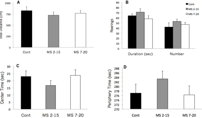
As adults, maternally separated pups have shown increased emotionality/anxiety behaviors (reviewed in Ladd et al., 2000). In the Elevated Plus Maze (EPM), MS animals spend more time in the closed arms, an indicator of increased emotionality in these animals (Daniels, Pietersen, Carstens, & Stein, 2004; Kalinichev, Easterling, Plotsky, & Holtzman, 2002; Madruga, Xavier, Achaval, Sanvitto, & Lucion, 2005; Mesquita, 2008). Similar results were found in the Open Field test (OF), where adult MS animals spent more time in the periphery of the arena showing less exploratory behaviors (Caldji, Francis, Sharma, Plotsky, & Meaney, 2000). In common, these two models are based on the conflict existing between the natural trend to explore a new environment (open arms in the EPM and the center of the arena and number of rearings in the OF) and its innate ability to explore unknown environments.
Depressive-like behaviors are commonly tested in the Forced Swimming Test. The immobility time spent by the rat is a good indicator for helplessness behavior. There is sufficient data to conclude the impact of this model in depressive-like behaviors, with neonatal separated pups demonstrating a significant increase in the immobility time compared to controls (Mesquita, 2008). These findings are corroborated by neurobiological changes in 5-HT system. Accordingly, the hippocampal contents of 5-HT and the raphe nucleus expression of 5-HTT mRNA were decreased in MS rats (Lee, Kim, Kim et al., 2007), young separated rats also showed increased serotonin turnover in the dorsal raphe nucleus (Mesquita et al., 2007). Pharmacology manipulations with Escitalopram have been proven to alleviate the depressive phenotype in adult rats maternally separated early in life (Khoury, Gruber, Mørk, & Mathé, 2006).
The majority of hippocampal granule neurons develops and maturates between postnatal days 1 and 21 (Amaral & Dent, 1981) and the peak of neurogenis overlaps the SHRP (Sapolsky & Meaney, 1986). Therefore, MS could modulate the normal maturation of hippocampal cells with memory and learning implications. Aisa et al. (2007; 2009) found that MS could induce significant decreases in markers of neuroplasticity in the hippocampal formation as well as the prevention of increases in NCAM expression, one plasticity marker that has been associated to some forms of stress effects on learning and memory. In the Morris Water Maze, a behavioral test for spatial memory assessment, MS increases the latency times in rats (Mesquita, 2008), showing the influence of early deprivation on spatial learning.
Among children, negative early life experiences such as maltreatment, have been associated to risk factor for aggression, violence and anti-social behaviors (Barnow & Freyberger, 2003; Fonagy, 2004). These findings were also found in primates and rodents, where increase in intermale aggression was detected in adult rats (Veenema, Blume, Niederle, Buwalda, & Neumann, 2006; Veenema, Bredewold, & Neumann, 2007) and also in juvenile pups (Veenema & Neumann, 2009) exposed to MS procedures.
As discussed above, abnormal social experiences during the neonatal period have been shown to alter the activity of neuropeptide systems, namely OT system. Furthermore, neurochemical synthesis and release changes of this neuropeptide may in turn mediate the regulation of social behaviors in adulthood (Ferris, 2005) such as partner preference formation, aggression, and maternal behavior (Bales and Carter, 2003). Therefore, early life stress (MS) may induce phenotype changes in social behaviors through oxytocinergic system.
1.6. Aims of the Study
The peak period of neurogenesis and denditric growth overlaps the SHRP in neonatal rats (Sapolsky & Meaney, 1986). Early life stress is a risk factor for the acquisition of (neuro)development milestones and future functioning if presented in this postnatal period in a chronically way. MS is a suitable animal model for understanding the potential mechanisms of environmental and developmental determinants of individual differences in stress response and future psychopathology (Holmes, le Guisquet, Vogel, Millstein, Leman, & Beizung, 2005). There is some consistent empirical evidence for the significant effects of MS in the behavior and
neuroendocrine phenotypes that last until adulthood. Even if CS role was commonly thought to have direct responsibilities in the abnormal phenotypes of early stressed pups, in fact these steroids probably affect the development of others neurochemical pathways influencing, in an indirect way, the abnormal reported behaviors of early MS pups. As many other interactions, CS have been proved to have disruptive functions on the OT production and release (Lukas, Bredewold, Neumann, & Veenema, 2010; Veenema et al., 2006; Veenema & Neumann, 2009).
In order to understand the mechanisms beyond MS impact on depressive-like behavior, anxious behavior, spatial memory performance and social behaviors, we explore the long-term impact of MS on the corticosteroid system, focusing the differential role of CS in two different time windows of development.
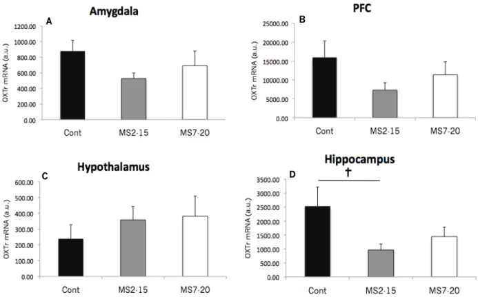
We expect that MS induce HPA axis disruption with increases of CS levels leading behavioral phenotypes changes in adulthood. However, CS are able to trigger changes in several neurotransmitters pathways, namely by region-specific changes in OTr mRNA expression which, in turn, might also contribute to variations in adult behaviors. The present study also tested the hypothesis that mRNA OT-r expression in some stress-sensitive brain areas could be affected by early life stress.
In summary, the present thesis aims to:
- Characterize the long-term consequences of stress exposure into two different developing periods, in terms of spatial memory performance, anxiety behaviors, depressive like-behaviors and social interactions.
- Evaluate long-term consequences of early life maternal deprivation in corticosteroid system. - Evaluate the early life stress consequences in oxytocinergic central pathway at adult age.
2. MATERIAL AND METHODS Animals
Wistar Han females (Charles River, Barcelona, Spain) during estrous were placed overnight with males in same cages under standard laboratory conditions (light/dark cycle 12/12 hours, 8:00 AM – 8:00PM, 22º C ambient
temperature; food and water ad libitum, cage size 60 cm x 40 cm x 20 cm). Males were removed from the cage
when a vaginal plug was confirmed; this day was designated as embryonic day 0 (E0) and the day of delivery as postnatal day 0 (PND 0). At the end of pregnancy (E22), all females were provided with nesting material and remain singly housed. Litters were delivered on gestation day 22; the size of each litter was adjusted to an average of 10 pups. The litters were randomly divided in 3 different groups: 2 maternal separation groups (MS2-15,
Separation procedure
Pups from MS2-15 group were daily separated from their mothers between the 2nd and the 15th post natal days.
Liters from each cage were separated from their dam for 360min (from 9.00 am to 3:00 pm) and kept together in the same plastic box, transferred to an adjacent room climatically controlled (37ºC). After the period of separation the pups returned to their home cages in the colony room. The same procedure was applied to the other experimental group; however for the MS7-20 animals, the period of separation occurred between the 7th and
the 20th post-natal days. Control animals where left undisturbed with their dams until weaning,
Behavioral tests
After weaning (P21) all animals were housed in groups of two and left undisturbed they reach 5 months of age when they were behaviourally tested. All behavioral tasks were conducted sequentially and performed during the light phase of the light/dark cycle.
Open Field
At the age of 5 months, free exploratory behavior of 14 MS2-15, 20 MS7-20, and 11 Control rats, was assessed
during 5 min in a white square open field (43.2 cm x 43.2 cm) surrounded with acrylic transparent walls (ENV – 515; MedAssociates, VT, USA). Room illumination was provided by a white bright light and the temperature was controlled. The session started with the animal placed in the center of the arena and, using a system of two 16 beam infra-red arrays connected to a computer, different parameters of emotional reactivity were measured and recorded: (a) time spent in the central area (a measure of anxious-like behavior); (b) total distance travel (a measure of general locomotor activity) (c) number and duration of rears (a measure of general exploratory behavior). The apparatus was cleaned with 10% ethanol and wiped between sessions.
Elevated Plus Maze
The Elevated Plus Maze was used to examine the anxious-like behavior. The maze has been validated and used as a measure of anxiety (Hogg, 1996; Rodgers and Dalvi, 1997a, 1997b) and reflects a conflict between the rat’s exploratory activity and its innate fear of height and exposed areas.
Animals were individually tested for 5 min in the experimental apparatus. This time interval was chosen due to decrease in avoidance and increase in fatigue behaviors after 5 to 10min of test (Pellow et al., 1985). The apparatus consisted of a plus-shaped platform elevated 72.4 cm above the floor. The maze consisted of two opposing arms (50.8 x 10.2) closed by a 40,6 cm-high side walls and two open arms (50.8 x 10.2) with no walls. Illumination was provided by a white bright light. In the beginning of the session the rats were placed in the centre of the maze and then allowed to explore either the open or closed arms of the maze. The time spent in the different arms of the maze was recorded using an infra-red beam system connected to a computer. The number of entries and time spent into the open arms were used as anxiety behavior indicators – more entries and higher
time spent in the open arms, less anxious behavior. The number of entries in the closed arms of the maze also served as a measure of the rat’s locomotor activity. The maze was cleaned and wiped between every animal with 10% ethanol.
Forced Swimming Test
In order to test the depressive like-behavior the learned helplessness was analyzed using the Forced Swimming Test. As described by Porsolt et al. (1977), the rats were placed individually in acrylic cylinders filled with water (25ºC) to a depth such the animals cannot reach the ground and had no solid support for their rear paws. The present test started with a 10 min pre-test session and, in the following day, animals were subjected to the same procedure for 5 min (test session). A video camera was used to record both of sessions. At the end of each test session, animals were dried and place under a heating lamp (15 min) before return to their home cages. The water was clean between each trial. Video recordings were later scored by two investigators blind to the experimental conditions. Three different parameters were analyzed: a) Immobility -that was considered as rats floating passively, displaying only small movements to keep its nose above the surface; b) activity was considered when the animal swim to escape from the cylinder; c) latency to immobility consisted to the time at which the animal give up swimming for the first time).
Morris Water Maze
The Morris Water Maze (MWM) consisted of a black tank with 170 cm in diameter and 50 cm in deep, filled with water at 22ºC, to a depth of 31 cm. A black platform (12 cm diameter, 30 cm high, invisible to rats) was placed in one of the 4 quadrants (that are divided by imaginary lines) below the surface of the water. The room was mild lighted and four extrinsic visual clues were glued in the room walls. Data related to the path swum and latency to reach platform were collected using video-tracking system fixed to the ceiling of the room (Viewpoint, Champagne au Mont d’Or, France).
A place learning task was performed in order to assess rats' ability to learn the position of the hidden platform. Animals were individually tested for 4 consecutive days (4 trials per day) and the platform was placed in the center of an arbitrary quadrant. Test session for all rats began with rodents being placed, facing the wall in the quadrant immediately at right of the one where the platform has been placed, completing a clockwise rotation in the remaining trials every day. The maximum duration of each trial was 120 sec and if the rat did not find the platform within this period, the experimenter guided the animal to the platform where it remains for 30 seconds. Between each trial all animals were dried before continuing the session. At the end of the four sessions, all rodents were dried with a heating lamp for 15min before placed at the home cage.
Social Interaction Task
The Social Interaction Test (SIT) was described by Field and Hyde (1978) as the first test using ethological sources of anxiety and a natural form of behavior as the dependent variables. Sensitive changes in anxiety
phenotype were found using this test (for a review, File & Seth, 2003). Thirty years later, SIT was extended to assess behaviors from different disorders (Tordjman et al., 2006), effects of pharmacological manipulations on behavioral phenotype (e.g. Bagdy, Graf, Anheuer, Modos, & Kantor, 2001; Bhattacharya & Mitra, 1992) or brain lesions and its implication on behavior (e.g. Duxon, Kennett, Lightowler, Blackburn, & Fone, 1997; Gonzalez, Andrews, & File, 1996). The original protocol's form (Field & Hyde, 1978) has been frequently modified according to the specificity of the research objectives, despite preserving the original objectives and measure parameters. In this way, we adapted Field and colleague protocol preserving its original bases.
The apparatus was constituted by an open arena of 100cm x 100cm x 40cm. The floor was from Morris Water Maze apparatus and sides were made of smooth black polyvinyl chloride (non-reflecting material). A video camera on the ceiling of the open arena was used to score the animals’ behavior. Lighting in the room and temperature was the same as the light phase at colony room.
Figure 2 – The Social Interaction Task apparatus
In the present social interaction protocol, the rats were singly housed for 5 days pior the test. This was because social isolation reliably increases the time spent in social contact and the stress-like responses (e.g., heart rate and blood pressure) (Sharp, Zammit, Azar, & Lawson, 2002). Originally, this period of housing isolation was stipulate in 5 days, but according Niesink and van Ree (1982) 4-7 days of individual housing was sufficient period to produce a maximal peak of interaction during the test. In our protocol, animals were singly housed 5 days prior the test. The cages were located together in racks so that auditory and olfactory contacts were maintained.
Two days prior the test, the animals were familiarized with the test arena. For that purpose, each animal was allowed to explore the apparatus during two 7-min trials (one per day) Between each trial, the arena was cleaned with 10% ethanol and wiped. In the day of the test, each rat was tested for social interaction with an unknown test partner (from another litter, not from experimental or control groups) that not differs by more 80g in weight.
Each trial of social interaction test had 7-min duration and was video recorded for posterior analysis. The arena was cleaned and wiped between each trial. For the present experiment 6 rats from each MS groups and 6 rats from control group were tested. The rats were randomly selected.
Social behavior was analyzed in terms of frequency and time and have been classified in 4 different categories: explorative behavior, non-aggressive (healthy social interaction), aggressive behavior and others (see Table 1). Two investigators blind to the experimental condition analyzed the videotapes off-line.
Table 1
Rodent's behaviors assed in the Social Interaction Test
Rearing The state in which rodent’ s body is vertically erected
sniffing the air
Exploration
Movement without social interaction Movements in arena, sniffing and inspecting the arena.
This includes stereotyped walking along specific routes
Investigation Sniffs at and investigates the other rat
Nosing Both rats meet nose to nose while stretching their body
slightly
Follow The rat follows the other rat, with a distance of 20cm or
less between the rats.
Approach The rat approach the other rat rapidly or changing the
route exhibited till then.
Grooming Grooming between the rats
Non-aggressive behaviors
Play behavior Any type of chase, spar, pin or wrestle without any type of
aggressive intention
Lateral threat The body is arched in a sideward posture towards the other
rat
Upright The rat is standing on its hind legs and his facing the
opponent
Stand over and lie under One rat is standing on top of the other rat, while the other is
lying on its back beneath the first rat Clinch (kicking, boxing, wrestling, agressive
grooming or biting Active fighting between the rats
Purse The rat runs after the other rat during a fight
Agressive behaviors
Escape The rat runs away from the other rat during a fight
Inactive Sitting quietly
Stationary stereotyped behavior The rat is stationary and performs circular head movements
o head weaving
Movement stereotyped behavior Body circling in the same position, digging, spontaneous
activity, jumping... (note: do not include exploration of the arena in stereotype routes)
Other type of behavior
Self-involved behaviors The rat is stationary and is self-grooming, scratching,
washing or involved in another type of self-injurious behaviors
Tissue preparation
All animals were sacrificed by rapid decapitation and blood samples were collected. The adrenal glands from all animals were also removed and weighted. The brain was removed and different areas were dissected using a microscope and following orientation marks provided by Paxinos and Watson (2005). Samples were snap-frozen by immersion in liquid nitrogen for posterior total RNA extraction.
Gene expression analysis
Total RNA was isolated from the left hippocampal formation, pre-frontal cortex, extended amygdaloid complex, and hypothalamus using Trizol (Invitrogen, Carlsbad, CA, USA). The RNA was then reverse transcribed into first-strand complementary DNA using the superscript first-first-strand synthesis system for reverse-transcription polymerase chain reaction (PCR) (Invitrogen) according to the manufacturer’s instructions (cycling parameters: 5min at 25ºC, 60min at 42ºC, and 5min at 85ºC).
Using a standard reverse transcriptase-polymerase chain reaction (iQ5 real-time PCR Detection System) protocol (Bio-Rad, Hercules, CA, USA), the expression levels of OT-r were assessed for the above described areas of Cont group (n=5), MS2-15 (n=5) and MS7-20 (n=5) animals and duplicates were performed for each sample. The
cycling parameters were 1 cycle of 95 °C for 15min, followed by 39 cycles of 94 °C for 15s, annealing temperature was 60°C for all genes analysed for 30s and 72 °C for 30s. Single acquisitions were done at the end of each annealing step. Primers: OT-r fw-5’-GTCAATGCGCCCAAGGAAGCTTC-3’ and rw-5’- CTCTGGCTTGAGCTGCGACGG-3’
Endocrine evaluation
Radioimmunoassay was performed using the blood samples collected between 1:00 and 3:00 p.m.; the interval between transferring animals from their undisturbed environment to decapitation was kept below 60s. Serum corticosterone levels were assessed by radioimmunoassay (RIA), using ImmuChemTM Corticosterone-125I kits (MP Biomedicals, LLC, Orangeburg, NY, USA). The CS concentration was determined using a γ counter. The minimum detectable dose is 7.7 ng/ml.
Statistical analysis
Data analysis were carried out using PASW 18.0 software (IBM® SPSS® Statistics) and statistical significance was accepted for p values ≤ 0.05. Whenever the homogeneity of variances or the normal distribution of the sample were not achieved, variables transformations were performed in the following sequence, till meet the criteria: Log10 , Square Root (SQRT) and Inverse (1/). Data are presented as mean ± SEM.
Biometric data (total body weight, thymus and adrenal glands weights) were statistically analyzed by one-way ANOVA (MS2-15 x MS7-20 x Cont). Tukey post-hoc test was used to analyze univariate significances.
The Elevated Plus Maze task was analyzed using one-way ANOVA with exception for “entry open arms” variable where Kruskal-Wallis test was performed followed by Mann-Whiney tests with Bonferroni correction. In the same behavioral test, “time spent in the open arms” variable was transformed by Log .