The maternal JAK/STAT pathway of
Drosophila
regulates embryonic
dorsal-ventral patterning
Departamento de Histologia e Embriologia, Universidade Federal do Rio de Janeiro, Rio de Janeiro, RJ, Brasil
E.S.S. Lopes and H.M. Araujo
Abstract
Activation of NFκB plays a pivotal role in many cellular processes such as inflammation, proliferation and apoptosis. In Drosophila, nuclear translocation of the NFκB-related transcription factor Dorsal is spatially regulated in order to subdivide the embryo into three primary dorsal-ventral (DV) domains: the ventral presumptive meso-derm, the lateral neuroectoderm and the dorsal ectoderm. Ventral activation of the Toll receptor induces degradation of the IκB-related inhibitor Cactus, liberating Dorsal for nuclear translocation. In addi-tion, other pathways have been suggested to regulate Dorsal. Signal-ing through the maternal BMP member Decapentaplegic(Dpp) inhib-its Dorsal translocation along a pathway parallel to and independent of Toll. In the present study, we show for the first time that the maternal JAK/STAT pathway also regulates embryonic DV patterning. Null alleles of loci coding for elements of the JAK/STAT pathway, hop-scotch (hop), marelle (mrl) and zimp (zimp), modify zygotic expres-sion along the DV axis. Genetic analysis suggests that the JAK kinase Hop, most similar to vertebrate JAK2, may modify signals down-stream of Dpp. In addition, an activated form of Hop results in increased levels of Cactus and Dorsal proteins, modifying the Dorsal/ Cactus ratio and consequently DV patterning. These results indicate that different maternal signals mediated by the Toll, BMP and JAK/ STAT pathways may converge to regulate NFκB activity in Drosophila.
Correspondence
H.M. Araujo
Departamento de Histologia e Embriologia, UFRJ CCS, Bl. F, Sala F2-031 Av. Brig. Trompowski, s/n 21949-900 Rio de Janeiro, RJ Brasil
E-mail: [email protected]
Presented at the XI Congresso Brasileiro de Biologia Celular, Campinas, SP, Brazil, July 15-18, 2004.
Research supported by FAPERJ and FIRCA/NIH TW001329-03 to H. Araujo, and a CNPq fellowship to E.S.S. Lopes.
Received June 1, 2004 Accepted September 23, 2004
Key words
•JAK kinase •Drosophila
•Embryonic patterning •hopscotch
•NFκB
Introduction
The establishment of the dorsal-ventral (DV) axis of the Drosophila embryo de-pends on processes acting during both oo-genesis and embryooo-genesis. DV asymmetry, first generated during oogenesis, is ultimately transmitted to the embryo in the form of a nuclear gradient of the NFκB-related trans-cription factor Dorsal. A series of maternally transcribed genes exert their effects during
homologue Cactus, releasing Dorsal for nuclear translocation (see references in Ref. 1). A ventral to dorsal nuclear gradient of Dorsal protein is thus formed, leading to the establishment of the three primary domains of the embryo: the ventral presumptive me-soderm, the lateral neuroectoderm and the dorsal ectoderm (6-12).
More recently, it has been shown that sig-naling through the tumor growth factor-ß (TGF-ß) family member decapentaplegic (dpp) also modifies the Dorsal gradient (13). Increased signaling through maternal dpp, the Droso-phila BMP2/4 orthologue (14), induces a shift towards more dorsal fates in the embryo. Con-versely, a decrease in the maternal dose of the BMP antagonist short gastrulation (sog; 11,15) induces a similar phenotype. It has been sug-gested that maternal dpp modifies the Dorsal gradient by regulating Cactus degradation through a Toll-independent pathway. The pres-ent model proposes that Dpp inhibits Cactus degradation, retaining Dorsal in the cytoplasm (13). The exact contribution of the signal-independent versus signal-dependent pathways of Cactus degradation to the establishment of the Dorsal gradient remains to be elucidated. However, there are indications that the activity of other genes may converge to regulate Dor-sal and Cactus, thus contributing to the estab-lishment of the embryonic DV axis.
Many different signals may regulate pro-teins of the NFκB/c-rel family. For instance, it has been shown in Drosophila that Dorsal may be phosphorylated (16-18) and that ven-tral signals modify Dorsal to regulate nuclear import independent of Cactus (19). Conversely, Cactus is regulated through several mechan-isms. As cited above, degradation of Cactus may follow through the Toll-dependent (20) or -independent pathways (21-23). In addition to phosphorylation regulated by ventral sig-nals, Cactus may also be phosphorylated by casein kinase II (22). Actually, proteins of the IκB family from vertebrates and invertebrates present several regulatory modules which are targeted by phosphorylation.
In an attempt to identify other molecules that regulate maternal NFκB/IκB signaling in Drosophila we have undertaken a directed screening for elements that modify a dorsal sensitized background. As a result of this analysis, we present evidence that elements of the JAK/STAT pathway also contribute to the establishment of the embryonic DV axis. Genetic data indicate that elements of this pathway may function by non-classical mechanisms to modify the Dorsal gradient. In addition, our results suggest that Hop-scotch, most similar to vertebrate Janus ki-nase JAK2 (24), regulates the total levels of both Cactus and Dorsal proteins.
Material and Methods
Fly stocks
Canton S (CS) was used as the wild type. All mutants, balancers and chromosomal markers used in this study were obtained from the Bloomington Indiana Stock Center, Bloomington, IN, USA. All embryos ana-lyzed in this study resulted from crosses between the maternal genotypes listed with male CS. For activation of the temperature-sensitive hop[Tum] allele females and re-sulting embryos were kept at 29ºC until em-bryo collection.
In situ hybridization
In situ hybridization was performed as described previously with antisense RNA probes (25) using full-length cDNAs as tem-plate.
Immunoblot analysis
Photoshop, by determining the intensity of white in negative images. To determine the ratio of Dorsal/Cactus, relative protein amounts were initially determined by divid-ing the intensity of Cactus or Dorsal autora-diographic bands by the corresponding Tu-bulin bands to obtain CacT and DlT. The values obtained were then used to define the normalized DlT/CacT used as the Dorsal/ Cactus ratio.
Results
Elements of the JAK/STAT pathway interact with Dorsal
DV territories in the Drosophila blasto-derm embryo can be visualized by in situ hybridization with specific RNA probes. In wild-type embryos, probes against the ven-tral nervous system defective (vnd) gene re-veal expression in the ventral-most third of the lateral neuroectoderm (8). The pattern is highly reproducible and forms a precise bor-der with the ventral presumptive mesobor-derm (8,13) (Figure 1A). However, in embryos laid by mothers whose dose of the dorsal (dl) gene was reduced to half (dl-/+) this border is not as precise, especially in an anterior domain where the head fold will later form (13). In a small percentage of these embryos (3%; Table 1) vnd expression totally invades the ventral territory at 30% egg length. Dor-sal is sensitive to gene dosage (26-28), and alterations in the dose of genes that interact with the Dorsal pathway modify the pen-etrance or expressivity of the dl-/+ pheno-type (13). Therefore, we have used the ma-ternal dl-/+ as our “sensitized background” to search for other genes that interact with the Dorsal pathway.
We have screened for elements of other pathways that have been shown to interact genetically or biochemically with NFκB/IκB in vertebrates or invertebrates. Null or hypo-morphic mutants for elements of the IL-1, IL-2 and calcium signaling pathways were
crossed to a dl mutant line and the embryos from mothers double heterozygous for these alleles were collected for in situ analysis (data not shown). Amongst the loci ana-lyzed, elements of the Drosophila JAK/STAT pathway consistently modified the dl-/+ phe-notype. Embryos derived from mothers double heterozygous for a null hopscotch allele (hop[2]) and a null dl allele (dl[15]) presented an increased penetrance of the dl-/+ phenotype (30%; Figure 1B and Table 1). A similar effect could be observed with mutant alleles of downstream elements of the JAK/STAT pathway such as a null allele of the Stat3/Stat5 homologue marelle (mrl) (24) and the PIAS negative regulator zimp (zimp; Table 1; 29). It may seem surprising that all three elements increase the penetrance
Figure 1. hopscotch regulates zygotic gene expression along the dorsal-ventral axis. Blastoderm stage embryos collected from wild-type (A) or hop[2]; dl/[15]/+ (B) mothers and processed for in situ hybridization with a vnd probe. Observe the ventral invasion of vnd
expression in B (arrow). Anterior is left, dorsal is up.
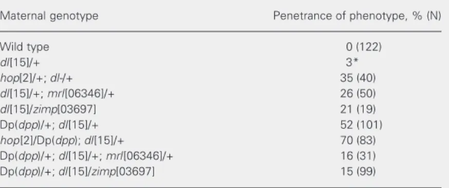
Table 1. Elements of the maternal JAK/STAT pathway alter embryonic gene expres-sion along the dorsal-ventral axis.
Maternal genotype Penetrance of phenotype, % (N)
Wild type 0 (122)
dl[15]/+ 3*
hop[2]/+; dl-/+ 35 (40)
dl[15]/+; mrl[06346]/+ 26 (50)
dl[15]/zimp[03697] 21 (19)
Dp(dpp)/+; dl[15]/+ 52 (101)
hop[2]/Dp(dpp); dl[15]/+ 70 (83) Dp(dpp)/+; dl[15]/+; mrl[06346]/+ 16 (31) Dp(dpp)/+; dl[15]/zimp[03697] 15 (99)
Embryos were collected from the maternal genotypes listed and processed for insitu
hybridization with a vnd probe. The penetrance of the dorsalized embryonic pheno-type revealed by ventral invasion of vnd expression (as shown in Figure 1B) is presented. Penetrance is defined as the percentage of embryos that present the phenotype. The total number of embryos analyzed for each condition is shown in parentheses. *Data taken from Ref. 13.
A
A
A
A
Using an activated hop allele to understand the mechanism of Hop action
In order to further test the relationship between the JAK/STAT and Dpp signaling pathways in the context of DV patterning, we used a temperature-sensitive gain-of-func-tion hop allele that is independent of up-stream receptor activation. This allele, termed hop[Tum] (for Tumoral), generates a hyper-activated JAK kinase at 29ºC (30,31). We intended to test if hop[Tum] could block a dorsalizing Dpp signal generated by an in-crease in the dose of dpp. Surprisingly, though, activation of signaling through hop by using the hop[Tum] allele also increased the dl-/+ phenotype (Table 2), i.e., it induced a dorsalized phenotype. As a dominant nega-tive form of hop was not available and as null hop clones would effect earlier develop-ment of follicles (32), we tested whether hop[Tum] and a duplication of dpp would present additive dorsalizing effects, suggest-ing that they alter DV patternsuggest-ing through different pathways.
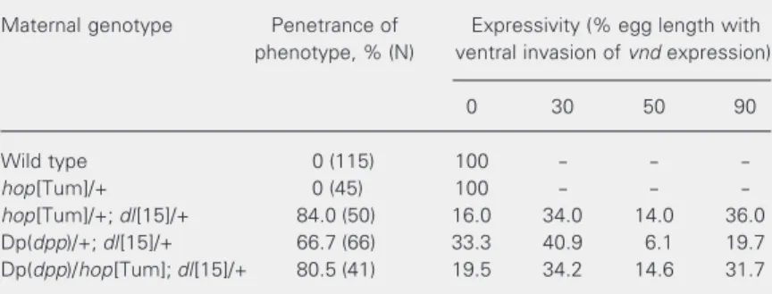
At 29ºC mothers double heterozygous for hop[Tum] and dl- generated 84% of dorsalized embryos, while mothers double heterozygous for Dp(dpp) and dl- generated 67% of dorsalized embryos (Table 2). Moth-ers triple heterozygous for hop[Tum], Dp(dpp) and dl- generated 80% of embryos with invasion of vnd expression in the ven-tral domain (Table 2, Figure 2). The pen-etrance of the phenotypes for the triple het-erozygotes could represent either average or equivalent effects of hop[Tum] and Dp(dpp) on DV patterning. Unfortunately, low vi-ability of the maternal genotypes resulted in a modest number of embryos analyzed, pre-cluding the use of statistical tests. Neverthe-less, our results argue against an additive effect of hop[Tum] and Dp(dpp). By analyz-ing the expressivity of the phenotypes for the above conditions, this can be seen more clearly (Figure 2, Table 2). While increasing the dose of dpp in dl-/+ mothers induced of the dl-/+ phenotype, especially
consider-ing that Zimp inhibits signalconsider-ing through Hop and Mrl. However, it should be kept in mind that this type of experiment, which relies on disturbance of the balance between different regulatory elements, does not necessarily reveal the direction of scored interactions.
We have shown that the Dpp pathway interacts with Dorsal. Alterations in the dose of maternal dpp by itself does not seem to modify the embryonic DV axis (13). How-ever, embryos derived from mothers con-taining a duplication of the dpp locus (Dp(dpp)) and heterozygous for a null dl allele show a great increase in the penetrance of the dl-/+ phenotype (Table 1) (13). In order to determine whether elements of the JAK/STAT pathway could modify the Dp(dpp)/+; dl-/+ phenotype we generated embryos from mothers triple heterozygous for Dp(dpp), dl- and alleles of hop, mrl or zimp. Interestingly, we could observe an ad-ditive effect of hop and Dp(dpp) on the embryonic phenotype, while alleles of mrl and zimp decreased the penetrance of the Dp(dpp)/+; dl-/+ phenotype (Table 1). Again, these experiments did not permit us to reach any conclusions regarding the direction of the interactions scored. However, they did suggest some dissociation between the ef-fects generated by hop versus mrl and zimp.
Table 2. The effects of maternal dpp and hop on embryonic gene expression are not additive.
Maternal genotype Penetrance of Expressivity (% egg length with phenotype, % (N) ventral invasion of vnd expression)
0 30 50 90
Wild type 0 (115) 100 - -
-hop[Tum]/+ 0 (45) 100 - -
-hop[Tum]/+; dl[15]/+ 84.0 (50) 16.0 34.0 14.0 36.0 Dp(dpp)/+; dl[15]/+ 66.7 (66) 33.3 40.9 6.1 19.7 Dp(dpp)/hop[Tum]; dl[15]/+ 80.5 (41) 19.5 34.2 14.6 31.7
ventral vnd invasion mostly in the anterior part of the embryo, inducing hop activity with or without a duplication of dpp ex-tended the ventral invasion of vnd expres-sion to more posterior regions of the em-bryos (Figure 2, Table 2). The distribution among expressivity classes was similar be-tween hop[Tum]/+; dl-/+ and hop[Tum]/ Dp(dpp); dl-/+.
These results indicate that, with activa-tion of the Hop kinase, Dpp can exert no further effect on DV patterning of the em-bryo. This would suggest that Dpp either acts directly to regulate Hop activity or re-quires Hop to regulate some downstream element of the maternal Dpp pathway.
Activated Hop modifies Cactus and Dorsal protein levels
Formation of the Dorsal gradient depends on a precise balance of Cactus and Dorsal protein levels. In addition to regulation of
Cactus degradation through the Toll-depend-ent and -independToll-depend-ent pathways, the level of Cactus protein depends on the amount of Dorsal protein. It has been shown that ge-netically modifying the dose of dl, and thus the amount of Dorsal protein, results in a concomitant alteration in the total level of Cactus (28). Therefore, one possible mech-anism for hop action would be to regulate the levels of Dorsal and Cactus. In a wild-type background, hop[Tum] does not alter the level of either protein (Figure 3A,B, lane 2). On the other hand, hop[Tum] induced an increase in the levels of both Cactus and Dorsal in a dl-/+ background (Figure 3A,B, lanes 3 and 4). Measurement of the protein band intensities indicated that the Cactus to Dorsal ratio was increased above that of the wild type (Figure 3, Legend), suggesting that the ventral invasion of vnd expression in embryos from hop/+; dl/+ mothers is a result of higher retention of Dorsal in the cyto-plasm by increased Cactus levels.
Figure 2. Embryonic phenotypes generated by activated Hop are variable. Blastoderm stage embryos were collected from hop[Tum]; dl[15]/+ mothers and processed for in situ hybridization with a vnd probe. At 29ºC the expressivity of the phenotype analyzed is variable, with vnd expression invading the ventral domain (arrows) at 30% (A), 50% (B) or 90% (C) egg length, with egg length determined from the anterior to the posterior end of the embryo. No invasion is defined as 0%. Anterior is left, dorsal is up.
Figure 3. Activated hop increases the levels of Dorsal and Cactus proteins. Total embryonic extracts were prepared from 0- to 1-h pre-blastoderm embryos, run on 8% SDS-PAGE and probed with antibodies against Dorsal (A) or Cactus (B). The same blot was subsequently probed for anti-Tubulin (lower panels) to assure that equivalent amounts of protein were loaded for each sample. Embryos were collected from wild-type (lane 1), hop[Tum]/ + (lane 2); hop[Tum]/+; dl[15]/+ (lane 3) or dl[15]/+ (lane 4) mothers. The relative amounts of Dorsal/Cactus were: 1.19 for wild type, 1.18 for dl[15]/+, and 0.92 for hop[Tum]/+; dl[15]/+.
α-Dorsal
α-Tubulin
1 2 3 4 1 2 3 4
α-Cactus
α-Tubulin
A
A
A
A
A
B
B
B
B
B
A
A
A
Discussion
hopscotch may signal through two different pathways to regulate embryonic DV patterning
The Drosophila JAK/STAT pathway plays several roles during development. These include segmentation, eye develop-ment, hematopoiesis and gametogenesis (see references in Ref. 33). In addition, an impor-tant role in the Drosophila immune response and blood cell proliferation has been de-scribed (reviewed in Ref. 34). Interestingly, this may be an evolutionarily conserved role as vertebrate JAK/STAT is fundamental in regulating proliferation and differentiation of hematopoietic stem cells and in the im-mune response (35). We describe here for the first time a role for JAK/STAT signaling in embryonic DV patterning.
All elements of the JAK/STAT pathway tested, hop, mrl and zimp, somehow altered vnd expression, indicating that classical ele-ments of the JAK/STAT pathway regulate embryonic DV patterning. However, while all three loci increased the penetrance of the dl-/+ phenotype, only hop could still induce an increase in this penetrance in the presence of a duplication of dpp. Even though these genetic tests do not reveal the direction of interactions, it is striking that hop versus mrl and zimp showed diverging effects only in the presence of Dpp. This could suggest that, in addition to hop acting on a classical path-way to activate mrl, it could also act inde-pendently on a pathway more related to dpp. For instance, Hop may be required to phos-phorylate some element of the signal-inde-pendent pathway of Cactus degradation, downstream of Dpp. It has been shown that the vertebrate JAK2 kinase is able to phos-phorylate IκB in response to erythropoietin (36). This results in increased degradation of IκB, with a consequent protection from ap-optosis. Interestingly, increased Hop activ-ity attained by using the
temperature-sensi-tive hop[Tum] allele increases the total amount of Cactus protein, a fact possibly due to altered degradation. Furthermore, Dpp also inhibits Cactus degradation (13). Fi-nally, Dp(dpp) does not increase the pen-etrance of the DV phenotype induced by hop[Tum], indicating that Hop activity is required downstream of Dpp to regulate Cac-tus levels. It should be pointed out, however, that our data do not address whether Cactus is a direct target of Hop, or whether it regu-lates the degradation of Cactus protein.
Other evidence that hop may exert an effect on DV patterning independently of mrl is the fact that hop[Tum] altered the levels of Cactus protein. It seems unlikely that mrl regulates the level of expression of cactus mRNA, especially if we consider that cactus is maternally transcribed and that no zygotic expression at the early embryonic stages has been described (16). Alternatively, mrl could regulate the stability of cactus mRNA or protein.
necessary to test this prediction.
The role of the JAK/STAT pathway in defining DV territories
Intersection between different signal transduction pathways results in a combina-torial control that may help fine-tune and integrate functional responses. Intersection between interleukin and NFκB pathways has been described in the regulation of nitric oxide synthase transcription (37), and of the type 2 Toll-like receptor (38). Intercommu-nication between interleukin and the TGF-ß pathway has also been reported. For instance, TGF-ß regulates the transcription of inter-leukin genes (39), and has an important role during the immune response. Another level of intersection between these elements may take part in the cytoplasmic environment as described during neuroprotection in verte-brates (36) and as suggested in the present study.
Our results suggest intersection between the Drosophila JAK/STAT, NFκB and TGF-ß family pathway elements. In spite of the clear genetic interactions between hop, dl and dpp, by examination of embryos derived from mothers with altered Hop signaling alone, we were unable to detect any effect on
expression patterns along the DV axis (Lopes E, unpublished results). This leads us to ask what role hop may play in DV patterning. Actually, it is quite hard to understand why mothers heterozygous for hop(2), hop[Tum] or Dp(dpp) had no effect on DV patterning by themselves, while they did so when the dose of dl was initially disturbed. One expla-nation would be that the Toll pathway is robust, accounting for subdivision of DV territories and their exact placement along the DV axis (40). Other pathways would have been co-opted to ensure that proper DV patterning takes place when direct signaling through Toll is disturbed. Certainly more experimentation will be necessary to test this hypothesis. It will also be very interesting to test the generality of the interactions de-scribed here in other model systems, and to assay the respective contributions of each of the interacting genes.
Acknowledgments
We are grateful to Silvania Nunes for fly food preparation and to the Bloomington Stock Center, Bloomington, IN, USA, for fly lines. We would also like to thank Dr. Ethan Bier (UCSD) for helpful discussions and intellectual support.
References
1. Morisato D & Anderson KV (1995). Signaling pathways that estab-lish the dorsal-ventral pattern of the Drosophila embryo. Annual Review of Genetics, 29: 371-399.
2. Schneider DS, Jin Y, Morisato D & Anderson KV (1994). A pro-cessed form of the Spatzle protein defines dorsal-ventral polarity in the Drosophila embryo. Development, 120: 1243-1250.
3. Stein D, Roth S, Vogelsang E & Nusslein-Volhard C (1991). The polarity of the dorsoventral axis in the Drosophila embryo is defined by an extracellular signal. Cell, 65: 725-735.
4. Morisato D (2001). Spatzle regulates the shape of the Dorsal gradi-ent in the Drosophila embryo. Development, 128: 2309-2319. 5. Morisato D & Anderson KV (1994). The spatzle gene encodes a
component of the extracellular signaling pathway establishing the dorsal-ventral pattern of the Drosophila embryo. Cell, 76: 677-688. 6. Steward R (1987). Dorsal, an embryonic polarity gene in Drosophila,
is homologous to the vertebrate proto-oncogene, c-rel. Science, 238: 692-694.
7. Thisse C, Perrin-Schmitt F, Stoetzel C & Thisse B (1991). Sequence-specific transactivation of the Drosophila twist gene by the dorsal gene product. Cell, 65: 1191-1201.
8. Mellerick DM & Nirenberg M (1995). Dorsal-ventral patterning genes restrict NK-2 homeobox gene expression to the ventral half of the central nervous system of Drosophila embryos. Developmental Biology, 171: 306-316.
9. Jiang J, Kosman D, Ip YT & Levine M (1991). The dorsal morphogen gradient regulates the mesoderm determinant twist in early Droso-phila embryos. Genes and Development, 5: 1881-1891.
10. Ip YT, Park RE, Kosman D, Bier E & Levine M (1992). The dorsal gradient morphogen regulates stripes of rhomboid expression in the presumptive neuroectoderm of the Drosophila embryo. Genes and Development, 6: 1728-1739.
gene. Genes and Development, 8: 2602-2616.
12. Doyle HJ, Kraut R & Levine M (1989). Spatial regulation of zerknullt: a dorsal-ventral patterning gene in Drosophila. Genes and Develop-ment, 3: 1518-1533.
13. Araujo H & Bier E (2000). sog and dpp exert opposing maternal functions to modify toll signaling and pattern the dorsoventral axis of the Drosophila embryo. Development, 127: 3631-3644. 14. Padgett RW, St Johnston RD & Gelbart WM (1987). A transcript
from a Drosophila pattern gene predicts a protein homologous to the transforming growth factor-beta family. Nature, 325: 81-84. 15. Ferguson EL & Anderson KV (1992). Localized enhancement and
repression of the activity of the TGF-beta family member, decapen-taplegic, is necessary for dorsal-ventral pattern formation in the
Drosophila embryo. Development, 114: 583-597.
16. Whalen AM & Steward R (1993). Dissociation of the dorsal-cactus complex and phosphorylation of the dorsal protein correlate with the nuclear localization of dorsal. Journal of Cell Biology, 123: 523-534.
17. Gillespie SK & Wasserman SA (1994). Dorsal, a Drosophila Rel-like protein, is phosphorylated upon activation of the transmembrane protein Toll. Molecular and Cellular Biology, 14: 3559-3568. 18. Drier EA, Huang LH & Steward R (1999). Nuclear import of the
Drosophila Rel protein Dorsal is regulated by phosphorylation.
Genes and Development, 13: 556-568.
19. Drier EA, Govind S & Steward R (2000). Cactus-independent regula-tion of Dorsal nuclear import by the ventral signal. Current Biology, 10: 23-26.
20. Wasserman SA (2000). Toll signaling: the enigma variations. Cur-rent Opinion in Genetics and Development, 10: 497-502.
21. Reach M, Galindo RL, Towb P, Allen JL, Karin M & Wasserman SA (1996). A gradient of cactus protein degradation establishes dorso-ventral polarity in the Drosophila embryo. Developmental Biology, 180: 353-364.
22. Liu ZP, Galindo RL & Wasserman SA (1997). A role for CKII phos-phorylation of the cactus PEST domain in dorsoventral patterning of the Drosophila embryo. Genes and Development, 11: 3413-3422. 23. Belvin MP, Jin Y & Anderson KV (1995). Cactus protein degradation
mediates Drosophila dorsal-ventral signaling. Genes and Develop-ment, 9: 783-793.
24. Hou XS, Melnick MB & Perrimon N (1996). Marelle acts down-stream of the Drosophila HOP/JAK kinase and encodes a protein similar to the mammalian STATs. Cell, 84: 411-419.
25. O’Neill JW & Bier E (1994). Double-label in situ hybridization using biotin and digoxigenin-tagged RNA probes. Biotechniques, 17: 870, 874-875.
26. Steward R (1989). Relocalization of the dorsal protein from the
cyto-plasm to the nucleus correlates with its function. Cell, 59: 1179-1188. 27. Rushlow CA, Han K, Manley JL & Levine M (1989). The graded distribution of the dorsal morphogen is initiated by selective nuclear transport in Drosophila. Cell, 59: 1165-1177.
28. Govind S, Brennan L & Steward R (1993). Homeostatic balance between dorsal and cactus proteins in the Drosophila embryo.
Development, 117: 135-148.
29. Betz A, Lampen N, Martinek S, Young MW & Darnell Jr JE (2001). A
Drosophila PIAS homologue negatively regulates stat92E. Proceed-ings of the National Academy of Sciences, USA, 98: 9563-9568. 30. Hanratty WP & Dearolf CR (1993). The Drosophila tumorous-lethal
hematopoietic oncogene is a dominant mutation in the hopscotch locus. Molecular and General Genetics, 238: 33-37.
31. Harrison DA, Binari R, Nahreini TS, Gilman M & Perrimon N (1995). Activation of a Drosophila Janus kinase (JAK) causes hematopoietic neoplasia and developmental defects. EMBO Journal, 14: 2857-2865.
32. McGregor JR, Xi R & Harrison DA (2002). JAK signaling is somati-cally required for follicle cell differentiation in Drosophila. Develop-ment, 129: 705-717.
33. Hombria JC & Brown S (2002). The fertile field of Drosophila Jak/ STAT signalling. Current Biology, 12: R569-R575.
34. Mathey-Prevot B & Perrimon N (1998). Mammalian and Drosophila
blood: JAK of all trades? Cell, 92: 697-700.
35. Hou SX, Zheng Z, Chen X & Perrimon N (2002). The Jak/STAT pathway in model organisms: emerging roles in cell movement.
Developmental Cell, 3: 765-778.
36. Digicaylioglu M & Lipton SA (2001). Erythropoietin-mediated neuro-protection involves cross-talk between Jak2 and NF-kappaB signal-ling cascades. Nature, 412: 641-647.
37. Ganster RW, Taylor BS, Shao L & Geller DA (2001). Complex regula-tion of human inducible nitric oxide synthase gene transcripregula-tion by Stat 1 and NF-kappa B. Proceedings of the National Academy of Sciences, USA, 98: 8638-8643.
38. Musikacharoen T, Matsuguchi T, Kikuchi T & Yoshikai Y (2001). NF-kappa B and STAT5 play important roles in the regulation of mouse Toll-like receptor 2 gene expression. Journal of Immunology, 166: 4516-4524.
39. Fargeas C, Wu CY, Nakajima T, Cox D, Nutman T & Delespesse G (1992). Differential effect of transforming growth factor beta on the synthesis of Th1- and Th2-like lymphokines by human T lympho-cytes. European Journal of Immunology, 22: 2173-2176.
![Figure 2. Embryonic phenotypes generated by activated Hop are variable. Blastoderm stage embryos were collected from hop[Tum]; dl[15]/+ mothers and processed for in situ hybridization with a vnd probe](https://thumb-eu.123doks.com/thumbv2/123dok_br/15809893.650895/5.918.87.832.643.786/embryonic-phenotypes-generated-activated-blastoderm-collected-processed-hybridization.webp)
