Escola de Engenharia
António André de Sousa Moreira
Candida bracarensis
virulence factors
Mestrado Integrado em Engenharia Biomédica
Ramo Engenharia Clínica
Trabalho efetuado sob a orientação da
Professora Doutora Mariana Henriques
e a co-orientação da
Nome: António André de Sousa Moreira
Endereço Eletrónico: [email protected] Cartão de Cidadão: 13384917
Título da Dissertação:
Candida bracarensis virulence factors Fatores de virulência de Candida bracarensis
Orientadora: Professora Doutora Mariana Henriques Co-orientadora: Doutora Cláudia Botelho
Ano de conclusão: 2013 Designação do Mestrado:
Mestrado Integrado em Engenharia Biomédica – Ramo Engenharia Clínica
É AUTORIZADA A REPRODUÇÃO INTEGRAL DESTA DISSERTAÇÃO APENAS PARA EFEITOS DE INVESTIGAÇÃO, MEDIANTE DECLARAÇÃO ESCRITA DO INTERESSADO, QUE A TAL SE COMPROMETE.
Universidade do Minho, ____/____/______
Várias foram as pessoas que contribuíram para a realização desta dissertação. Sem a colaboração de todas elas, quer de uma forma, quer de outra, os objetivos propostos não teriam sido alcançados e este trabalho não teria sido concretizado. Deste modo, quero aproveitar esta secção para expressar o meu sincero agradecimento a essas mesmas pessoas.
À Professora Mariana Henriques, por toda a dedicação e empenho que demonstrou ao longo do meu trabalho, através das suas orientações e do seu constante incentivo, e por ter acreditado nas minhas capacidades. Agradeço-lhe também o facto de ter partilhado comigo todos os seus conhecimentos, assim como os seus sábios conselhos. Para mim, foi um gosto e um privilégio ter tido a oportunidade de ter trabalhado com a professora.
À Doutora Cláudia Botelho, pela sua simpatia, disponibilidade e pelos seus conselhos e sugestões que me ajudaram a atingir todos as metas programadas. A sua partilha de conhecimento também foi uma mais-valia para mim, pelo que não posso deixar de lhe agradecer: “O meu muito obrigado!”.
À Doutora Sónia Silva, pela amizade, carinho, boa disposição e por ser a excelente pessoa que é. A ti, agradeço também toda a ajuda e o excelente apoio prestado no laboratório e não só... Sónia, de facto, por mais que escreva, nunca conseguirei agradecer-te por tudo. Como diriam os críticos: “És !”.
Ao Carlos Tiago Alves, pelo seu companheirismo, assim como também pela sua ajuda e conselhos. E claro, pela sua responsabilidade e imensa boa disposição que contagia e estimula qualquer um.
À Professora Paula Sampaio e à Professora Célia Pais, do Departamento da Biologia da Universidade do Minho, por terem cedido as estirpes com as quais pude trabalhar e por terem confiado em mim para dar continuidade às investigações sobre C. bracarensis.
Agradeço também ao Departamento de Engenharia Biológica da Universidade do Minho por ter cedido as suas instalações para a realização do meu trabalho experimental.
Hugo, pelo fantástico ambiente de trabalho que me proporcionaram, pela interajuda, pelo apoio e, sobretudo, pelo convívio. Quero deixar também uma palavra de carinho à Doutora Ana Oliveira pela sua ajuda, pela sua constante e imensa boa disposição, pela sua simpatia e por todos os momentos hilariantes protagonizados por nós os dois. Todos esses momentos acabaram por ser um estímulo para que tudo o resto parecesse mais simples.
A todos os meus colegas de Engenharia Biomédica, pela interajuda, por todos os convívios, por me terem proporcionado momentos únicos e inesquecíveis, pela boa disposição, pelas alegrias e por terem ajudado a tornar-me numa pessoa melhor e mais capaz de atingir os meus objetivos.
Por último, e não menos importante, quero agradecer aos meus Pais e à minha Família, pelo apoio emocional e financeiro, pela paciência nos dias menos bons, pelo conforto e pelo estímulo. Eles farão sempre parte da minha vida…
The preset work was developed under the project PTDC/SAU-MIC/119069/2010.
Abstract
Candida bracarensis is a rare Candida species, which was recently discovered during an epidemiological study of candidiasis performed in Portugal. Initially, C. bracarensis was identified as C. glabrata, but detailed analyses showed differences between both species. However, there is still little information about C. bracarensis and, for that reason, this dissertation had the general aim to study three strains of the that species (NCYC 3133, CNM-CL-7030 and 153 MT) mainly in terms of its
virulence factors and to compare them with C. glabrata ATCC 2001 and C. albicans ATCC 90028. The first objective was to characterize the three strains of C. bracarensis, concerning its colony and cell morphology and its growth kinetics. The results showed that, on CHROMagar, all strains presented white colonies, unlike C. albicans and C. glabrata. It was also verified that C. bracarensis species formed elliptic or oval cells, with an intermediate size comparatively to the other studied species, and that were unable to produce hyphae or pseudohyphae. In the growth kinetics, all species presented similar behaviours between them, but different specific growth rates.
In addition to filamentous structures, other virulence factors of C. bracarensis were studied. The ability of each strain to adhere and form biofilms was assessed and, in this sequence, it was showed that C. bracarensis adhered in higher extension, in RPMI, and that its biofilms presented the lowest total biomass values. Additionally, the biofilm matrix composition was analysed, but few differences were detected between the studied species. Hydrolytic enzymes secretion was also assessed by growing the Candida strains on agar medium supplemented with BSA (proteases), egg (phospholipases) or blood sheep (haemolysins). C. bracarensis NCYC 3133 was able to secrete proteases, like C. albicans, but unlike C. glabrata. In addition, all C. bracarensis strains were able to produce haemolysins, but not phospholipases.
Lastly, the antifungal resistance of C. bracarensis in planktonic cells and in biofilms (pre-formed and during its formation) was analysed. In both situations, strains NCYC 3133 and 153 MT
were the most and least resistant species to amphotericin B, respectively. In contrast, all C. bracarensis strains showed high susceptibility to fluconazole, mainly in terms of planktonic cells. The effect of these antifungal agents in the enzymatic activity of C. bracarensis was also evaluated. Both agents caused a significant increase in the proteolytic activity, but in different Candida species.
In conclusion, this study clarified some significant differences between C. bracarensis and C. glabrata, which were initially mistakenly, particularly concerning differences found in virulence traits and antifungal resistance.
Resumo
Candida bracarensis é uma espécie rara de Candida, que foi descoberta recentemente num estudo epidemiológico realizado em Portugal. Inicialmente, foi identificada como C. glabrata, mas análises detalhadas mostraram diferenças entre elas. No entanto, ainda há pouca informação sobre C. bracarensis e, por essa razão, esta dissertação teve como objetivo geral estudar três estirpes desta espécie (NCYC 3133, CNM-CL-7030 e 153 MT), principalmente em termos dos seus fatores de
virulência, e compará-las com a C. glabrata ATCC 2001 e C. albicans ATCC 90028.
O primeiro objetivo foi caracterizar as três estirpes de C. bracarensis, no que se refere à sua morfologia de colónia e celular e à sua cinética de crescimento. Os resultados mostraram que, em CHROMagar, todas as estirpes apresentaram colónias brancas, ao contrário de C. albicans e C. glabrata. Verificou-se também que as espécies de C. bracarensis formaram células elípticas ou ovais, com um tamanho intermédio comparativamente às outras espécies estudadas, e que não foram capazes de produzir hifas ou pseudohifas. Na cinética de crescimento, todas as espécies apresentaram comportamentos semelhantes, mas diferentes taxas de crescimento específico.
Numa segunda fase, mediu-se a capacidade de cada estirpe para aderir e formar biofilmes e, nesta sequência, mostrou-se que C. bracarensis aderiu em maior extensão em RPMI, e que os seus biofilmes apresentaram os valores de biomassa total mais baixos. Adicionalmente, analisou-se a composição da matriz de biofilme, mas poucas diferenças foram detetadas entre as espécies. A secreção de enzimas hidrolíticas também foi medida por crescimento em meio agar suplementado com BSA (proteases), ovo (fosfolipases) ou sangue de carneiro (hemolisinas). C. bracarensis NCYC 3133 foi capaz de segregar proteases, como C. albicans, contrariamente à C. glabrata. Além disso, todas as estirpes de C. bracarensis foram capazes de produzir hemolisinas, mas não fosfolipases.
Por último, analisou-se a resistência antifúngica da C. bracarensis em células planctónicas e em biofilmes (pré-formados e durante a sua formação). Em ambas as situações, as estirpes NCYC 3133 e 153 MT foram as espécies mais e menos resistentes à anfotericina B, respetivamente. Em
contraste, todas as estirpes de C. bracarensis mostraram alta suscetibilidade ao fluconazol, sobretudo em termos de células planctónicas. O efeito destes agentes antifúngicos na atividade enzimática de C. bracarensis também foi avaliado. Ambos os agentes causaram um aumento significativo na atividade proteolítica, mas em diferentes espécies de Candida.
Em conclusão, este estudo clarificou algumas diferenças significativas entre C. bracarensis e C. glabrata, que inicialmente eram confundidas, nomeadamente no que diz respeito à sua virulência
Table of contents
Acknowledgments ... iii
Abstract... v
Resumo ... vii
Table of contents ... ix
List of abbreviations ... xiii
List of figures ... xv
List of tables ... xix
Chapter 1 | General introduction ... 1
1.1. Contextualization and aims... 3
1.2. Candida species ... 4
1.2.1. Discovery ... 4
1.2.2. Phenotypic characteristics ... 6
1.2.3. Pathogenicity and virulence factors ... 9
1.2.3.1. Adhesion ... 9 1.2.3.2. Biofilm formation ... 11 1.2.3.3. Enzymes production ... 12 1.2.3.4. Filamentous growth ... 14 1.3. Candida bracarensis... 15 1.3.1. Discovery ... 15
1.3.1.1. Cases reported to date... 15
1.3.2. Characteristics ... 16
1.3.3. Candida bracarensis versus other species ... 17
1.4. Antifungal resistance ... 19
1.4.1. Antifungal susceptibility testing... 19
1.4.2. Resistance mechanisms to antifungal agents ... 20
1.4.2.1. Polyenes ... 21
1.4.2.2. Azoles ... 22
1.4.3. Resistance mechanisms of fungal biofilms ... 25
1.4.3.1. Physiological state ... 26
1.4.3.2. Cell density ... 26
1.4.3.3. Overexpression of drug targets ... 26
1.4.3.4. Drug efflux pumps ... 27
1.4.3.5. Extracellular matrix ... 27
1.4.3.6. Persister cells ... 28
1.4.3.7. Stress ... 28
Chapter 2 | Candida bracarensis general characteristics ... 29
2.1. Introduction ... 31
2.2. Materials and methods ... 32
2.2.1. Organisms ... 32
2.2.2. CHROMagarTM Candida identification ... 32
2.2.3. Epifluorescent microscopy ... 32
2.2.3.1. Media and growth conditions ... 32
2.2.3.2. Calcofluor white staining ... 33
2.2.4. Growth kinetics ... 33
2.3. Results ... 34
2.3.1. Candida bracarensis identification through CHROMagarTM Candida ... 34
2.3.2. Candida bracarensis morphology analysed by epifluorescent microscopy ... 34
2.3.2.1. Six hours of growth in YPD medium ... 35
2.3.2.2. Twenty four hours of growth in YPD, RPMI and in an hyphae inductor medium . 36 2.3.3. Growth kinetics of Candida bracarensis strains ... 39
2.4. Discussion ... 42
Chapter 3 | Candida bracarensis virulence factors ... 47
3.1. Introduction ... 49
3.2. Materials and methods ... 50
3.2.1. Organisms ... 50
3.2.2. Media and growth conditions ... 50
3.2.3. Adhesion and biofilm formation ... 50
3.2.3.1. Colony forming units enumeration ... 50
3.2.4.1. Extraction method ... 51
3.2.4.2. Protein and carbohydrate quantification... 51
3.2.5. Proteolytic and phospholytic activity ... 52
3.2.6. Haemolytic activity ... 52
3.2.7. Statistical analysis of data ... 53
3.3. Results ... 54
3.3.1. Adhesion ability of Candida bracarensis strains ... 54
3.3.2. Biofilms formation ability of Candida bracarensis strains ... 55
3.3.3. Extracellular biofilm matrix composition of Candida bracarensis strains ... 56
3.3.4. Proteolytic, phospholytic and haemolytic activity by Candida bracarensis strains... 58
3.4. Discussion ... 60
Chapter 4 | Candida bracarensis antifungal resistance ... 67
4.1. Introduction ... 69
4.2. Materials and methods ... 71
4.2.1. Organisms ... 71
4.2.2. Planktonic cells susceptibility tests to antifungal agents ... 71
4.2.3. Biofilm cells susceptibility tests to antifungal agents ... 71
4.2.4. Effect of antifungal agents in the proteolytic, phospholytic and haemolytic activity production ... 72
4.2.5. Statistical analysis of data ... 72
4.3. Results ... 73
4.3.1. Candida bracarensis planktonic cells susceptibility to antifungal agents ... 73
4.3.2. Candida bracarensis biofilm cells susceptibility to antifungal agents ... 74
4.3.2.1. During biofilm formation ... 75
4.3.2.2. Pre-formed biofilms ... 76
4.3.3. Effect of antifungal agents in the proteolytic, phospholytic and haemolytic activity production ... 78
4.4. Discussion ... 81
Chapter 5 | Final conclusions and future perspectives ... 89
5.1. Concluding remarks ... 91
Appendices ... 107 Appendix A– Calibration curves ...109
List of abbreviations
Abs AbsorbanceAIDS Acquired Immune Deficiency Syndrome ALS Agglutinin like sequence gene
ANOVA Analysis of variance
ATCC American Type Culture Collection BMD Broth microdilution
BSA Bovine serum albumin
CDR Complementarity-determining region gene CFUs Colony forming units
CHROMagar Chromogenic agar medium
CLSI Clinical and Laboratory Standards Institute CLSM Confocal laser scanning microscopy CV Crystal violet
DNA Deoxyribonucleic acid ECM Extracellular matrix EPA Epithelial adhesin gene Epa Epithelial adhesin protein ERG Ergosterol gene
EUCAST European Committee on Antimicrobial Susceptibility Testing FBS Fetal bovine serum
FKS Fungal glucan synthase gene
GPI Glycosylphosphatidylinositol-anchor protein HLP Haemolysin like protein gene
ITS Internal transcribed spacer MAPK Mitogen-actived protein kinase MDR Multidrug-resistance gene
MIC Minimum inhibitory concentration NCAC Non-Candida albicans Candida PBS Phosphate-buffered saline PCR Polymerase chain reaction
PLB Phospholipase B gene PVC Polyvinyl chloride
rDNA Recombinant deoxyribonucleic acid RNA Ribonucleic acid
RPMI Roswell Park Memorial Institute medium RTT Riding theory test
SAP Secreted aspartyl proteinase gene Saps Secreted aspartyl proteinase proteins SDA Sabouraud dextrose agar
SDB Sabouraud dextrose broth UMP Uridine monophosphate USA United States of America YM Yeast malt
List of figures
Chapter 1Figure 1.1 - The growth forms of Candida species: growth as yeasts (a), production of true hyphae (b)
and pseudohyphae (c). Adapted from (13). ... 6
Figure 1.2 - A section of the C. albicans cell wall by transmission electron microscopy and scheme about the arrangement of the major components of cell wall (19). ... 7
Figure 1.3 - Colonies of Candida species grown for 48 h on CHROMagar at 37° C: (a) C. albicans; (b) C. glabrata; (c) C. parapsilosis and (d) C. tropicalis. Adapted from (22). ... 8
Figure 1.4 - Representation of interaction between yeast cells mediated by fungal adhesins. Generally, in this process, active confirmation of the adhesins is achieved by action of calcium ions (30). ... 10
Figure 1.5 - Model of biofilm development in Candida albicans. Adapted from (37). ... 11
Figure 1.6 - Confocal laser scanning microscopy (CLSM) image that shows the invasion of C. tropicalis 75 in human oral epithelium, after 24 h of incubation. Adapted from (50). ... 14
Figure 1.7 - Morphology of C. bracarensis 153 MT cells grown in YM broth after 3 days at 30°C (Bar, 10 µm). This image was obtained by differential interference contrast microscopic (4). ... 17
Figure 1.8 - (a) PCR profiles (with primer T3B) obtained to C. glabrata CBS 138T (1); Kluyveromyces delphensis PYCC 2899 (2); strain 153 MT (3) and strain NCYC 3133 (4). (b) Phylogenetic tree derived from the alignment of 26S rDNA D1/D2 region sequences. It is represented only bootsrap percentages (1000 replicates) of 50% or greater. Bar, 1% nucleotide sequence divergence (4). ... 18
Figure 1.9 - Targets for antifungal therapy. Adapted from (66). ... 21
Figure 1.10 - Chemical structure of amphotericin B. Adapted from (68). ... 21
Figure 1.11 - Chemical structure of fluconazole. Adapted from (68). ... 23
Figure 1.12 - Chemical structure of flucytosine. Adapted from (64). ... 24
Figure 1.13 - General structure for echinocandins (65). ... 25
Chapter 2
Figure 2.1 - Identification of C. albicans ATCC 90028, C. glabrata ATCC 2001, C. bracarensis NCYC 3133, C. bracarensis CNM-CL-7030 and C. bracarensis 153 MT species by CHROMagar. ... 34
Figure 2.2 - Epifluorescent microscopy images of Candida species stained with calcofluor white after 6 h of growth in YPD medium: (a) C. albicans ATCC 90028, (b) C. glabrata ATCC 2001, (c) C. bracarensis NCYC 3133, (d) C. bracarensis CNM-CL-7030 and (e) C. bracarensis 153 MT. ... 35
Figure 2.3 - Growth curve (Log10 (cells/ml) vs time(h)) of C. albicans ATCC 90028, C. glabrata ATCC
2001, C. bracarensis NCYC 3133, C. bracarensis CNM-CL-7030 and C. bracarensis 153 MT, in (a)
YPD and (b) RPMI medium. ... 40
Chapter 3
Figure 3.1 - (a) Number of viable cells of Candida species per cm2 (mean ± standard deviation) and
(b) absorbance values of CV solutions (Abs570nm/cm2) (mean ± standard deviation) obtained from 2 h
of adhesion of Candida species in different culture media. Statistically different compared to YPD medium (*p<0.05; ***p<0.001). ... 54 Figure 3.2 - (a, b) Number of viable cells of Candida strains and (c, d) absorbance values of CV solutions per cm2 of biofilm formed in YPD and RPMI for 24 and 48 h by different Candida species.
Error bars represent standard deviation. Statistically different compared to 24 h biofilm (*p<0.05; **p<0.01; ***p<0.001). ... 55 Figure 3.3 - Images showing the effect of secreted (a) proteases, (b) phospholipases and (c) haemolysins by Candida species (C. tropicalis ATCC 750, C. albicans ATCC 90028, C. glabrata ATCC 2001, C. bracarensis NCYC 3133, C. bracarensis CNM-CL-7030 and C. bracarensis 153 MT). The
opaque halo of degradation around the colonies grown on agar medium supplemented with (a) BSA, (b) egg and (c) sheep blood is visible. ... 58
Chapter 4
Figure 4.1 - Number of viable cells of Candida strains in the presence of different concentrations of amphotericin B. Error bars represent standard deviation. Statistically different compared to respective control (*p<0.05; **p<0.01; ***p<0.001). ... 73
Figure 4.2 - Number of viable cells of Candida strains in the presence of different concentrations of fluconazole. Error bars represent standard deviation. Statistically different compared to respective control (**p<0.01; ***p<0.001). ... 74 Figure 4.3 - (a, b) Number of viable cells of Candida species (c, d) and absorbance values of CV solutions per cm2 of biofilm in the presence of (a, c) amphotericin B and (b, d) fluconazole, during its
formation. Error bars represent standard deviation. Statistically different compared to respective control (*p<0.05; **p<0.01; ***p<0.001)... 75 Figure 4.4 - (a, b) Number of viable cells of Candida species (c, d) and absorbance values of CV solutions per cm2 of biofilm in the presence of (a, c) amphotericin B and (b, d) fluconazole, after 24 h
of growth. Error bars represent standard deviation. Statistically different compared to respective control (*p<0.05; **p<0.01; ***p<0.001)... 77
Appendices
Figure A.1 - Calibration curve of BSA. ... 109 Figure A.2 - Calibration curve of glucose. ... 109
List of tables
Chapter 1Table 1.1 - Frequency, virulence and clinical associations of some Candida species (11) ... 4
Table 1.2 - Candida species involved in human infection (1) ... 5
Table 1.3 - Characteristics of C. glabrata, C. parapsilosis and C. tropicalis biofilm (10) ... 12
Table 1.4 - Cases reported of Candida bracarensis ... 16
Chapter 2 Table 2.1 - Epifluorescent microscopy images of Candida species stained with calcofluor white after 24 h of growth in different culture media ... 37
Table 2.2 - Morphological characteristics of C. albicans ATCC 90028, C. glabrata ATCC 2001, C. bracarensis NCYC 3133, C. bracarensis CNM-CL-7030 and C. bracarensis 153 MT species, after 24 h of growth in different media culture ... 39
Table 2.3 - Specific growth rate (µ) of C. albicans ATCC 90028, C. glabrata ATCC 2001, C. bracarensis NCYC 3133, C. bracarensis CNM-CL-7030 and C. bracarensis 153 MT species ... 41
Chapter 3 Table 3.1 - Protein and carbohydrates contents (mg/g of biofilm dry weight) extracted from biofilms matrices formed in YPD and RPMI for 24 and 48 h by different Candida strains. The values are means ± standard deviations ... 57
Chapter 4 Table 4.1 - Effect of amphotericin B (AMB) and fluconazole (FLU) on the enzymatic activity of the Candida species ... 79
Table 4.2 - Effect of amphotericin B and fluconazole in secretion of proteases by Candida species (the opaque halo of degradation around the colonies grown on agar medium supplemented with only BSA (control) and with BSA and amphotericin B or fluconazole is visible) ... 80
Chapter 1
1.1. Contextualization and aims
This dissertation focuses on a study carried out concerning a rare and relatively recent Candida species – Candida bracarensis (1). Candida is a type of yeast that causes various infections in humans and with different infection profiles in the different regions of the world (2). The incidence of these infections has significantly increased in last two decades, particularly within health units. The major consequences associated to this situation are morbidity and mortality in hospitalized patients (3).
The increase of the number of infections acquired in the health units, commonly designated by nosocomial infections, can be attributed to several factors. Recent developments in surgical interventions, the increase of the number of immunodeficient patients (such as cancer patients and patients hospitalized in intensive care or postsurgical units) and the increase of the antibiotics/antifungal resistance are some these factors (4, 5).
Concerning to antifungal resistance, it is important to note that this phenomena is considered a big threat to human health (6). It is caused mostly by the intensive use of antifungal/antibiotics (circa 100,000-200,000 tonnes per annum worldwide, according to estimate made in 2002), originating a reduction in the efficiency of therapies to the infections treatment and a increase of the complications (7).
In this context, biomedical engineers play an important role. They gather strategies to prevent these infections, such as the establishment of a set of standards of hygiene and sterilization and the development of materials less liable to the emergence of pathogenic microorganisms. They should also sensitize health professionals to the use of these materials and investigate new therapies in the treatment of infections, taking into account the resistance mechanisms that have already been assumed by several pathogenic strains.
Therefore, the aims of this project are the study of general characteristics (Chapter 2) and potential virulence factors (Chapter 3) of the three Candida bracarensis strains – NCYC 3133, CNM-CL-7030 and 153 MT – and the analysis of susceptibility of these three strains to two types of
antifungal agents – amphotericin B and fluconazole – on both planktonic and sessile cells (Chapter 4). The results obtained from the three Candida bracarensis strains will be also compared with reference strains of Candida albicans and Candida glabrata.
1.2.
Candida
species
From all the pathogenic fungi, Candida species are the most common, mainly in human infections (5).
The genus Candida comprises about 200 species, where Candida albicans strain is the best known and the most prevalent, in normal and disease conditions (1, 8). In a study conducted by Calderone et al. (9), C. albicans was detected in 80% of isolates from human candidiasis. Nevertheless, in the last two decades, and thanks to the improvements in the diagnostic methods and the emergence of molecular genetics techniques, new Candida species have been reported in human fungal infections – Non-Candida albicans Candida (NCAC) species, specifically Candida tropicalis, Candida glabrata, Candida parapsilosis (5, 10). In Table 1.1, it is possible to find more information about the most frequent Candida species.
Table 1.1 - Frequency, virulence and clinical associations of some Candida species (11)
Species Frequency Virulence Clinical associations
C. albicans 42% - 65% High Most common in all settings
C. tropicalis 11% - 25% High Cancer
C. glabrata 7% - 15% Low Cancer
C. parapsilosis 7% - 18% Variable Plastic devices, hyperalimentation
C. krusei 1% - 4% Low Cancer
C. lusitaniae 1% - 2% Low Cancer
1.2.1. Discovery
In 1665, Pepys reported the first type of candidiasis, a case of “thrush”. But, there have never been references to the relationship between the pathogen and this disease until 1846. In that year, and through a scientific approach to study thrush, Berg proved that this disease was caused by a fungus. After this discovery, other authors did interesting observations: in 1849, in addition to the oral infections, Wilkinson observed also the appearance of the aphthae in the sexual organs, caused probably by dimorphic fungus and, in 1862 Mayer reported six cases of vaginal thrush. Other forms of candidiasis were reported, including cutaneous and systemic disease (9).
Nevertheless, the identification of the fungus responsible for the thrush was not easy. Langenbeck was the first to observe a fungus associated with the thrush, but the identification of this organism was not correct. Later, the organism isolated by Langenbeck was classified as a species of
Sporotrichum by Gruby, in 1842, and reclassified as Oidium albicans by Robin, in 1847. Other incorrect identifications, such as the species of Monilia, were also proposed (9).
However, in 1923, Berkhout and collaborators concluded that the fungus responsible for thrush lesions did not match Monilia species. Then, a new generic name was indicated by Berkout: Candida, a name derived from toga candida, a Latin phrase used by candidates, for the Roman Senate, to classify a white robe worn. In this sequence, in 1954, the binomial Candida albicans was officially approved as the nomen conservandum in the Eighth Botanical Congress (9).
Since then, further taxonomic, biochemical and microscopic studies about Candida albicans have been conducted. In the last 50 years, new study methodologies of Candida have been adopted, mainly those related to molecular biology. The appearance of new identification techniques have also contributed to the emergence of new species of Candida (NCAC species) (9, 12). Currently, there are approximately 200 known Candida species that are involved in human infection. In Table 1.2, it is possible to see some common, less common and rare species of genus Candida known until this moment (1).
Table 1.2 - Candida species involved in human infection (1)
Commom species Less commom species Rare species
Candida albicans Candida glabrata Candida tropicalis Candida parapsilosis Candida krusei Candida guilliermondii Candida lusitaniae Candida kefyr Candida dubliniensis Candida famata Candida inconspicua Candida lipolytica Candida metapsilosis Candida norvegensis Candida orthopsilosis Candida pelliculosa Candida rugosa Candida zeylanoides Candida blankii Candida bracarensis Candida catenulate Candida chiropterorum Candida ciferri Candida eremophila Candida fabianii Candida fermentati Candida freyschussii Candida haemulonii Candida intermedia Candida lambica Candida magnoliae Candida membranaefaciens Candida nivariensis Candida palmioleophila Candida pararugosa Candida pseudohaemulonii Candida pseudorugosa Candida pintolopesii Candida pulcherrima Candida thermophila Candida utilis
1.2.2. Phenotypic characteristics
The genus Candida corresponds to a heterogeneous group of yeasts (13). The colonies of these microorganisms, in Sabouraud dextrose agar (SDA) medium, present an oval shape, a cream colour and can have a bright, dry, smooth or wrinkled texture. Moreover, in standard conditions, this yeast grows as budding cells (blastoconidia), yielding spherical or oval cells (approximately 2-5 × 5-7 µm) (5). Some Candida species, such as C. albicans and C. dubliniensis, have also the ability to produce a filamentous type of growth: true hyphae and/or pseudohyphae (14, 15).
The difference between hyphae and pseudohyphae is related to the way they are formed (Figure 1.1). Pseudohyphae are formed from yeast cells or from hyphae by budding. This new structure remains attached to the parent cell and elongates, forming filaments with constrictions and without septa (internal cross walls). Regarding to true hyphae, they are formed from yeast cells or from hyphae branches. The development of these filaments begins in the outgrowths of the yeast cell (germ tubes), followed by an elongation and a branching of these same structures. Septa are also created to limit the hyphae of fungi (5, 13). Sometimes, hyphae forms are not visible during yeast growth in liquid culture or on an agar surface, which suggests that the formation of these biologic structures is triggered by contact with a plastic or metal surface, for example (16).
(a) (b) (c)
Figure 1.1 - The growth forms of Candida species: growth as yeasts (a), production of true hyphae (b) and pseudohyphae (c). Adapted from (13).
Due to their ability to produce hyphae and/or pseudohyphae, C. albicans and C. dubliniensis are considered polymorphic microorganisms (5, 14). On the contrary, C. glabrata is described as non-polymorphic because it does not produce growth filaments, growing only as blastopore. Interestingly, in an initial phase and due to this situation, C. glabrata was classified in the genus Torulopsis. Later, in 1978, it was verified that the ability to produce hyphae and/or pseudohyphae was not a decisive factor in the distinction of members of the genus Candida. Moreover, other Candida species are able to form filaments as C. parapsilosis that can produce pseudohyphae, but no true hyphae and C.
tropicalis that forms true hyphae and was described, in some cases, as producer of pseudohyphae (5).
Concerning Candida cell wall (Figure 1.2), Chattaway (17) demonstrated that the composition of Candida albicans cell wall is practically constant in different media and temperatures. In addition, it was shown that the composition of the yeast and hyphal cell walls also does not vary qualitatively, but quantitatively for some specific components, such as chitin (18).
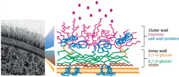
Figure 1.2 - A section of the C. albicans cell wall by transmission electron microscopy and scheme about the arrangement of the major components of cell wall (19).
In fact, the cell wall of Candida is composed by diverse components: lipids, proteins and carbohydrates (chitin, glucans and mannan). Lipids are the minor component of the cell wall of fungi in general, including C. albicans. Phospholipomannan is perhaps the most important lipid because it reacts with antibodies specific to ß-1,2-oligomannosides. Furthermore, it is also possible to find proteins in the cell wall that assume important and varied roles, such as the degradation of large impermeable molecules facilitating the internalization of nutrients, and the involvement in the establishment of the pathogens in the host. Some of these proteins are covalently associated to mannose polymers (mannan), forming mannoproteins that constitute the fibrillar outer layer of the cell wall. Mannan is an interesting component that confers immunodominant properties to the cell and promotes the adherence of the fungus to epithelial cells. Regarding chitin, this is one of the most insoluble natural products which, in conjunction with ß-1,3-glucans, belongs to the mannoprotein outer layer. Glucans are the most abundant polysaccharides present in the cell wall of Candida, which does not have α-glucans, but only ß-glucans (ß-1,3 and ß-1,6 glucans, for example) (19, 20).
(3.3-3.7%) and mannose (24.3-28.9%), and a larger amount of chitin (4.1-4.3%) and glucans (10-11.2% [ß-1,3]; 25.1-27.3% [ß-1,6]) than C. glabrata (21).
The different Candida species can be easily distinguished by other factors: colony color on chromogenic agar medium (CHROMagar), yeast size and biochemical and genetic characteristics (5). On CHROMagar (Figure 1.3), C. albicans presents blue-green colonies, C. tropicalis dark-blue colonies, C. glabrata white or pink-purple colonies, whereas C. parapsilosis has white colonies (13, 22). Regarding yeasts’ size, and according to Calderone (13), C. albicans cells (4-6 × 6-10 µm) are bigger than cells of C. tropicalis (4-8 × 5-11 µm), C. parapsilosis (2.5-4.0 × 2.5-9.0 µm) and C. glabrata (1-4 µm). In this sequence, C. glabrata cells are the smallest in comparison with the other species already mentioned (12).
Figure 1.3 - Colonies of Candida species grown for 48 h on CHROMagar at 37° C: (a) C. albicans; (b) C. glabrata; (c) C. parapsilosis and (d) C. tropicalis. Adapted from (22).
In relation to biochemical characteristics of these species, it is important to refer that C. albicans has the ability to ferment or assimilate a large group of sugars with the exception of sucrose, while the NCAC species ferment and assimilate specific sugars: glucose and trehalose, in the case of C. glabrata, and sucrose and maltose, in the case of C. tropicalis, for example. It is also described that C. parapsilosis does not have the ability to ferment maltose (5).
From the genetic point of view, there are also differences between some NCAC species and C. albicans. In fact, C. glabrata and C. lusitaniae present a haploid genome, while C. albicans and
(a) (b)
several NCAC species (for example, C. dubliniensis, C. tropicalis and C. parapsilosis) have a diploid genome. Moreover, it is also referenced that C. tropicalis presents a higher similarity to C. albicans and a lower one to C. glabrata (5, 23).
1.2.3. Pathogenicity and virulence factors
Virulence factors are all those factors associated to metabolic pathways and that directly interact with the host tissue causing damages and infection: the ability to adhere and form biofilm, the secretion of hydrolytic enzymes (e.g. proteases, phospholipases and haemolysins) and the ability to produce filamentous forms that facilitate the invasion of the host tissue, for example (24, 25). Then, the pathogenicity will depend on the several of virulence factors that the microorganism has (5).
Investigations about virulence factors of Candida species have shown that Candida albicans is the most pathogenic organism in the Candida group (26, 27) and that it has the highest levels of virulence factors (8).
1.2.3.1. Adhesion
The adhesion of a microorganism to a host tissue and/or medical device surface is the first event in Candida infection, giving origin to biofilm formation (10). Adhesion is an essential phenomenon because it contributes to the persistence of the pathogen in the host and to the establishment of infection (5). However, the adhesion is dependent on several factors such as the cell wall proteins, that are recognized by the receptors present on epithelial, endothelial and foreign-body surfaces (for example, fibronectin, fibrinogen and vitronectin), and cell surface physicochemical properties (28).
In the Candida species, there are specialized cell wall proteins in the adhesion process – the adhesins. Fungal adhesins (Figure 1.4) present a common three-domain structure: the C-terminal part (composed by a glycosylphosphatidylinositol (GPI)-anchor and by addition links to the cell wall), the N-terminal part (composed by a carbohydrate or peptide binding domain) and the large middle domain (that contains multiple serine- and threonine-rich repeats). These glycoproteins promote the interactions of the yeast cells with each other, mammalian host tissues and inert surfaces, mainly in biofilm formation (29, 30).
albicans have been reported (such as EAP1, INT1 and HWP1 genes) (29, 31). In relation to adhesins of C. glabrata, they are encoded by the EPA (epithelial adhesin) genes, where their expression is induced by the presence of nicotinic acid (10, 32). There are few studies about Epa proteins, but it is already known that Epa1p is a calcium-dependent lectin, whose deletion of EPA1 alone reduces adherence in vitro and that EPA6 expression does not happen in vitro, but in vivo during urinary infection (10, 33). About the adhesins of other Candida species, there is still little information (5, 10).
Figure 1.4 - Representation of interaction between yeast cells mediated by fungal adhesins. Generally, in this process, active confirmation of the adhesins is achieved by action of calcium ions (30).
Concerning cell surface physicochemical properties, it is necessary to refer cell surface hydrophobicity. Some studies have reported that there is a correlation between adhesion and surface hydrophobicity of Candida cells, but there are other studies that show the opposite (5). In 2001, Panagoda et al. (34) verified the existence of an association between the initial adhesion of C. parapsilosis cells and the hydrophobicity of acrylic surfaces. Nevertheless, in 2007, Camacho et al. (35) demonstrated that there was no correlation between adhesion of Candida species to siliconized latex catheters and the cell hydrophobicity alone. Another study about adhesion of NCAC species to silicone in the presence of urine, developed by Silva et al. (36), also showed that C. glabrata, C. parapsilosis and C. tropicalis were hydrophilic, a fact that did not influence the adhesion of these strains. They even concluded that zeta potential was not primarily responsible for the adhesion. Furthermore, some authors have considered that the extent of surface roughness is an important factor in the adhesion of microorganism to surfaces (36).
1.2.3.2. Biofilm formation
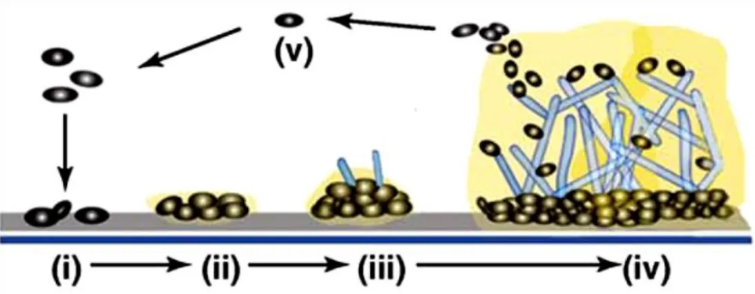
Biofilm is the most prevalent growth form of microorganisms and is defined as a community of cells attached to a surface and embedded in an extracellular matrix (ECM), that is composed by substances produced by microbial cells (37, 38). It is a severe virulence factor of Candida species, because it presents a complex and heterogeneous three-dimensional structure that prevents the penetration of substances/drugs, which confers antifungal resistance, and attenuates immune responses from host beneath cells contained in matrix. This architecture also promotes the influx of nutrients, the secretion of waste products and the establishment of microniches, allowing the maturation of biofilms (10, 39).
The formation of these biologic structures (Figure 1.5) can be explained in five stages. Initially, yeast cells are adsorbed (i) and adhere (ii) to the surface followed by the formation of the basal layers of yeast colonies, the initial formation of hyphae (in the cases where Candida species have this ability) and the production of ECM surrounding the cells and hyphae (iii). Finally, it occurs the maturation of the biofilm, that results in the association of several microcolonies and emergence of water channels that allow circulation of nutrients and other substances (iv), followed by the dispersal of cells biofilm (v) (37). However, the formation and the architecture of mature biofilms depend on the species, strain, substrate properties (such as medium composition) and growth conditions (for example, pH and oxygen) (10, 39).
Figure 1.5 - Model of biofilm development in Candida albicans. Adapted from (37).
There are some differences between the biofilms produced by Candida albicans and by NCAC species (39). Due to the dimorphism of C. albicans (ability to grow in the yeast form and in the hyphae form), its biofilms are distinguished by the presence of two distinct layers (Figure 1.5 (iv)): thin (basal yeast layer) and thicker (less compact hyphal layer) (10, 40). It is important to highlight
ECM composition of C. albicans biofilms, Al-Fattani et al. (38) showed that this contains a high level of carbohydrates (39.6%, including 32.2% glucose) and a low level of proteins (5.0%), hexosamines (3.3%), phosphorus (0.5%) and uronic acid (0.1%).
Concerning NCAC species biofilms, there are also differences between C. glabrata, C. parapsilosis and C. tropicalis, as it can be observed in Table 1.3 (10). In Sabouraud dextrose broth (SDB) or another rich culture medium, C. glabrata forms lower biofilms, compared to C. parapsilosis and C. tropicalis, but its biofilm structure is more compact (43). As for C. parapsilosis, its biofilm structure is thinner, less organized and composed essentially by aggregated blastopores. C. parapsilosis has still a greater ability to produce biofilm on materials with plastic surface, such as catheters (10). Finally, C. tropicalis is described as a prolific biofilm producer and it has ability to produce extensive biofilms in polyvinyl chloride (PVC) catheter and polystyrene surfaces. A dense network of yeast cells and hyphae and/or pseudohyphae characterizes its biofilm structure (10, 44).
Table 1.3 - Characteristics of C. glabrata, C. parapsilosis and C. tropicalis biofilm (10)
Species Biofilm formation Biofilm structure Matrix composition
C. glabrata Less biofilm growth Compact monolayer or multilayer
High level of carbohydrates and proteins
C. parapsilosis Robust biofilm growth Discontinuous monolayer or multilayer
High level of carbohydrates and low levels of proteins
C. tropicalis Robust biofilm growth Discontinuous compact monolayer
Low level of carbohydrates and proteins
The ECM composition of NCAC species biofilms also varies. C. glabrata presents a biofilm matrix composed by high quantities of carbohydrates and proteins, while the biofilm matrix of C. parapsilosis has high levels of carbohydrates, but low amounts of proteins. Moreover, biofilm matrix of C. tropicalis contains high levels of hexosamine – the major component –, low quantities of carbohydrates and proteins, phosphorus and uronic acid (10, 38).
1.2.3.3. Enzymes production
Candida species produces and release hydrolytic enzymes to the environment, such as secreted aspartyl proteinases (Saps), phospholipases, and haemolysins. These enzymes promote the destruction of host tissues, constituting important virulence factors (5).
Secreted aspartyl proteinases
Saps are enzymes that facilitate invasion of host tissues through the degradation of mucosal components (collagen, keratin and mucin) and important immune proteins (antibodies, complements and cytokines). Studies have demonstrated that C. albicans, C. parapsilosis and C. tropicalis are good producers of Saps (5, 44-46). In C. albicans, ten SAP genes, which act in the adhesion (of this species), tissue damage and immune responses evasion of host, encode Saps. It is also necessary to refer that there are SAP genes of C. albicans more specific for mucosal (SAP1-3) and systemic (SAP4-6) infections and that proteinases activity varies with pH (45). In relation to C. parapsilosis, three genes have been identified (SAPP1-3), but two of these genes are not characterized. In addition, C. tropicalis Saps are encoded by four genes (SAPT1-4), but only Sapt1p is duly characterized. About C. glabrata, there is only a study that indicates that this species produces proteinase, but the proteinase type is unknown (5).
Phospholipases
Phospholipases hydrolyse phospholipids into fatty acids and cause host cell membrane damage, facilitating the invasion of host tissues by exposition of receptors to adhesion. The production of four types of phospholipases by C. albicans has been reported (phospholipase A, B, C and D). Other studies concerning C. albicans phospholipases allowed the detection of the presence of extracellular products related only to PLB1 and PLB2 genes. The PLB1 has been indicated as the gene that has an important role in the secretion of phospholipase B by C. albicans (45).
According to several studies, NCAC species also produce extracellular phospholipases, but in lower quantities than C. albicans (5). However, C. tropicalis has been indicated as the producer of low levels of extracellular phospholipases (44). Besides, there are few studies about the production of these enzymes by C. tropicalis and C. parapsilosis and there are not any investigations concerning C. glabrata phospholipase production (5).
Haemolysins
Haemolysins are responsible for the degradation of haemoglobin and by extracting the elemental iron from host cells. It is through haemoglobin degradation that microorganisms (such as Candida species) develop growing in host tissues, because they use this compound as a source of iron (5, 47).
Several authors have reported that C. albicans and some NCAC species produce haemolysins after 48 hours of incubation (44, 48, 49). It is known that C. albicans, through iron, produces a haemolytic factor that allows the release of haemoglobins by erythrocytes lyses. However, the production of haemolysins in vitro by C. glabrata, C. parapsilosis and C. tropicalis causes partial or total erythrocyte lyses. Moreover, the HLP (haemolysin like protein) gene has also been indicated as responsible for the haemolytic activity of C. glabrata (5, 47).
1.2.3.4. Filamentous growth
As already mentioned in section 1.2.2., there are some Candida species that produce hyphae and/or pseudohyphae. These biologic structures facilitate the invasion of pathogenic microorganism in healthy tissue of host and increase resistance to phagocytosis (5).
In 2006, Jayatilake et al. (8) has shown that C. albicans with no hyphae presented a lower ability to invade tissue compared to other Candida species with which presented hyphae. In addition, studies developed by Silva et al. (50, 51), about the influence of filamentous forms on host tissue invasion, indicated interesting results. They revealed that the filamentous forms of C. tropicalis were able to invade a human oral epithelium (Figure 1.6) (50) and that the invasion of C. parapsilosis in this same tissue is not related to pseudohyphae formation, contrary to what happens with C. albicans and C. tropicalis (51). Relatively to C. glabrata, it has been described as non-invasive in studies of in vitro invasion by Candida species, where reconstituted human epithelium was used. This situation has been explained by the inability of C. glabrata to generate filamentous forms (10).
Figure 1.6 - Confocal laser scanning microscopy (CLSM) image that shows the invasion of C. tropicalis 75 in human oral epithelium, after 24 h of incubation. Adapted from (50).
1.3.
Candida bracarensis
Candida bracarensis is a rare Candida species, which was discovered recently (1, 4).
1.3.1. Discovery
In 2004, following an epidemiological study of candidiasis in the north of Portugal, one strain of Candida (153 MT) was collected in one of the clinical isolates from a vaginal exudate. This strain
was initially identified as C. glabrata, but later, some studies showed differences between the strain 153 MT and C. glabrata (4).
Moreover, a second strain (NCYC 3133, formerly NCYC D3411) was found in a search for similar sequences in the GenBank sequence database. This strain, collected from a blood culture from a patient in a United Kingdom hospital, presented a high sequence similarity with the 26S rDNA D1/D2 region of 153 MT (only a single nucleotide was different) (4).
Strains 153 MT and NCYC 3133 were then analysed by standard chemotaxonomic methods
and by polymerase chain reaction (PCR) fingerprinting with primer T3B, with the purpose of clarifying the taxonomic position of new Candida species. Throughout this analysis, it was verified that the two isolates presented similar physiological and molecular characteristics with C. glabrata and Kluveromyces delphensis (at the present Nakaseomyces delphensis). Though, the level of differences between the two isolates and these two last microorganisms was considered large enough for the emergence of a new Candida species – Candida bracarensis. Out of curiosity, the choice of the species name comes from Bracara Augusta, the roman name of Braga (Portuguese city) where strain 153 MT was isolated (4).
In the sequence of this discovery, new cases of C. bracarensis have been reported. Some authors, such as Bishop et al. (52) and Warren et al. (53), have considered the strain 153 MT as
control in the identification of new cases of C. bracarensis.
1.3.1.1. Cases reported to date
To author’s best knowledge, there are only few cases of Candida bracarensis reported. In the Table 1.4, it is possible to see information about these cases.
Table 1.4 - Cases reported of Candida bracarensis
Publication
Year Isolate [References] Source Region/Country
2006 153 MT * (4, 54) Vaginal exudates from patient with
candidiasis Braga, Portugal 2006 NCYC 3133** (4, 55) Blood culture from patient central
venous catheter United Kingdom 2008 C. bracarensis HOP-15*** (52) Stool from patient with AIDS USA 2008 Cagl-78 (52) Stool from oncology patient (with
acute myelogenous leukaemia) USA 2008 Cagl-112 (52) Pelvic abscess from patient with
perforated diverticulitis USA 2008 Cagl-121 (52) Throat from oncology patient (with
anaplastic large-T-cell lymphoma) USA 2008 CNM-CL-7030 (56) Catheter exudate of a critically ill
patient Spain
2009 C. bracarensis no. 1 (57) Sputum USA
2009 C. bracarensis no. 2 (57) Blood culture USA
2009 CNM-CL-7326 (56) Pleural liquid from a patient who
underwent thoracic surgery Spain 2009 CNM-CL-7380 (56) Blood culture from a patient with a
haematological disease Spain 2010 Clinical isolate 1 (53) Blood culture from
immunocompromised patient Canada Clinical isolate 2 (53)
2010 Two isolates (58, 59) Various clinical samples India
* Equivalent strain designations: NCYC 3397, NCYC D3853T, CECT 12000T, CBS 10154T, NRRL Y-48270T, C. bracarensis type strain (TS).
** Equivalent strain designation: NCYC D3411. *** Equivalent strain designation: NRRL Y-27794.
As for the isolates mentioned in the Table 1.4, it is necessary to refer that the isolates Cagl-78, Cagl-112 and Cagl-121 were identified by peptide nucleic acid fluorescence in situ hybridization probes and were then confirmed by sequencing of the D1/D2 region (52). In addition, the two isolates from Canada were not initially identified as C. glabrata by the API 20C system, unlike what happened with other studies. This fact suggests that C. bracarensis has been misidentified as C. glabrata (53). Today, commercial identification kits can already easily distinguish these two Candida species such as the Glabrata RTT test (Fumouze Diagnostics, Levallois Perret, France) (1).
1.3.2. Characteristics
Candida bracarensis is a pathogenic and anamorphic yeast (1). The cells of this species have a spherical shape (3.0–3.5 µm) or an elliptical one (3.0-4.0 x 4.0-4.5 µm). In Figure 1.7, it is possible
to see that the cells in this Candida species may appear singly or grouped. Still in its morphology, it was not detected the production of pseudohyphae and true hyphae, under the coverglass in Dalmau plate culture on cornmeal agar after 21 days at 25°C, nor the asexual (ballistospores, arthrospores, endospores, chlamydospores) and sexual spores (ascospores and teliospores) (4, 54, 55). These fungal cells also divide by budding, which is multipolar, and not by fission (54, 55).
Figure 1.7 - Morphology of C. bracarensis 153 MT cells grown in YM broth after 3 days at 30°C (Bar, 10 µm). This image was obtained by differential
interference contrast microscopic (4).
On Yeast malt (YM) agar, C. bracarensis produces cream/pale yellow, shiny, smooth and butyrous colonies with an entire margin, whereas on CHROMagar Candida medium (at 37°C), these colonies are white (4, 52, 53, 60). Furthermore, in broth medium, this Candida species is not flocculent (54, 55).
As for its fermentation process, this yeast has the ability to ferment glucose and trehalose. For this reason, C. bracarensis assimilates some carbohydrates (glucose, sucrose and α,α-trehalose), glycerol, D-glucono-1,5-lactone and D-gluconate. As a nitrogen source, this Candida specie only assimilates the L-lysine (4, 54, 55).
1.3.3.
Candida bracarensis
versus other species
As previously mentioned in point 1.3.1., strains 153 MT and NCYC 3133 were characterized
by Correia et al. (4) in order to clarify its taxonomic position. The study revealed similarities of these two isolates with C. glabrata and K. delphensis, similarities that we will now see.
In the PCR fingerprinting with primer T3B, it was detected (Figure 1.8 (a)) that the strains 153 MT and NCYC 3133 presented similar profiles with one another, but different profiles with C.
glabrata and K. delphensis (4).
Through the neighbour-joining method, based in the alignment of 26S rDNA D1/D2 region sequences, and PAUP* version 4.0b8 software package, it was possible to build the phylogenetic tree represented in Figure 1.8 (b). The sequence similarity values were also estimated and it was verified that between C. glabrata and K. delphensis, there are 92% of similarities and that strain 153 MT is
94.8% similar to C. glabrata and 93.8% with K. delphensis. However, this phylogenetic analysis was also enlarged to the comparison between entire ribosomal internal transcribed spacer (ITS) region sequences. In this case, strain 153 MT revealed 65% similarities with C. glabrata, 68% with K.
delphensis and 96.9% with NCYC 3133 (4).
(a) (b)
Figure 1.8 - (a) PCR profiles (with primer T3B) obtained to C. glabrata CBS 138T (1); Kluyveromyces delphensis PYCC 2899 (2); strain 153 MT (3) and strain
NCYC 3133 (4). (b) Phylogenetic tree derived from the alignment of 26S rDNA D1/D2 region sequences. It is represented only bootsrap percentages (1000 replicates) of 50% or greater. Bar, 1% nucleotide sequence divergence (4).
Another factor that distinguishes C. bracarensis from C. glabrata and K. delphensis is compounds assimilation. While C. bracarensis assimilates L-lysine as a sole nitrogen source, C. glabrata does not utilize this compound or utilizes it in small amounts. Concerning the K. delphensis, the distinction with C. bracarensis is also easy because strains 153 MT and NCYC 3133 assimilate
1.4. Antifungal resistance
As already mentioned, infections by Candida species have increased significantly, causing high morbidity and mortality in immunocompromised patients. In the origin of this phenomenon is the increase of antifungal resistance (3, 61, 62). Several factors can explain the former situation: the increase of intrinsically resistant species, the accumulation of mutations that cause resistance, the swap of mobile resistance components and the excessive use of drugs to treat infections. So, the understanding of mechanisms associated to antifungal resistance is indispensable to counter actual trend (6).
Antifungal resistance can be characterized by microbiologic or clinical resistance, or by both. Concerning microbiologic resistance, this happens when the growth of the pathogen is inhibited by a higher dosage of antifungal concentration connected/related to the susceptibility breakpoint for that same microorganism (61, 63). This resistance can be intrinsic (primary) or acquired (secondary). The first is found in fungi without prior exposure to the antifungal agent, while acquired resistance occurs in the strains, previously classified as susceptible, after exposure to the drug. Generally, in this last case, the resistance appears associated to altered gene expression (61).
When there is a failure in the administration of an antifungal agent (with in vitro activity against the pathogen), this resistance is considered of clinical origin. This failure is due to a combination of several factors originating from the administered drug, host and type of pathogen (61).
Resistance is only classified as microbiologic and clinical when normal concentrations of antifungal do not inhibit the pathogens and when the minimum inhibitory concentrations (MICs) are situated in an interval propitious to the development of the resistance mechanisms or where there are not studies about the clinical success of antifungal agent against the respective microorganism (63).
Regarding the consequences of antifungal resistance, it is noteworthy the economic problems and the elevated MICs, associated to the increase of invasive fungal infections in high-risk patients during the antifungal therapy and prophylaxis (7, 63). It is also important to refer that the level of severity of consequences depends on the biology and population size of the pathogen, the antifungal properties and the resistance mechanisms acquired by the respective strain (6).
1.4.1. Antifungal susceptibility testing
susceptibility testing of yeasts and filamentous fungi, and by the European Committee on Antimicrobial Susceptibility Testing (EUCAST), for testing Candida and Aspergillus species (61).
The CLSI, apart from the broth microdilution (BMD) testing, has presented other MIC methods for antifungal susceptibility testing such as disk diffusion testing for yeasts. This last method is more convenient, simpler, more economical and more suited for water-soluble antifungals (such as fluconazole and voriconazole) than the BMD (63).
Before proceeding, it is important to clarify the MIC concept: it is the lowest drug concentration for which it is verified a significant reduction (50% or 90%) of growth of a microorganism. This value is quantified from the measuring of a strain growth in contact with a range of drug concentrations, during a period time and according to a specific standard (64).
However, MICs values do not always reflect the clinical response to antifungal therapy, because there are always complex interactions between the pathogen and host that are unquantifiable (64). In fact, in 90% of cases, the antifungal therapy is successful against infections caused by susceptible strains, while the therapy used to treat infections caused by resistant strains, it has only success in 60% of cases. Furthermore, MICs are not always considered the best measure of resistance because, in fungal infections exposed to the antifungal agent, it has been detected some activity against conidia and not against filamentous structures such as hyphae (61).
1.4.2. Resistance mechanisms to antifungal agents
The main antifungal agents currently used are polyenes (ergosterol disruptors), azoles (fungal ergosterol synthesis inhibitors), flucytosine (nucleic acid synthesis inhibitor) and echinocandins (glucan synthesis inhibitors). These antifungals are classified in function of their targets in the antifungal therapy (Figure 1.9) (65).
Figure 1.9 - Targets for antifungal therapy. Adapted from (66).
1.4.2.1. Polyenes
Polyenes are fungicidal agents and have the broadest spectrum of action against fungi species. These agents act within fungal membrane where, in conjunction with ergosterol (the main sterol present in the fungal cell membranes), form porin channels. In turn, these channels increase membrane permeability, leading to leakage of cellular constituents and, consequently, loss of cellular function and cell death (61, 65, 67).
Nystatin and amphotericin B (Figure 1.10) are examples of polyenes isolated from Streptomyces species. The first drug, discovered in 1950, is recommended to the treatment of oropharyngeal candidiasis, despite not being absorbable after oral administration. In what concerns amphotericin B, this antifungal is used in cases of serious and invasive Candida infections and against Blastomyces dermatitidis, Coccidioides immitis, Cryptococcus neoformans, Histoplasma capsulatum, Paracoccidiodes brasiliensis and Sporotrichium species (5, 65). However, amphotericin B has some disadvantages such as toxicity to mammalian cells, since it causes nephrotoxicity, and low blood potassium levels (65, 68).
Nevertheless, recently, some resistance to amphotericin B has been described. The resistance to this polyene is detected for a MIC superior or equal to 1 µg/ml. Various causes can explain the resistance to polyenes, namely, mutation in the ERG3 (the gene responsible for ergosterol biosynthesis), which leads to formation and to accumulation of other sterols in the plasmatic membrane of the fungal cell; and mutation in ERG11 and in ERG6 (genes that produce lanosterol 14α-demethylase and that intervene in normal membrane function, respectively) (5, 61, 69, 70). Either the increase catalase activity or the decrease of susceptibility to oxidative damage also contribute to resistance to amphotericin B (61).
1.4.2.2. Azoles
Azoles constitute the largest group of the antifungals class used clinically and, generally, present a fungistatic activity against Candida species (5, 71). The main function of these agents is to block the ergosterol biosynthesis pathway, through the inhibition of lanosterol 14α-demethylase (an enzyme responsible for converting lanosterol to ergosterol) and, in some fungi species, of the inhibition of ∆22-desaturase step (68, 69). In this sequence, ergosterol is depleted and the fungal membrane structure undergoes some changes, in terms of permeability and fluidity, as well as its functions (for example, the non-production of the enzymes necessary for cell wall synthesis). Thus, all these phenomena lead to the inhibition of fungal growth (63, 68).
Fluconazole (Figure 1.11) and itraconazole are the main azoles clinically used, but there are others such as voriconazole and posoconazole, for example. The fluconazole is a drug that emerged in 1994, to treat vaginal candidiasis, and currently, it is extensively used in the treatment of superficial and invasive infections caused by various Candida species, such as C. albicans, C. parapsilosis and C. tropicalis. Itraconazole is another azole that has anti-Aspergillus activity and that can be administered in patients infected with Candida species susceptible or resistant to fluconazole, except for C. glabrata. However, fluconazole is better than itraconazole, specifically in the treatment of acute cryptococcal infection and cryptococcal meningitis, in patients with Acquired Immune Deficiency Syndrome (AIDS). In relation to voriconazole, it has fungicidal activity against Candida (including C. albicans and C. glabrata), Aspergillus and Fusarium species. The patients treated with voriconazole can present some secondary effects such as hallucinations, encephalopathy, skin rash among others. Finally, there is posoconazole, which emerged in 2006, to treat invasive Aspergillus and Candida infections. Posoconazole presents fungistatic activity mainly against some NCAC species
and a molecular structure similar to itraconazole. The main secondary effects of this last antifungal are gastrointestinal (5, 65).
Figure 1.11 - Chemical structure of fluconazole. Adapted from (68).
Concerning resistance to azoles, there are four main mechanisms described to Candida species. In each resistant strain, it is possible to find more than one active mechanism. The first is associated with activation of two types of efflux pumps, encoded by either CDR or MDR genes (two genes that belong to gene families of transporters). This activation leads to decreased antifungal concentration in the site of action, within the fungal cell, therefore causing the resistance. Studies have proven the involvement of the CDR or MDR genes in resistance of C. albicans (CDR1, CDR2, and MDR1), C. glabrata (CgCDR1 and CgCDR2) and C. dubliniensis (CdCDR1 and CdMDR1) to azoles. The second mechanism involves point mutations in ERG11 (gene that encodes for the target enzyme), which decreases the binding capacity between azoles and target enzymes. However, this situation can be aggravated if there is an overexpression and upregulation of the altered target enzymes – the third mechanism. In this case, the azole agents are overwhelmed and, subsequently, the therapeutic drug concentrations are insufficient to treat the infected patient. Nevertheless, it is important to highlight that this mechanism is not yet considered the most responsible for resistance to azoles. Finally, the fourth mechanism is related to the development of bypass pathways, caused by mutations in ERG3 gene. This altered gene prevents the formation of 14α-methyl-3,6-diol, a toxic component necessary to death of the cell, and, subsequently, it originates functional membranes, denies the effects of azoles and inhibits the fungal cells growth (61, 63, 69).
1.4.2.3. 5-Flucytosine
5-Flucytosine is an antifungal agent that inhibits DNA and protein synthesis. This drug, initially developed as an anticancer agent, is internalised by cytosine permease and, once inside the
acid, by UMP pyrophosphorylase. Afterwards, 5-fluorouridylic acid is phosphorylated and incorporated into RNA molecules, affecting both the DNA and protein synthesis (5, 65).
The spectrum of activity of flucytosine (Figure 1.12) is restricted once that, currently, it is used to treat infections caused by Candida species, Cryptococcus species and Aspergillus species (5).
Figure 1.12 - Chemical structure of flucytosine. Adapted from (64).
Furthermore, the prevalence of primary resistance to this antifungal is low because it is not used as first option in clinic. Though, resistance mechanisms to flucytosine are associated above all to mutations in cytosine permease, cytosine deaminase or in UMP pyrophosphorylase (61). These mutations interfere in intracellular enzymatic steps of action mechanism of 5-flucytosine, causing resistance. The increase in the production of pyrimidines can also contribute to resistance to this agent (5).
1.4.2.4. Echinocandins
Echinocandins (Figure 1.13) is another class of antifungal agents. These are responsible for the inhibition of the enzyme ß-1,3-D-glucan synthase, that in turn leads to the inhibition of the biosynthesis of ß-1,3-D-glucan, an important constituent of the fungal cell wall (63). Consequently, the disruption of cell wall structure occurs, which results in the osmotic instability and cell death. The toxicity of these antifungal is low because their target is not present in the mammalian cells (65). There are three types of echinocandins: caspofungin, micafungin and anidulafungin. They have been used to treat infections caused by Candida species, including C. albicans, C. glabrata and C. tropicalis (5).
Nevertheless, C. albicans and NCAC species have revealed some resistance to echinocandins. This resistance is induced by mutations in the two important regions of FKS1 (HS1 and HS2), the gene responsible for the encoding of the major and presumed catalytic subunit of ß-1,3-D-glucan synthase complex. It is important to refer that, into C. glabrata, the resistance can also
Figure 1.13 - General structure for echinocandins (65).
1.4.3. Resistance mechanisms of fungal biofilms
The first study about the resistance of Candida biofilms to antifungal agents was published in 1995 (72). Since then, several authors have presented studies about fungal biofilms resistance mechanisms, but some of these are not yet fully understood (5).
Biofilms exhibit greater antifungal resistance in comparison with planktonic cells (circa 10-1000 times more) (16). While in planktonic cells, the resistance is due mainly to genetic mutations, as already mentioned above, in biofilms, the resistance results from the physical structure and density of population, for example, regardless of the existence of changes in the genes (71).
Thus, antifungal resistance in biofilms is a complex and multifactorial mechanism. Factors as physiological state, cell density, overexpression of drug targets, efflux pumps, ECM, persister cells and stress, can lead to resistance into biofilms (Figure 1.14) (71).