Universidade de Lisboa
Faculdade de Ciências
Departamento de Química e Bioquímica
DEVELOPMENT OF NANOMATERIALS FOR CULTURAL HERITAGE
CONSERVATION. EVALUATION ON THE DEGRADATION STATE OF ANTIQUE
COINS BY SPECTROSCOPIC TECHNIQUES
Maria Inês Baião Ramos de Oliveira
Dissertação
Mestrado em Química
Especialização em Química-Analítica
Tese orientada por:
Professora Doutora Maria da Estrela Borges de Melo Jorge
Professora Doutora Maria Luisa de Carvalho Dias de Sousa Leonardo
i | P a g e
A
CKNOWLEDGMENTS
First of all I would like to express my sincere gratitude to my supervisors Professors Maria Estrela Jorge (Centro de Química e Bioquimica, Faculdade de Ciências da Universidade de Lisboa (CQB-FCUL)) and Maria Luísa Carvalho (Centro de Física Atómica, Universidade de Lisboa (UL) and Faculdade de Ciências e Tecnologias da Univerisade Nova de Lisboa (FCT-UNL)) for the guidance, learning opportunities and the friendship.
To Drs. Sofia Pessanha and Marta Manso thank you so much for your generous help and for providing an amazing working environment.
I would like to thank to Mário Costa for providing the working material and for the numismatic support.
I also offer my appreciation to my Italian supervisors Professors Piero Baglioni and Rodorico Giorgi for the opportunity of working in the Consorzio Interuniversitario per lo Sviluppo dei Sistemi a Grande Interfase (Università degli Studi di Firenze (CSGI-UniFi)). I would like to thank Professor Giorgi for his, support and understanding.
I also would like to thank to Drs David Chelazzi and to Giovanna Poggi for their time and assistance.
I have learnt a great deal from those whom I had the pleasure of working with either in Portugal or in Italy and, for sure, I will take all off you in my heart and memory.
i i i | P a g e
A
BSTRACT
This work is presented in two complementary parts covering the two main fields on the study of cultural heritage.
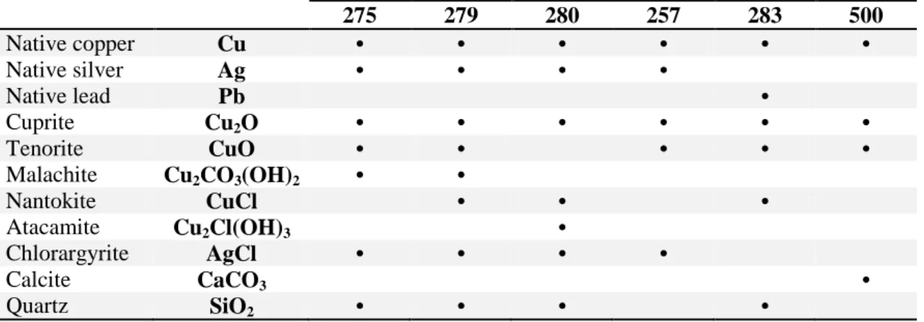
The first one is focussed on the analysis of a set of six dinheiros from the first Portuguese dynasty (13rd and 14th centuries). The coins were analyzed using μ-Energy Dispersive X-ray Fluorescence in order to evaluate their elemental composition, allowing to confirm in four of the coins a billon alloy (copper/silver) and in the remaining two a reduced content of silver. This fact is compatible with the social and economic Portuguese reality during D. Fernando I reign. The coins were further evaluated by means of X-ray Diffraction identifying natives copper and silver, as well as some of their degradation products, such as cuprite, tenorite, malachite, nantokite and atacamite and on the numisms with high silver content, chlorargyrite. By making use of Scanning Electron Microscopy/Energy Dispersive Spectroscopy the morphological analysis on the coins surface was performed evidencing the corroded surface and allowing to identify different morphologies corresponding to copper/copper oxides and silver/silver chloride phases.
On the second part, several calcium carbonate (CaCO3) and calcium hydroxide functionalized nanoparticles were synthesized, purified and physicochemical characterized, in order to study their application by dispersion, on manuscripts and its similar for conservation purposes. The effect of different percentages of the functionalizing agent (dodecanoic acid) was investigated. Results from the visual and turbidimetry analyses showed that the dispersions were not stable for the proposed end. By making use of Attenuated Total Reflectance and X-ray Diffraction was possible to conclude that the acid is present on the final CaCO3 product. Scanning Electron Microscopy allowed to prove the morphological changes on the powders particles and made clear the shortage of nano-objects. The followed route is not effective regarding the synthesis of nanoparticles which may justify the instability of the dispersions.
Key words: corrosion, coins, nanoparticles, XRF, XRD, SEM/EDS, turbidimetry,
v | P a g e
R
ESUMO
Este trabalho é composto por duas partes distintas mas complementares e que diferem apenas na base em estudo. Abrangem-se assim dois dos campos fundamentais no estudo do património cultural, nomeadamente, a avaliação do estado de conservação de um conjunto de moedas antigas, seguido pela proposta de um método para a preservação do papel e seus similares.
A primeira parte da investigação foca-se na análise de um conjunto de seis dinheiros datados da primeira dinastia Portuguesa (séculos XIII e XIV), particularmente dos reinados de D. Sancho II (1223-1248); D. Afonso III (1248-1279); D. Dinis (1279-1325); D. Pedro I (1357-1367) e D. Fernando I (1367-1383). Este trabalho baseia-se na avaliação do estado de conservação dos numismas, por meio da identificação e caracterização dos produtos que constituem a sua patina bem como as alterações morfológicas a que a sua superfície esteve sujeita devido à sua interação com o ambiente circundante. Tenta-se ainda propor um mecanismo de corrosão porém devido à falta de informação sobre o local da descoberta, o estado do enterro/desenterro ou mesmo das datas relacionadas com qualquer um destes processos, esta investigação é um desafio tanto a nível do estudo da sua camada de corrosão como a nível do património cultural.
De salientar que os dinheiros foram as primeiras moedas portuguesas, cunhadas em liga de bolhão (maioritariamente constituída por cobre, seguido de prata e alguns elementos químicos minoritários). Estas moedas estiveram em circulação, em Portugal, desde meados do século XII até, aproximadamente, 1502. Mais ainda, patina é a camada que reveste a superfície do metal, no seu estado puro ou em liga, sendo constituída por todos os compostos químicos, nomeadamente produtos de corrosão e outros elementos exógenos formados e mantidos na superfície do objeto, alterando a sua cor e textura. Urge considerar o sistema liga/patina/ambiente como um sistema global, em que a patina resulta das interações físico-químicas entre a liga e o ambiente, ao longo do tempo.
A ação combinada de técnicas analíticas não destrutivas como a espectrometria de micro-Fluorescência de raios-X de Energia Dispersiva (μ-EDXRF), a Difração de raios-X (XRD) e a Microscopia Eletrónica de Varrimento acoplada com a técnica de Espectroscopia Dispersiva de raios-X (SEM/EDS) permitem identificar a natureza dos produtos da patina formados no conjunto de moedas estudadas, contribuindo para compreender e preservar a história das sociedades e ajudar à preservação dos mesmos. Do ponto de vista macroscópico e por meio de análise visual pôde concluir-se que, de um modo geral, as superfícies das moedas se encontravam bem definidas e preservadas, dado o seu período de origem. Todas as moedas apresentavam uma coloração preta-acastanhada e, em alguns casos, uma coloração acobreada nas zonas de alto-relevo. Exceção feita à face de uma das moedas que apresentou, pontualmente, sinais típicos de corrosão do tipo “couve-flor”. Vários tipos de patinas foram identificados, a saber uma camada amarelo-acastanhado nas áreas de baixo-relevo e, localmente, colorações verde (baixo-relevo) e vermelho-acastanhado (cobrindo a superfície) em todas e/ou alguns dos numismas.
As moedas foram analisadas usando μ-EDXRF de modo a quantificar a sua composição elementar. Com recurso a esta técnica foi possível proceder à confirmação da liga de bolhão em todas as moedas notando-se porém uma redução do teor de prata
v i | P a g e
nas duas moedas datadas do reinado de D. Fernando I. Este facto é comprovado pela realidade social e económica à época, onde era já conhecida a depreciação do metal. Por meio da técnica de XRD foi possível identificar cobre (Cu), prata (Ag) e chumbo (Pb) nativos bem como alguns dos seus produtos de degradação, nomeadamente cuprite (Cu2O), tenorite (CuO), malaquite (Cu2CO3(OH)2), nantoquite (CuCl) e atacamite (Cu2Cl(OH)3) e, nos dinheiros com teor de prata mais elevado, cloroargirite (AgCl). As moedas foram ainda analisadas com recurso à técnica SEM/EDS tendo sido possível observar a morfologia das suas superfícies. Estas apresentavão sinais característicos de corrosão tendo sido identificadas, maioritariamente, fases ricas em cobre/óxidos de cobre e prata/cloreto de prata.
A interação ente o ambiente e o material metálico tornou-se assim evidente permitindo assinalar a presença de elementos exógenos à liga, tais como silício, cloro, cálcio, fósforo, potássio, e, possivelmente, ferro e mercúrio. Os resultados de μ-EDXRF permitiriam ainda avançar para a possível constituição da liga através da identificação de cobre e prata, bismuto, ouro e, provavelmente, antimónio, chumbo e ferro como parte do minério. A evolução da camada de corrosão pode ser assumida como tendo início aquando da formação da cuprite e, em concordância com a interface metal/meio, a sua consequente conversão noutros compostos químicos. O significativo teor de ião cloreto (Cl-) registado, quer através das técnicas de análise elementar quer pela identificação dos seus produtos de degradação (CuCl, AgCl e Cu2Cl(OH)3), pode indicar a exposição a um ambiente marinho (rico em iões Cl-). Estes resultados são compatíveis com a geografia costeira de Portugal. Mais ainda, os iões Cl- induzem geralmente um processo de corrosão autocatalítica em artefactos ricos em cobre, o que pode explicar o facto de duas das moedas com teores de Cl- mais elevados, serem aquelas que apresentam maiores sinais de corrosão. Finalmente é importante notar que outros produtos de patina, como por exemplo compostos amorfos, nomeadamente, outros óxidos, fosfatos ou sulfatos, poderão também estar presentes na superfície das moedas.
A segunda parte do presente trabalho tem como base a síntese, purificação e caracterização físico-química de várias nanopartículas de carbonato de cálcio (CaCO3) e hidróxido de cálcio (Ca(OH)2) funcionalizadas com ácido dodecanoíco (DA), com o objetivo de estudar a sua aplicação, por dispersão, em manuscritos e seus similares com o propósito da sua conservação.
O efeito de diferentes percentagens de agente funcionalizante bem como diversos solventes orgânicos apolares e as condições de realização das dispersões foram estudados; tendo-se concluído que o solvente que apresentou os resultados mais satisfatórios foi o ciclohexano, quando sujeito ao maior número de ciclos ultrassónicos aplicados. Por outro lado os resultados da análise visual e da turbidimetria, técnicas usadas para avaliar a estabilidade das dispersões, mostraram que estas não são estáveis para o fim proposto uma vez que as dispersões obtidas se demonstraram instáveis por um período de tempo estipulado de 18 h a 24 h. Embora o ciclohexano tenha sido o solvente que apresentou os resultados mais promissores outros solvents, além dos estudados, deveriam ser testados. Com base nestes resultados, os estudos seguintes foram realizados apenas para os pós de CaCO3 compostos por percentagens de 0.0% e 5.0% de DA.
Com recurso às técnicas de Refletânica Total Atenuada (ATR) e XRD verificou-se a presença do ácido no produto final de CaCO3 funcionalizado com 5.0% de DA. Mais ainda, por combinação dos resultados de ambas as práticas foi possível identificar calcite e aragonite no produto sem funcionalização (0.0% DA), ao passo que, no produto hidrofóbico (5.0% DA) foram identificadas fases relativas à calcite, vaterite
v i i | P a g e
e aragonite bem como do sal do ácido carboxílico (Ca(C12H23O2)2). Finalmente, o SEM permitiu observar alterações morfológicas resultantes da introdução do agente funcionalizante bem como provar a não existência de nanopartículas. Em qualquer um dos pós obtidos a carência de nanomateriais tornou-se evidente tendo sido possível identificar aquilo que parecem ser clusters de nano-objectos.
A reduzida quantidade de nanopartículas provocou uma redução drástica no número de partículas passíveis de dispersão pois devido ao seu tamanho, estas não têm capacidade de penetrar (tão) profundamente no interior da superfície do papel tornando-se pouco eficazes contra a acidez interna da base. Pode assim concluir-se que a reação, segundo as condições específicas aplicadas, embora eficaz na produção dos pós sintetizados se mostrou pouco eficiente para produzir nanopartículas, justificando assim a instabilidade das dispersões obtidas.
Palavras chave: corrosão, moedas, nanopartículas, XRF, XRD, SEM/EDS,
i x | P a g e
I
NDEX
Acknowledgments ... i Abstract ... iii Resumo...v Index... ix Figure Index ...xvTable Index ... xix
Acronyms and Abbreviations List ... xxi
1 Introduction ...1
1.1 COINS ... 1
1.1.1 Coin history and social impact ... 2
1.1.2 Chemical composition of coins ... 3
(i) Copper...4
(ii) Silver...4
(iii) Minor elements...5
1.1.2.1 Provenance ... 6
1.1.2.2 Manufacturing ... 6
1.1.3 Coin deterioration ... 7
1.1.3.1 Coin patina ... 7
1.1.3.2 Usually compounds and their formation reactions ... 8
1.1.3.3 Patina structural aspects ... 11
1.1.3.4 Patina and the corrosive environment ... 12
(i) Exposure to humidity and air pollutants...13
(ii) Exposure to marine environments...14
(iii) Soil exposure...15
1.1.3.5 Corrosion inhibitors ... 16
x | P a g e
1.2.1 Paper history and social impact ... 18
1.2.2 Chemical composition of paper... 19
1.2.2.1 Cellulose ... 19
1.2.2.2 Lignin ... 20
1.2.3 Paper deterioration ... 21
1.2.3.1 Manufacturing and storage ... 21
1.2.3.2 Acid catalyzed degradation of cellulose ... 22
1.2.3.3 Alkaline degradation of cellulose ... 22
1.2.3.4 Oxidation of cellulose ... 23
1.2.3.5 Biological degradation of cellulose ... 24
1.2.3.6 Iron-gall ink corrosion ... 24
1.2.4 Paper conservation ... 25
1.2.4.1 Deacidification agents ... 25
1.2.4.2 Calcium carbonate and calcium hydroxide nanoparticles ... 26
(i) Carbonates...26
(ii) Hydroxides...27
(iii) Calcium carbonate and calcium hydroxide nanoparticles functionalization...27
2 Techniques ...31
2.1 Coins visual analysis: a macro scale morphologic approach ... 31
2.2 Nanoparticles dispersions analysis ... 31
2.3 Turbidimetry ... 32
(i) Equipment and measurements...32
2.4 Attenuated Total Reflectance ... 33
(i) Equipment and measurements...33
2.5 X-ray techniques ... 34
2.5.1 X-ray Fluorescence ... 34
x i | P a g e
(ii) Qualitative and quantitative analysis...40
(iii) Measurements...40
2.5.2 X-ray Diffraction ... 41
(i) Equipment and measurements...42
2.6 Scanning Electron Microscopy / Energy Dispersive X-ray Spectroscopy...43
(i) Equipment and measurements...44
Evaluation on the degradation state of antique coins by spectroscopic techniques 3 Experimental Part ...47
3.1 Coins visual analysis: a macro scale morphologic approach ... 47
3.2 μ-Energy Dispersive X-ray Fluorescence ... 47
3.3 X-ray Diffraction ... 47
3.4 Scanning Electron Microscopy / Energy Dispersive X-ray Spectroscopy...48
4 Results Presentation and Discussion ...49
4.1 Coins visual analysis: a macro scale morphologic approach ... 49
4.2 μ-Energy Dispersive X-ray Fluorescence ... 52
4.2.1 Limit of Detection ... 54
4.2.2 Coins analysis ... 55
4.3 X-ray Diffraction ... 59
4.3.1 Coins analysis ... 59
4.3.2 Coin 275: a particularly case of study ... 63
4.4 Scanning Electron Microscopy / Energy Dispersive X-ray Spectroscopy...64
4.4.1 Surface analysis ... 64
4.4.1.1 Identified phases ... 66
4.4.2 Scraped edges ... 72
x i i | P a g e
Development of nanomaterials for Cultural Heritage Conservation
6 Experimental Part ...79
6.1 Synthesis of calcium carbonate and calcium hydroxide nanoparticles . 79 6.1.1 Material and Reactants ... 79
6.1.2 Procedure ... 79
6.2 Characterization of calcium carbonate and calcium hydroxide nanoparticles...80
6.2.1 Nanoparticles dispersions analysis ... 80
6.2.2 Turbidimetry... 81
6.2.3 Attenuated Total Reflectance ... 81
6.2.4 X-ray Diffraction ... 81
6.2.5 Scanning Electron Microscopy ... 81
7 Results Presentation and Discussion ...83
7.1 Nanoparticles´ visual dispersions analysis ... 83
7.1.1 Number of ultrasonic cycles dependence ... 83
7.1.2 Other dispersions (nonane and 1-butanol influence) ... 84
7.1.3 Nanoparticles dispersions conclusions ... 84
7.2 Turbidimetry ... 84
7.2.1 CaCO3 dispersions... 85
7.2.2 Ca(OH)2 dispersions ... 86
7.2.3 Turbidimetry conclusions ... 87
7.3 Attenuated Total Reflectance ... 87
7.4 X-ray Diffraction ... 88
7.5 Scanning Electron Microscopy ... 90
8 Conclusions ...93
9 Future Work and Significance ...95
x i i i | P a g e
Annexes ... i
Annexe I: Definitions ... iii
Annexe II: Statistics for analytical chemistry and method evaluation ... iv
A.II.1Mean ... iv
A.II.2 Standard deviation, standard deviation of the mean and relative standard deviation ... iv
A.II.3 Uncertainty of the Limit of Detection ... v
A.II.4 Method precision ... v
Annexe III: Detailed Results Presentation ... vi
A.III.1μ-Energy Dispersive X-ray Fluorescence ... vi
A.III.2 X-ray Diffraction ... xii
A.III.3 Scanning Electron Microscopy / Energy Dispersive X-ray Spectroscopy ... xviii
Annexe IV: Experimental Part ... xxii
A.IV.1 Nanoparticles syntheses ... xxii
A.IV.2 Nanoparticles dispersions ... xxiii
Annexe V: Detailed Results Presentation ... xxiv
A.V.1 Turbidimetry ... xxiv
x v | P a g e
F
IGURE
I
NDEX
Figure 1.1 – Patina evolution scheme on copper artefacts along time and
environment of exposure. ………...………...………… 12
Figure 1.2 – Cellulose structure: (a) two unlinked molecules of ß-D-glucose and (b) cellobiose. ………...………...………... 19
Figure 1.3 – Structural model of softwood lignin [59]. ... 20
Figure 2.1 – Optical arrangements of turbidimetry [80]. ... 32
Figure 2.2 – ATR principle. Internal reflections through an ATR crystal. 33 Figure 2.3 – Schematics of X-ray Fluorescence phenomena. ... 34
Figure 2.4 – X-ray emission lines, particularly K and L radiation and its respective notations according Siegbahn and IUPAC rules. ………. 35
Figure 2.5 – Absorption and dissipation (in all angles) of electromagnetic radiation. ………...………...………... 36
Figure 2.6 – Compton (a) and Rayleigh (b) scattering effects. ……… 36
Figure 2.7 – Spectrum artifacts and characteristic signals from the used equipment. ………...………...………... 39
Figure 2.8 – Illustration of Bragg´s Law [94]. ... 42
Figure 2.9 – Geometry of the Bragg–Brentano diffractometer [92]. ... 42
Figure 2.10 – Schematic figure on Scanning Electron Microscopy [98]. ... 44
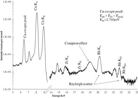
Figure 4.1 – µ-EDXRF spectra and images obtained on 283 coin for pt.1 (blue line) and pt.2 (red line). Copper quantification on both points. ………. 53
Figure 4.2 –Copper and silver contents (wt.%) (a) and silver contents (wt.%) (b) variations on the studied set of dinheiros. ………...………... 56
Figure 4.3 - µ-EDXRF spectrums and images obtained on G1 (275, 279, 280 and 257). ………...………...………... 57
Figure 4.4 - µ-EDXRF spectrums and images obtained on G2 (283 and 500). .... 58
Figure 4.5 – Stacked XRD patterns obtained on the studied set of dinheiros Principal peak identification. ………...………... 59
Figure 4.6 – Main compounds found on patina layers of the studied coins. Possible patina evolution with time and environment of exposure and its possible formation reactions. ………...………...……….. 62
Figure 4.7 – Possible stratification scheme of species in a Cu corrosion film on marine rich environment. ………...………... 62
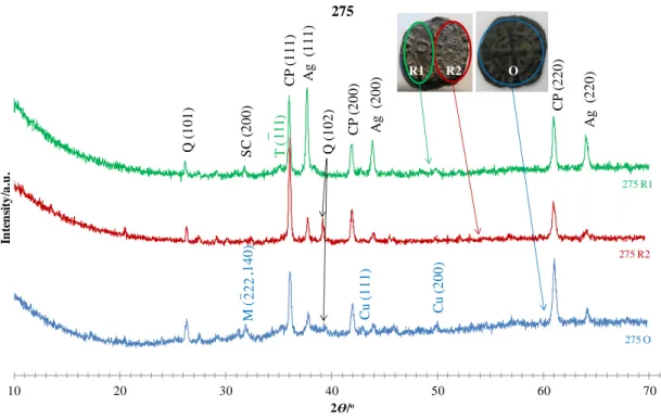
Figure 4.8– Powder XRD patterns obtained for 275 coin on both obverse (blue line denoted by 275 O) and reverse faces, particularly, the visibly corroded area (“cauliflower” – red line denoted by 275 R2) and the rest of the coin reverse (green line denoted by 275 R1). ………...………... 63
Figure 4.9 – SEM images on the surface of five of the studied coins. ... 65
Figure 4.10 - SEM images on the coins surfaces regarding compact (279 (a) and 257) and porous (275, 279(b), 283 and 500) patina layers. ... 66
Figure 4.11 – SEM/EDS copper phase examples obtained for 275, 279, 283 and 500. ………...………...………...………… 67
Figure 4.12 – SEM/EDS copper (1) and silver (2) phases obtained for 257. ... 68
Figure 4.13 – SEM/EDS silver phase examples obtained for 279 and 280. ... 69
Figure 4.14 – SEM/EDS silicon phase obtained for 275, 279 and 283. ... 70
Figure 4.15 – SEM/EDS calcium phase obtained for 280 and 500. ... 70
x v i | P a g e Figure 7.1 – CaCO3 and Ca(OH)2 nanoparticles dispersions in cyclohexane from
0.0% to 5.0% DA after 6 ultrasound cycles, 10 min each. ... 83
Figure 7.2 – Stacked turbidimetry results for the CaCO3 with 0.0, 0.5, 1.5, 2.0 and 5.0% DA dispersions in cyclohexane. ... 85
Figure 7.3 – Stacked turbidimetry results for the Ca(OH)2 with 0.0, 0.5, 1.5, 2.0 and 5.0% DA dispersions in cyclohexane. 86 Figure 7.4 – ATR stacked spectra for CaCO3 with 0.0% DA (a) and 5.0% DA (b) powders. ………...………... 87
Figure 7.5 – Stacked XRD patterns obtained for CaCO3 0.0% (a) and 0.5% DA (b) powders. Principal peak identification. …………...…...……….. 89
Figure 7.6 – SEM images on CaCO3 with 0.0% (a, b, c) and 5.0% (d, e, f) DA powders. ………...………...………... 90
Figure A.III.1 – µ-EDXRF spectra obtained on 275 coin. ………... vi
Figure A.III.2 – µ-EDXRF spectra obtained on 279 coin. ………... vii
Figure A.III.3 – µ-EDXRF spectra obtained on 280 coin. ………... vii
Figure A.III.4 – µ-EDXRF spectra obtained on 257 coin. ………... viii
Figure A.III.5 – µ-EDXRF spectra obtained on 283 coin. ………... viii
Figure A.III.6 – µ-EDXRF spectra obtained on 500 coin. ………... ix
Figure A.III.7 – Stacked XRD patterns obtained for 279 coin on both obverse (279 O) and reverse (279 R) faces. ....………..………... xiii
Figure A.III.8 – Stacked XRD patterns obtained for 280 coin on both obverse (280 O) and reverse (280 R) faces. ....………..………... xiv
Figure A.III.9 – Stacked XRD patterns obtained for 257 coin on both obverse (257 O) and reverse (257 R) faces. ....………..………... xv
Figure A.III.10 – Stacked XRD patterns obtained for 283 coin on both obverse (283 O) and reverse (283 R) faces. ....………..………... xvi
Figure A.III.11 – Stacked XRD patterns obtained for 500 coin on both obverse (500 O) and reverse (500 R) faces. ....………..………... xvii
Figure A.III.12 – SEM/EDS copper phase obtained on 275 coin. ……… xviii
Figure A.III.13– SEM/EDS copper phase obtained on 279 coin. ………. xviii
Figure A.III.14 – SEM/EDS copper phase obtained on 283 coin. ……… xviii
Figure A.III.15 – SEM/EDS copper phase obtained on 500 coin. ……… xviii
Figure A.III.16 – SEM/EDS copper phase obtained on 283 coin. ……… xix
Figure A.III.17 – SEM/EDS silver phase obtained on 279 coin. ……….. xix
Figure A.III.18 – SEM/EDS silver phase obtained on 280 coin. ……….. xix
Figure A.III.19 – SEM/EDS silicon phase obtained on 275 coin. ……… xix
Figure A.III.20 – SEM/EDS silicon phase obtained on 279 coin. ……… xix
Figure A.III.21 – SEM/EDS silicon phase obtained on 283 coin. ……… xx
Figure A.III.22 – SEM/EDS calcium phase obtained on 280 coin. ……….. xx
Figure A.III.23 – SEM/EDS calcium phase obtained on 500 coin. ……….. xx
Figure A.III.24 – SEM/EDS lead phase obtained on 283 coin. ……… xx
Figure A.III.25 – SEM/EDS of non-scraped edge obtained for 257 coin. ……… xx
Figure A.III.26 – SEM/EDS of manual scraped edge obtained for 257 coin. ….. xxi
Figure A.III.27 – SEM/EDS of non-scraped edge obtained for 500 coin. ……… xxi
Figure A.IV.1 – Effects of the weight ratio of DA on the CaCO3 particles´ contact angle, obtained at 20° C with a Ca(OH)2 concentration of 5 wt.% [68]. ... xii
Figure A.V.1 – Turbidimetry results for the CaCO3 with 0.0% DA dispersion in cyclohexane. ....………..………...………..……... xxiv
Figure A.V.2 – Turbidimetry results for the CaCO3 with 0.5% DA dispersion in cyclohexane. ....………..………...………..……... xxv
x v i i | P a g e Figure A.V.3 – Turbidimetry results for the CaCO3 with 1.5% DA dispersion in
cyclohexane. ....………..………...………..……... xxv
Figure A.V.4 – Turbidimetry results for the CaCO3 with 2.0% DA dispersion in
cyclohexane. ....………..………...………..……... xxv
Figure A.V.5 – Turbidimetry results for the CaCO3 with 5.0% DA dispersion in
cyclohexane. ....………..………...………..……... xxv
Figure A.V.6 – Turbidimetry results for the Ca(OH)2 with 0.0% DA dispersion
in cyclohexane. ....………..………...………..……. xxvi
Figure A.V.7 – Turbidimetry results for the Ca(OH)2 with 0.5% DA dispersion
in cyclohexane. ....………..………...………..……. xxvii
Figure A.V.8 – Turbidimetry results for the Ca(OH)2 with 1.5% DA dispersion
in cyclohexane. ....………..………...………..…… xxvii
Figure A.V.9 – Turbidimetry results for the Ca(OH)2 with 2.0% DA dispersion
in cyclohexane. ....………..………...………..…… xxvii
Figure A.V.10 – Turbidimetry results for the Ca(OH)2 with 5.0% DA dispersion
x i x | P a g e
T
ABLE
I
NDEX
Table 1.1 - Set of the studied dinheiros and its respective kingdom and date,
schematic figures and mint houses [1, 7]. ....………..………... 2
Table 1.2 – List of reported elements in antique and native copper and their
potential uses in achaeometallurgy. ……...…………...……….. 6
Table 1.3 – Most commonly crystalline copper compounds found in patina of
Cu/Cu-alloys artefacts. .……….. 9
Table 1.4 – Examples of some recent copper corrosion inhibitors. ...………… 17
Table 4.1 - Observations on the macro scale general appearance on the studied
set of dinheiros. ...………. 50/51
Table 4.2 – Macro scale morphologic characteristics on the studied set of
dinheiros. ………...………...………...………... 52
Table 4.3 – LOD values obtained by µ-EDXRF on CRM 32X SN7 (wt.%). ….... 54
Table 4.4 – Copper and silver contents (wt.%) on the studied set of dinheiros. ... 55
Table 4.5 – Comparison between the silver contents in literature (legal content
and the results from Guerra et al. [12]) and the results obtained in the present analysis for the coins dated from D. Afonso III, D. Dinis, D. Pedro I and
D. Fernando I. …….………...………...………... 57
Table 4.6 – Correlation between the identified peaks on XRD patterns and the
respective coins. ...………..………… 60
Table 4.7 - SEM/EDS obtained for 257 scraped and non-scraped edges and for
500 non-scraped edges. ...………. 72
Table 7.1 – Time of analyses for the various cyclohexane dispersions of CaCO3
and Ca(OH)2. …….……….……….……….………..… 84
Table 7.2 – Turbidimetry results comparison for the CaCO3 with 0.0% to
5.0% DA dispersions in cyclohexane, respectively. ………... 85
Table 7.3 – Turbidimetry result comparison for the Ca(OH)2 with 0.0% to
5.0% DA dispersions in cyclohexane, respectively. ………... 86
Table 7.4 – Peak list identification on the ATR spectra obtained for CaCO3
0.0% and 0.5% DA powders. ………...………...………... 88
Table A.III.1 - µ-EDXRF quantification obtained on 275 coin. ………... x
Table A.III.2 - µ-EDXRF quantification obtained on 279 coin. ………... x
Table A.III.3- µ-EDXRF quantification obtained on 280 coin. ………….……... x
Table A.III.4 - µ-EDXRF quantification obtained on 257 coin. ………... x
Table A.III.5 - µ-EDXRF quantification obtained on 283 coin. ………... x
Table A.III.6 - µ-EDXRF quantification obtained on 500 coin. ………... x
Table A.III.7 – Detailed and statistical information about the µ-EDXRF
quantifications on the set of the studied dinheiros (n.q. - non-quantified). ……… xi
Table A.III.8 – Powder XRD patterns peak attribution for 275 coin obverse (O). xii
Table A.III.9 – Powder XRD patterns peak attribution for 275 coin the visibly
corroded area (“cauliflower” –R2). ………...…. xii
Table A.III.10 – Powder XRD patterns peak attribution for 275 coin reverse
(R1). ……….………...……... xii
Table A.III.11 - Powder XRD patterns peak attribution for 279 coin on both
obverse (O) and reverse (R) faces. ……….………..…. xiii
Table A.III.12 - Powder XRD patterns peak attribution for 280 coin on both
x x | P a g e Table A.III.13 - Powder XRD patterns peak attribution for 257 coin on both
obverse (O) and reverse (R) faces. ……….………..…. xv
Table A.III.14 - Powder XRD patterns peak attribution for 283 coin on both
obverse (O) and reverse (R) faces. ……….………..…. xvi
Table A.III.15 - Powder XRD patterns peak attribution for 500 coin on both
obverse (O) and reverse (R) faces. ……….……..…. xvii
Table A.IV.1 – CaO mass (g) and ethanol:DA volume (dm3) measured for each
reaction. ………..…... xxii
Table A.IV.2 – Masses of CaCO3 and Ca(OH)2 used in the cyclohexane
dispersions. …..………..…… xxiii
Table A.IV.3 – Masses of 5.0% DA CaCO3 and Ca(OH)2 used in nonane,
nonane+1-butanol and cyclohexane+1-butanol dispersions. ………. xxiii
Table A.V.1 – Powder XRD patterns peak attribution for CaCO3 0.0% and
5.0% DA powders. ………..….. xxviii
Table A.V.2 – Powder XRD patterns peak attribution for CaCO3 5.0% DA
x x i | P a g e
A
CRONYMS AND
A
BBREVIATIONS
L
IST
APT - 5-(3-aminophenyl)-tetrazole BTAH – Benzotriazole
DA – Dodecanoic acid
DMTD - 2,5-Dimercapto-1,3,4-thiadiazole EDS – Energy Dispersive Spectroscopy LOD – Limit of Detection
MMPB - 3-((2-mercaptophenyl)imino)butanoate RH - Relative Humidity
RSD - Relative Standard Deviation SEM – Scanning Electron Microscopy XR – X-ray(s)
XRD – X-ray Diffraction XRF – X-ray Fluorescence
μ-EDXRF – micro-Energy Dispersive X-ray Fluorescence
XRD Crystal systems abbreviations:
A - aragonite (orthorhombic structure of CaCO3) AT – atacamite (Cu2Cl(OH)3)
C – calcite (rhombohedral structure of CaCO3) CL – calcium laurate (Ca(C12H23O2)2)
CP – cuprite (Cu2O)
M – malachite (Cu2CO3(OH)2) N – nantokite (CuCl)
Q – quartz (SiO2)
SC – chlorargyrite (AgCl) T – tenorite (CuO)
1 | P a g e
1 I
NTRODUCTION
The thorough knowledge of an archaeological object with its various aspects is a precondition to any applied research. Investigations involving chemical and physical non-destructive instrumental methods contribute greatly to understand and preserve the history of societies.
The present work will be presented in two separate but complementary parts. The first approach (Evaluation on the degradation state of antique coins by spectroscopic techniques) is based on the assessment on the conservation state of antique Portuguese coins by identification and understanding of its corrosion products and surface morphologic changes. While the second part (Development of nanomaterials for Cultural Heritage Conservation) is result of the synthesis, purification and physical-chemistry characterization of several calcium carbonate (CaCO3) and calcium hydroxide (Ca(OH)2) nanoparticles hydrophobically functionalized with dodecanoic acid (DA), to application on manuscripts for conservation purpose.
In consequence the two main fields on the study of cultural heritage are covered: the investigation on the state of conservation of an object and a proposal to a conservation mode. In this particular case the studied objects are different but the approach is highlighted.
Annexe I presents a list of important definitions and concepts that are going to be use
on this work.
1.1 COINS
Coins are particular and important findings during archaeological investigations as source of documentation, understanding and knowledge of people and their societies.
Once these objects play an important role in any national cultural heritage, this study represents a very important step in the first Portuguese dynasty billon coins characterization. It is focus on a set of six dinheiros dated from the 13rd and 14th centuries, namely from D. Sancho II (1223-1248); D. Afonso III (1248-1279); D. Dinis (1279-1325); D. Pedro I (1357-1367) and D. Fernando I (1367-1383). Due to the limited knowledge, in particular the lack of information about the place of find, state of burial/dig up or even its related dates (the numisms could have been buried, submerged or even exposed to other kind of environments) this investigation is a challenge on both corrosion science and cultural heritage. Is thus
2 | P a g e
justified the importance of a detailed analysis in order to gain intensive information about chemical composition, nature of the patina and corrosion features on the coins.
1.1.1 Coin history and social impact
The invention of coins resumes to the 650/700 b.C. and since that time they have been used as a medium of exchange. These metal objects are standardized in weight and produced in large quantities at a mint (coinage house).
Numismatics1 and the study of the coins´ chemical composition (surface and bulk) give important information about the culture, economy and science of a society [1, 2, 3]. For example, the study on the coin depreciation (by reducing the precious metal content) can be correlated to the economic decline as evidence on periods of economic difficulties [4, 5].
Portuguese dinheiros
Portugal's independence was proclaimed by D. Afonso Henriques on 25 July 1139, being the first Portuguese coins (dinheiros) issued by him. Dinheiros (minted in billon alloy) were the currency of Portugal from the late 12nd century until, approximately, 1502.
Various kinds of coins were introduced in the Portuguese culture during the first dynasty. Around 1200, a gold currency was produced and a century later a silver coin was introduced [6].
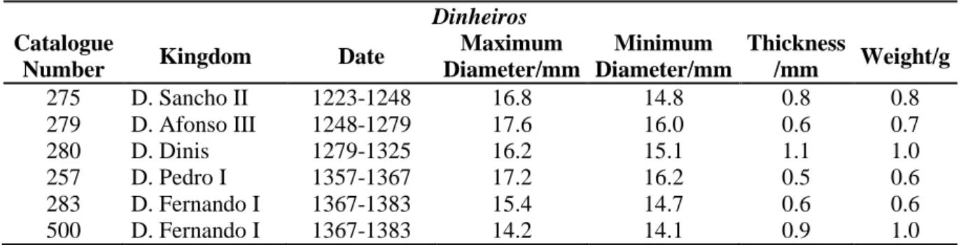
The set of the studied dinheiros, their respective kingdom, date, schematic figures and the mint houses are presented on Table 1.1.
Table 1.1 - Set of the studied dinheiros and its respective kingdom and date, schematic figures and mint houses [1, 7].
# King Date Dinheiro Legend Mint
4th D. Sancho II 1223-1248 SANCIVS REX
PORTVGAL
Braga and Lisbon
5th D. Afonso III 1248-1279 ALFONSVS
REX/PORTVGAL Coimbra and Lisbon 6th D. Dinis 1279-1325 D.REX PORTVGL/ALGARBII Lisbon Casa da Moeda 8th D. Pedro I 1357-1367 P.REX PORTVGL/ALGARBII Lisbon
9th D. Fernando I 1367-1383 FERNANDVS REX
PORTVGALI ET ALGARBI
Portugal and Spain
1
3 | P a g e
D. Sancho II known as the “Cowled One” was born in Coimbra, in 1209, and his death is reported to 1248 in exile (Toledo). He ascended to the throne in 1223 and was deposed by the Holy See due his political and personal ideas, in 1245. Sancho II introduced a great development in minting once the coinage necessity grew due the increasing of the commercial transactions [1].
The “Bolognese” or D. Afonso III became king of Portugal in 1248. He was born in Coimbra (1212) and died in Lisbon (1279). Afonso III extended his domain conquering the southward to Algarve and gave Portugal its definitive boundaries. This king produced several legislation regulating minting alloys, weights and alterations in coins [1].
D. Dinis, the “Husbandman”, was born in Lisbon in 1261, ascended to the throne in 1279, and died in Santarém in 1325. During his time, the land was colonized and cultivated; industry grew stronger and the external commerce expanded protected by an organized navy and defended by treaties and protective laws [1]. With D. Dinis the first Portuguese university (“Estudo Geral”) was established in Lisbon (1290) being later transferred to Coimbra (1308) [8]. As a consequence, Latin gave way to Portuguese language and started to be used on official documents and diplomas.
D. Pedro I known as the “Just” or the “Cruel” was born in 1320 (Coimbra), died in 1367 (Estremoz) and acceded to the throne by death of his father (D. Afonso IV) in 1357. During his reign the economic situation of the country improved and prospered. Despite its political internal instability, trade was carried on the most important ports of Europe [1].
D. Fernando I known as the “Handsome” (Coimbra, 1345 – Lisbon, 1383) ascended to the throne in 1367 being the last king of the first Portuguese dynasty. Fernando I was involved in unending and unprofitable wars against the Castile which created enormous economic and financial difficulties. In fact, the coinage reflected his irregular and dissolvent administration. He changed and created new types and denominations to mask the weakening and debasement of the alloy (impoverishing in metal) in relation to the inflated nominal values it represented [1].
After the death of D. Fernando I, Portuguese people reacted against his Castilian succession and required the protection of the “Master of the Order of Avis”, who acceded to the throne in 1385, founding the second Portuguese dynasty.
1.1.2 Chemical composition of coins
Compositional information constitutes a fundamental tool in the study of antique coins. Studying the elemental composition pattern of the alloy, its fineness and debasement, can
4 | P a g e
provide valuable information either to provenance studies or for investigations of manufacturing processes, as well as identification of authenticity [4, 5, 9-11].
Billon was one of the most important and common alloys used for minted coins since the ancient Greece. However, only a few studies were made on Portuguese antique coins (Guerra et al. [10, 12], Martins et al. [13-15] and Mata et al. [16, 17]) being the most notorious conclusion that these coins were mostly constituted by copper (approximately 90.0%), silver (around 8.0%) and some minor elements.
(i) Copper
Copper (Cu) occurs naturally as native Cu or alloyed with other elements. In both states it forms a large group of industrially important materials owing to its appealing visual appearance, excellent electrical and thermal conductivities, mechanical workability and resistance [18-20].
Native copper identification with silver and locally arsenic as major impurities is highly desirable in artefacts because it is generally held that the working metals began with the use of pure copper [21]. According to Pernicka [21] the presence of volatile metals such as mercury (Hg) could provide chemical evidence for the presence of native copper; however this element can also be absorbed on copper from groundwater during the burial, inducing inaccurate conclusions.
Historically, the main working processes on this element were the cold working process2; annealing3, smelting4 and re-melting5. It is important to retain that the ores were generally inhomogeneous showing zoning effects, and some elements could be lost during any of these processes. Even more, after the smelting process finding alloying copper with other metals was very common. Numerous copper alloys exist, many with important uses. Apart from billon; brass is a copper-zinc alloy and bronze usually denotes copper-tin alloys yet can refer to any alloy of copper (e.g. aluminium bronze). The metal composition of bronze alloys and its deterioration processes has been examined numerous times [2, 5, 9, 17, 21-30].
(ii) Silver
Silver (Ag) and its alloys have been used in many objects of cultural heritage due to its luxuriousness, luster and intrinsic value. Even more, the surface enrichment of archaeological copper alloys, either intentional or due to corrosion processes, has been known for many years and it is of main interest for a chronological assignment [31, 32].
2-5 The definitions are presented on Annexe I.
5 | P a g e
The application of a layer of silver onto the surface of a low noble metal (silver surface enrichment) was possible by using chemical, mechanical, thermo-mechanical or physical treatments [5, 33]. All of this processes varied greatly from time to time and place to place. Particularly the presence of areas in the coin with higher content of silver is a normal behaviour in billon alloys due to the low solubility of silver in copper, and vice versa, at room temperature. Based on Martins and Martins [15] the solubility of Cu in Ag is of 8.0% at 780° C, and practically zero at room temperature. During cooling, the system separates each component in the pure state with the same reticular structure as the one in the supersaturated solid solution; so the formation of rich areas of silver dispersed in copper matrix depends on the rate of cooling and thickness of the sample.
Silver was not the only metal applied to the surface of the coins in order to give them a rich and shine appearance. Other intentional plating elements were carried out with antimony or arsenic, for example.
(iii) Minor elements
There are a number of factors which can give rise to systematic variations in the concentration levels of minor elements in antique copper alloys, including, for example, the mineralogy of the copper ore source; partition, volatilisation and contamination of trace elements (from various sources) during the smelt between metal and slag phases; the deliberate addition of other metals (e.g. tin or lead) to the finished metal; co-smelting of different ores to produce alloys directly; the poly-metallic ore sources that would naturally create alloys in the smelt; and changes in chemistry through melting, re-melting and working of the metal [25].
Elements such as lead, tin, antimony or zinc, nickel and cobalt, gold or even silver can be present on the surface of copper alloy coins [5, 10, 21, 23, 28, 34]. Lead (Pb) due to its compact structure and large atoms forms a good alloy with other elements in particular with copper [23]. Concerning the presence of this element on the coin surface, it is related to its very low solubility in copper and its low melting temperature with respect to that of the copper matrix, which causes the formation of fine particles dispersed throughout the copper [5]. Generally, less amount of lead is an effective evidence for a good refining process [21, 34]. Ingo et al. [5] put forward the hypothesis that the presence of antimony (Sb) could have been enhanced by the craftsmen, firstly during the blank production thus inducing the occurrence of the inverse segregation phenomenon on the outermost region.
6 | P a g e
The hypothesis that minor elements concentrations should be a guide to the provenance of antique metals has been around for more than a hundred years and explored for decades [10, 21]. The content of these elements can be governed by ore composition or related to the smelting process. In fact changes on minor elements levels give important information about the ore provenance [12].
1.1.2.1 Provenance
The most difficult question in numismatics is the attribution of provenance [10]. Either the mine is known and the goal is to correlate the objects to it, or the mine is unknown and the target it to determine which mine was exploited to make the objects. However, the place of origin/burial is of great importance to understand the mechanism of the phenomena responsible for the deterioration.
Based on literature [15, 21, 25] it is possible to elaborate Table 1.2 which presents a list of elements that were deliberately alloyed with copper as well as those stemming from the ore extraction. Only a very limited range of trace elements are directly related to the provenance of the ore. Even more, in earlier times it cannot be assumed that deliberate alloying occur.
Table 1.2 – List of reported elements in antique and native copper and their potential uses in achaeometallurgy.
Technology Provenance and/or technology Provenance
B, Ba, Be, Cr, Cs, Ga, Ge, Hf, Li, Mn, Mo, Na, Nb, Rb, S, Sc, REEc,
Sr, Ta, Ti, Th, U, V, W, Y, Zr
Alb, As, Cab, Cda, Clb, Co, Feb, In, Hga,b, Kb, Mgb, Pb, Re, Sib,
Sb, Se, Te, Tla Au, Ag, Bi, Ir, Ni,
Os, Pd, Pt, Rh, Ru Sn > ca. 1% Zn and Pb> ca. 5% Sn < ca. 1% Zn and Pb< ca. 5% a
Only applicable with native copper. b Soil contamination. c Rare Earth Elements.
1.1.2.2 Manufacturing
Coin minting obeys to several laws of the monetary system (national and/or worldwide) and its values are linked to a monetary standard. Moreover, there is the intent to produce objects that will last and can be easily distinguished from one another.
Ferraro Vaz [1] presents a resume about the antique Portuguese coins manufacturing process. According to this author, when a coin is hammered or milled the metal is prepared in disks and then submitted to the minting by manual pressure, marking them with the letters and figures (usually different) in both faces (obverse and reverse).
7 | P a g e
Studies presented by Guerra [10] show that varying the ratio of some minor elements with respect to the major ones (usually silver, copper or gold) may indicate a deliberate addition of a specific element to the alloy, meaning a change in the manufacture technology.
1.1.3 Coin deterioration
Studies on corroded metallic archaeological artefacts are of great importance since they can improve knowledge in the field of the long-term corrosion phenomena providing help, to scientists and conservators, in order to control and stop the process of deterioration of historic metals in museums and in selecting their ideal storage conditions; or even on its historical and archaeological classification [11, 17, 28]. However metals are generally difficult materials to analyze once they might have been re-melted and reused in new minting or for producing other objects [10].
Coins are very sensitive samples to the effect of deterioration processes. Their state of conservation depend on normal wearing processes before and after burial, being the extent and depth of the corrosion phenomena closely dependent on the corrosive environment (chemical composition, pH, resistivity, etc) and other non-negligible parameters, such as the type of electrolyte and the alloy microstructure, historical periods, metallurgical techniques or even the kind and size of the artefact [2, 15, 28, 29].
Based on these assumptions the interaction of archaeological artefacts with the neighbouring environment becomes very important in the field of preservation and corrosion. Most studies of different environmental conditions (soils [28, 35, 36], atmosphere [37- 43] and marine exposure [18, 28, 44, 45]) tried to establish the correlation between the artefacts chemical composition, its surroundings and their patinas structures.
Apart from corrosion occurring in air and wet soil it has to be considered that such leaching phenomena can also be caused by chemical treatments of the objects. In organic or inorganic acidic solutions the less noble constituents of the alloy are dissolved, while the nobler components, e.g. silver or gold, are enriched at the surface [32].
1.1.3.1 Coin patina
Patina is a coating of all chemical compounds such as corrosion products (namely, oxides, carbonates, sulphides and sulphates, nitrites and nitrates, phosphates, etc.) and other exogenous elements (silicon, calcium, etc.) formed and retained on the surface of the pure or alloyed metal, changing its surface texture and colour [6, 40, 46]. It can be produced and changed by chemical processes, wear, polishing, age, or, principally, due exposure to
8 | P a g e
atmospheric elements (air, rain, soil, etc.). As that it is important to consider the alloy/patina/environment as a global system in which the patina must be regarded as the result of the physicochemical interactions between the alloy and the environment, with time [29].
Patinas are built up during relatively short periods of time (6-50 years) with respect to the total conservation environment duration (hundreds to thousands of years). Once formed, patina layer is relatively stable and it becomes a permanent part of the object acting as a protective barrier, attenuating or eliminating the corrosive phenomena [13, 40, 41]. Copper patinas are generally regarded as aesthetically pleasing to the point of being purposely deposited on the metal surface by artists and metalworkers.
1.1.3.2 Usually compounds and their formation reactions
Corrosion of archaeological artefacts is studied through the analyses of its patina products. The knowledge of its constituents is the first step to understand the corrosion mechanism. Since the morphology of the patinas, their adhesion and degree of porosity are dependent on the climatologically conditions, nature and level of pollutants, each metal behaves in a different way and forms specific compounds that reflect the chemical properties of the metal and the environment to which it is exposed.
Initially the interface metal/medium is formed by cuprite (Cu2O) (Reaction 1.1) [13, 15, 27, 40, 41, 46]. This compound is also the dominating phase that constitutes about half of the total patina mass [40] and it growths proceeds for years, centuries, or millennia, and may reach thicknesses of the order of several tens of micrometers [13].
Reaction 1.1
Since Cu2O suffers an increasing rate of nucleation with increasing the relative humidity (RH), due the higher quantity of adsorbed water clusters that acts as nucleation sites, its reaction formation is RH dependent [43]. Reaction 1.2 shows the cuprite oxidation, noticing that it does not take place any further in the later stage of the corrosion [39, 46]. Moreover, oxidation of cuprite under atmospheric conditions must be slower than that of copper otherwise the intermediate cuprite would not exist. Reaction 1.4 represents the balanced redox reaction for the anodic and cathodic reactions in oxygen presence (Reactions
1.2 and 1.3, respectively):
Reaction 1.2
Reaction 1.3
9 | P a g e
Cuprite is formed on the surface and later converted into other chemical compounds in agreement with the environment. Hydroxides and hydrated compounds of Cu (II) could be formed after the long-term (<1000 years) due the interaction between the coins, its first patina layer and the corrosion environment [27].
Table 1.3 presents a resume on the most commonly crystalline copper compounds
found and studied over different patinas on copper and copper alloys artefacts.
Table 1.3 – Most commonly crystalline copper compounds found in patina of Cu/Cu-alloys artefacts.
Name Formula Colour Reference
Cuprite Cu2O
Red-brownish.
Becomes black over time. [13, 15, 27, 40, 41, 46]
Tenorite CuO Steel-grey, black [13]
Malachite Cu2CO3(OH)2 Green [13, 15, 27]
Atacamite Cu2Cl(OH)3 Green [15, 27, 41, 46]
Brochantite Cu4SO4(OH)6 Green [15, 41, 46]
Azurite Cu3(CO3)2(OH)2 Blue [27]
Nantokite CuCl Colourless, greyish to green [13]
Paratacamite Cu4Cl2(OH)6 Green [13, 41]
Antlerite Cu3SO4(OH)4 Green [40]
Posnjakite Cu4SO4(OH)6.H2O Blue [41, 46]
Langite Cu4SO4(OH)6.2H2O Blue [40]
Gerhardtite Cu2NO3(OH)3 Green [40]
Chalcopyrite CuFeS2 Yellow [30]
Chalcocite Cu2S Black [15]
Patina colours are directly connected to the nature of the corrosive environment and not to the composition of the alloy. However, they cannot be considered as a valid criterion for providing a quick identification of typical corrosion products.
Depending on the environment, tenorite (CuO) (Reaction 1.5), malachite (Cu2CO3(OH)2) (Reaction 1.6), atacamite (Cu2Cl(OH)3) (Reaction 1.7) and brochantite (Cu4SO4(OH)6) (Reaction 1.8) are the principal patina constituents on copper-silver alloys, being the three last ones only compatible with oxidizing conditions; while compounds as cuprite (Cu2O), chalcocite (Cu2S) and nantokite (CuCl) (Reaction 1.9) are formed under typical reducing conditions, unless the conditions varies imposed by organic and/or inorganic matter in the medium.
Reaction 1.5
Reaction 1.6
Reaction 1.7
1 0 | P a g e
Reaction 1.9
In fact what usually happens is the inter-conversion of cuprite, generally by its oxidation, into several different compounds according to the different environmental conditions. Beyond those exposed above, for example, azurite (Cu3(CO3)2(OH)2) (Reaction
1.10) or paratacamite (Cu4Cl2(OH)6) (Reaction 1.11) formation reactions:
Reaction 1.10
Reaction 1.11
Due Cu2O oxidation, pH of the aqueous layer increases, while the formation of copper sulphates (e.g. brochantite or posnjakite) and the presence of weak atmospherics acids (e.g. formic, acetic, oxalic and/or carboxylic acids) act as buffers making the pH reaming at its equilibrium for the sulphates formation, increasing the time of reaction [46]. Reaction 1.12 shows the reaction on brochantite stability domain. Even more, posnjakite is a hydrated form of brochantite and could be formed as a precursor of it. Studies made by Fitzgerald et al. [46] show that pH increases until tenorite is formed on the surface of cuprite and stifled further oxidation. Brochantite dissolution and the formation of tenorite under these conditions are given by Reaction 1.13.
Reaction 1.12
Reaction 1.13
There exists a critical SO2 deposition rate above which brochantite does not form and existing patinated surfaces will dissolve [46].
Some of these compounds can be used at screening for the coins “survival” for so many years. Even more, they can be associated with particular environments. Generally, malachite presence suggests that the object could be buried (in soil); brochantite has normally origin in the atmosphere contact; atacamite and paratacamite suggest the presence of chlorides in the place of burial (maybe near seawater) and chalcocite the presence of anaerobic reducing environments [15, 26].
Finally, is important to notice that noble metals (e.g. Ag) accelerate corrosion, while the most catholically active ones (e.g. Pb, Sn) protect it during a certain period [13]. Silver does not react readily with the oxygen at room temperature. However, as shown before, adsorbed water layers into the oxide structure promotes irregularities that allow the penetration of corrosive ions. In consequence silver may suffer corrosion due to local action cells according to the Reactions 1.14 to 1.16 [14, 15]:
1 1 | P a g e
Reaction 1.14
Reaction 1.15
Reaction 1.16
Moreover, silver can also suffer attack by Cu (II) ions in the presence of chloride ions [15] as Reaction 1.17 shows:
Reaction 1.17
The silver chloride, although insoluble in water, does not grow as a protective coating on the surface, so this reaction is rendered decisively corrosive in the copper enriched areas being induced by oxygen and humidity [15].
1.1.3.3 Patina structural aspects
Divalent metal ions such as Cu2+ commonly form compounds with layered structures. Copper patina, typically, consists of two distinct layers: a 5-15 µm inner layer of essentially continuous cuprite and an external, porous layer, about 5-40 µm of basic copper sulphates, chlorides and carbonates [14, 17, 39, 40, 46]. However, it can continue to grow during many years or even decades of exposure and may reach a thickness of the order of several tens of micrometers [10, 40]. These layers are held together by sulphate groups, hydrogen bonds and weak Cu-O bonds and, in spite of the structural resemblance between the compounds, the fact is that the phases have different cell symmetry [40]. For example, native silver and native copper, cuprite, nantokite and chlorargyrite present a cubic crystal system; atacamite is orthorhombic; and malachite, tenorite, brochantite and azurite present a monoclinic cell symmetry. This structural resemblance suggests that one phase can act as seed crystal for the formation of a subsequent phase.
Figure 1.1 shows a scheme for the general patina evolution on copper artefacts,
displaying the formation of different compounds as a function of exposure time in different environments. One consequence of the gradual evolution of patina is that it gradually becomes less soluble and, hence more resistant to atmospheric corrosion [40].
1 2 | P a g e Figure 1.1 – Patina evolution scheme on copper artefacts along time and environment of exposure. Robiola et al. [28] established a new structure patina classification of copper based alloys (particularly, Cu-Sn alloys), when exposed to an oxygenated corrosive medium. In resume, they assert that the corrosion patterns can be differentiated into two categories, according to patina colours, aspects and state of preservation of the original size of the object (i.e. the original surface limit) and based on its microstructures identification and characterization. Both categories can be observed on one and the same artefact. He et al. [27] later added that this double-structured deposit consist of an inner layer of Cu (I) salts and an external layer of Cu (II) compounds, which are depended on the history and the elemental composition of the object. Basically, Type I or “even” surface patinas build up under a mild corrosion condition and in a relatively short period of time being very protective and strongly influenced by the presence of incorporated soil components. While Type II or “coarse” surface patinas are thicker patinas formed when the original surface has been destroyed or deformed by severe corrosive attacks during the early stage of exposure.
As the bronze corrosion process leads to a preferential dissolution of copper ions into the environment, exogenous elements (textiles or leather, insects, woods or even pure organic matters) can be entrapped in the corrosion layers or mineralised [29].
1.1.3.4 Patina and the corrosive environment
The importance of exploring the influence of environmental conditions on atmospheric corrosion rates of metals is justified by prediction of future corrosion rates as well as to control its degradation. Changes in land use, new large industries, afforestation, highway
Cu4SO4(OH)6.2H2O
Cu2O
Amorphous
copper sulfate Cu
4SO4(OH)6
Cu4SO4(OH)6.H2O Cu3(SO4)(OH)4
Flaking through volume expansion. Cu2Cl(OH)3 CuCl Cu2NO3(OH)3 Cu2CO3(OH)2 CuO Cu(OH)2 2 3 CO 3 2/NO NO 2 4 2/SO SO Cl
1 3 | P a g e
engineering or lowering the groundwater table are some human factors which contribute for a change in the different environments.
(i) Exposure to humidity and air pollutants
Exposures to humidity or air pollutants create difficult multiphase systems to study. Several corrosive species and corrosion products that interact with each other are present and all of them vary in amount and time. Even more, humidity, precipitation and wind play an important role on the atmospheric pollutants transportation and deposition on the object.
Electrochemical corrosion of copper is dependent on the type of metal as well as the pollutants present in the system. Once the metal dissolution occurs in presence of an electrolyte provided by atmospheric precipitation or by adsorption of water molecules on the surface of the corrosion layer, relative humidity plays a central role among the climatic factors. Especially since the presence of corrosive species which attract water vapour become soluble above a critical RH, lead to a sharp increase in the corrosion rate [39].
Among the most abundant contaminants are sulphates, nitrates and nitrites, ozone, chlorides, carbonates, hydrogen ions, ammonium, metal ions, atmospheric particles and also organic compounds.
Generally and based on literature [39, 42, 43] ozone (O3) has the strongest effect on the corrosion of copper followed by nitrogen dioxide and sulphur dioxide (NO2 and SO2, respectively). Even more, O3 and NO2 led to a uniform corrosion attack while in the presence of SO2 a locally attack can be observed [39].
Ozone is recognised to be a potential corrosion accelerator in corrosion research originating metal loss and hindering the passivation layer formation. This occurs possibly due O3 strong oxidative power and because it can produce hydroxyl radical, which is balanced by the metal dissolution [39]. Moreover, the exposure to ozone led to the formation of a considerable amount of water-soluble nitrites and to copper sulphite species oxidation, forming copper sulphates (CuSO4.xH2O) and increasing the Cu2O formation [39, 43].
When exposed to NO2 the major species formed on the copper surface are: Cu2O and gerhardtite; moreover, when copper sulphide species are present this pollutant is able to oxidize them to CuSO4.xH2O [13, 38, 39, 42, 43]. In water rinses nitrate dominates over nitrite [43]. Formation of gerhardtite can be a result of nitric acid (HNO3) and dinitrogen pentoxide reactions [38, 39]. Due to the high water solubility of HNO3 this pollutant dissolves easily in the adlayer and the chemical reaction on the metal surface is enhanced leading to a decreased surface resistance.
1 4 | P a g e
Sulphates can be supplied directly from rain water; via adsorption and oxidation of sulphur dioxide within the aqueous layer; or from aerosols [46]. However, deposition rates depend greatly on the nature and geometry of the surface. Adsorption of SO2 on copper surfaces is strongly influenced by RH [46]. The interaction between a water covered metal surface and SO2 can be described by Reactions 1.18 to 1.21 sequence:
Reaction 1.18
Reaction 1.19
Reaction 1.20
Reaction 1.21
After deposition and dissolution of SO2 into the water layer (Reaction 1.18), a
bisulphite ion (HSO3-) is formed by hydrolysis of sulphur dioxide (Reaction 1.19) producing a metal-sulphito surface complex ( ), through an exchange mechanism with the hydroxylated metal oxide surface ( ) (Reaction 1.20). Liquid metal-sulphito complexes ( ) subsequently detach from the metal-surface (M) and precipitate as solid corrosion products (Reaction 1.21) [42].
The principal compounds found in copper patinas when exposed to SO2 rich environments are cuprite, basic copper sulphates and sulphites [13, 39, 40, 43].
When sulphur- or chlorine-containing atmospheric species are more dominant, the patina formation reaction sequence becomes more complex and involves more copper patina constituents [40].
Conventional atmospheric parameters that affect copper generally affects silver too comprising weathering factors (temperature, moisture, radiation, wind velocity, etc.), air pollutants and aerosols. In presence of a strong oxidizer, silver rate corrosion increases [37].
(ii) Exposure to marine environments
Marine environments, such as coastal or near-coastal countries as Portugal, are chloride (Cl-) rich environments.
Urban atmosphere patinas are, in general, more adherent and uniform, than the ones exposed to chloride-rich environments. These last patinas are generally heterogeneous and present flaking and scaling, which allows localized corrosion; however, in the marine atmosphere patinas the degree of adhesion increases with time [18, 41, 44]. Zhang et al. [44] concluded that flaking mechanism associated to these loosely adherent patina layers is connected with the formation of nantokite.
![Table 1.1 - Set of the studied dinheiros and its respective kingdom and date, schematic figures and mint houses [1, 7]](https://thumb-eu.123doks.com/thumbv2/123dok_br/19187227.948147/28.892.102.800.757.1094/table-studied-dinheiros-respective-kingdom-schematic-figures-houses.webp)