Printed version ISSN 0001-3765 / Online version ISSN 1678-2690 http://dx.doi.org/10.1590/0001-3765201620150484
www.scielo.br/aabc
Lipolytic response of adipose tissue and metabolic adaptations to long
periods of fasting in red tilapia (
Oreochromis
sp., Teleostei: Cichlidae)
WaLTer Dias Junior1
, amanDa m. Baviera2
, neusa m. Zanon3
, viCTor D. GaLBan3
, maria anTonieTa r. GarófaLo3, CeLio r. maChaDo3, eLisa f.L.C. BaiLão4 and isis C. KeTTeLhuT3
1
Laboratório de Fisiologia e Bioquímica Toxicológica,Faculdade de Enfermagem, Universidade Estadual de Goiás/ UEG, Campus de Ceres, Rua Lucas Marcelino dos Santos, Qd34 Lt03, 76300-000 Ceres, GO, Brasil 2
Departamento de Análises Clínicas, Faculdade de Ciências Farmacêuticas de Araraquara, Universidade Estadual Paulista Júlio de Mesquita Filho/UNESP, Rua Expedicionários do Brasil, 1621, 14801-136 Araraquara, SP, Brasil
3
Departamento de Bioquímica, Faculdade de Medicina de Ribeirão Preto, Universidade de São Paulo/ FMRP-USP, Av. Bandeirantes, 3900, Monte Alegre, 14049-900 Ribeirão Preto, SP, Brasil 4
Faculdade de Farmácia, Universidade Estadual de Goiás/UEG, Câmpus Henrique Santillo, Campus CCET, BR 153, 30105, Fazenda Barreiro do Meio, 75132-903 Anápolis, GO, Brasil
Manuscript received on August 7, 2015; accepted for publication on December 7, 2015
aBsTraCT
Adaptive changes of carbohydrate and lipid metabolism induced by 7, 15, 30, 60, 90, 150 and 200 days of fasting were investigated in red tilapia (Oreochromis sp.). Plasma glucose, lactate and free fatty acids (FFA) levels, liver and muscle glycogen and total lipid contents and rates of FFA release from mesenteric adipose tissue (MAT) were measured. Plasma glucose levels showed significant differences only after 90 days of fasting, when glycemia was 34% lower (50±5mg.dL-1) than fed fish values (74±1mg.dL-1
), remaining relatively constant until 200 days of fasting. The content of liver glycogen (»15%) in fed tilapia fell 40% in 7 days of food deprivation. In 60, 90 and 150 days of fasting, plasma FFA levels increased 49%, 64% and 90%, respectively, compared to fed fish values. In agreement with the increase in plasma FFA, fasting induced a clear increase in lipolytic activity of MAT incubated in vitro. Addition of isobutylmethylxanthine (cAMP-phosphodiesterase inhibitor) and isoproterenol (non selective beta adrenergic agonist) to the incubation medium induced a reduction of lipolysis in fasted fish, differently to what was observed in mammal adipose tissue. This study allowed a physiological assessment of red tilapia response to starvation.
Key words: adipose tissue, adrenoreceptor, lipolysis, liver glycogen.
Correspondence to: Walter Dias Junior E-mail: [email protected]
inTroDuCTion
The ability to use carbohydrate for energy varies
greatly among fish species and it is generally related
to the feeding habitats of the species (NRC 2011).
In contrast, fish present a relative efficiency in lipid
and protein digestion (Clements and Raubenheimer
2006). In this way, in general fish use preferentially
1994, Machado et al. 1989, Nagay and Ikeda 1972). Fish lipids are found scattered in hepatic tissue, muscle (main red muscle) and in mesenteric tissue involving the gut (Machado et al. 1989), and these site of deposition are species-dependent (Weil et al. 2013). Adipose tissue is distributed in the abdominal cavity located periviscerally in many
fish species, including tilapia, and the morphology
and distribution of adipose cells in visceral fat and muscle of tilapias are still not clear (Albalat et al. 2005).
Despite the presence of appreciable amounts of organized fat tissue in several species, information
about the regulation of the lipolytic activity in fish
species when compared to mammals are sparse (Albalat et al. 2005). Mammals lipolytic hormones, like catecholamines and glucagon, did not change fatty acids mobilization on adipose tissue
from fish, amphibian and reptiles (Farkas 1967,
Migliorini et al. 1992). However, Migliorini et al. (1992) found that the in vitro lipolytic activity of
Hoplias malabaricus adipose tissue was markedly increased in the presence of cAMP analogues or xanthine derivatives, which inhibit cAMP-phosphodiesterase, increasing the intracellular concentration of cAMP. Moreover, Magnoni et al. (2008) found that the in vivo lipolysis was inhibited after norepinephrine administration (0.45nmol. Kg-1) in rainbow trout for 10 minutes.
Many studies were carried out in fish to investigate the mechanism by which catecholamines modify lipolysis via direct activation of adipocyte alpha and/or beta-adrenoceptors. Previous studies demonstrated the role of beta-adrenoceptors in
lipolysis inhibition or stimulation in fish, indicating
a novel phenomenon and a different mechanism from that described in mammals, since in these animals the beta-adrenoceptors stimulation results just in lipolysis increase (Magnoni et al. 2008, Van Raaij et al. 1995, Van Den Thillart et al. 2001, Vianen et al. 2002). But the participation of adrenoceptors
in lipolytic response of fish adipose tissue appears
to be variable and dependent on fish species. In
Oreochromis mossambicus (common tilapia) and in
Cyprinus carpio (carp) beta1-adrenoceptors inhibit and beta2-adrenoceptors stimulate lipolysis in adipose tissue (Van Den Thillart et al. 2001, Vianen et al. 2002). Furthermore, Vianen et al. (2002) showed the participation of beta3-adrenoceptors in the inhibition of free fatty acids (FFA) mobilization from adipose tissue in tilapia. A fall in plasma FFA levels after norepinephrine action in C. carpio was observed (Van Raaij et al. 1995, Van den Thillart et al. 2001), but alpha2-adrenoceptor antagonist blocked the norepinephrine effect (Van Den Thillart et al. 2001). Vianenet al. (2002) found that norepinephrine induces a reduction of adipocyte FFA release in O. mossambicus and that the addition of phentolamine (alpha1- and alpha2-adrenoceptor antagonist) did not affect this mechanism.
It has been described that external factors could
also regulate the lipolysis activity in fish. Fasting
increased the lipolysis activity of gilthead seabream visceral adipocytes starved for 11 days (Albalat et al. 2005). In rainbow trout, it was observed a visceral fat depletion and a reduced expression of genes involved with triacylglycerol breakdown in 6 week of fasting (Kittilson et al. 2011). Fish, in general, have such ability to withstand long periods of fasting (Navarro and Gutiérrez 1995, Vigliano et al. 2002), since prolonged drought periods, reproductive process or prey availability are responsible for the natural process of fasting (Caruso et al. 2010, Pérez-Jiménez et al. 2012).
Farmed fish may also experience fast situations
imposed by routine procedures in aquaculture, as those used to avoid risks of overproduction (Krogdahl and Bakke-McKellep 2005).
Tilapias are the ninth most important aquaculture species group and the second most
important fish group species in terms of weight of
production worldwide. The name tilapia includes many species of the Oreochromis Genus. These
they have been introduced into most tropical and subtropical countries (Boyd 2004). The Brazilian
aquaculture is the second (Chile is the first one) in
South American production, supported basically by production chains of shrimp and tilapia culture. Brazil has genetically improved tilapia strains to better adaptation to local climatic conditions (Scorvo Filho et al. 2010).
Due to the large number of controversial
re-sults concerning fish lipolysis regulation, and the
commercial importance of tilapia culture in Brazil, whose drought periods are prolonged, the objec-tive of this work was to investigate the metabolic adaptation of red tilapia to long periods of fasting. It was investigated the fatty acid mobilization in vivo and the lipolytic response in vitro to alpha and beta adrenoceptors of the mesenteric adipose tis-sue. Agonists and antagonists inhibitors of cAMP, forskolin, phosphodiesterase (isobutylmethylxan-thine), theophyline, and activators of adenylate cy-clase were also investigated. Blood glucose and lactate levels, glycogen and lipids content in liver and white muscle were also measured.
maTeriaLs anD meThoDs
maintenanceof animalS
This study agrees with Ethical Principles in Animal Research adopted by Brazilian College of Animal Experimentation (COBEA) and was approved by the Ethical Committee for Animal Research (CEEA) of Universidade de São Paulo (USP).
Adult male red tilapias (Oreochromis sp.), weighting 400-500 g were supplied by Aquaculture Center, Usina São Geraldo, Sertãozinho, São Paulo, Brazil. Fish were acclimated in 8 aquaria of
250 L (10 fish/aquarium) with indoor recirculation
system, equipped with a mechanical and biological
filtering system, in 12:12 h (light:dark) photoperiod,
controlled temperature at 28±1 oC for at least
two weeks before experiments. All aquaria were
connected and linked to same filtering system. The
water of this system was maintained with 5 mg O2.L-1 and pH ~7.0. All the experiments were done between 8:00-10:00 h.
The fish were kept without food for 7, 15, 30,
60, 90, 150 and 200 days (n=10 for each fasting period). For control, fish were daily fed for 7, 15, 30, 60, 90, 150 and 200 days (n=10 for each feeding period) at 2% of body weight per day with commercial pelleted fish food (Laguna) 10 mm
diameter (protein: 28%; lipids: 6%; fibers: 10%;
minerals: 9%; Ca++: 3%; P: 0.5%; moisture: 8%). The data presented in this paper about the control
fish were obtained by a mean of all the feeding
periods, since a statistical difference between all the control groups used in this work was not observed using ANOVA.
bodyand tiSSue ParameterS obtainment
The fish were sacrificed by rapid spinal transection
at cervical level and, then, the animals were eviscerated for removal of liver, gonads, stomach, gut and mesenteric adipose tissue (MAT), which were immediately weighted to get somatic indices. The condition factor was determined using the formula: (BW/SL3) x 100, where BW is body weight and SL is standard length. Hepatosomatic index was determined using the formula: (liver weight/ fish weight) x 100. Gonadosomatic index was
determined using the formula: (Gonad weight /fish
weight) x 100. Stomach and gut somatic index was determined using the formula: (Stomach and gut
weight /fish weight) x 100. MAT somatic index was determined using the formula: (MAT weight /fish
weight) x 100.
PlaSma metabolite levelS determination
Blood samples of live fish were collected from
the caudal vein into ice-cooled, heparin-flushed
glycogenand total liPidS contentin liverand
muScle determination
After the fish sacrifice, liver and white muscle
fragments, removed from left side of body fish at the anal fin level, were taken to immediate measure
of glycogen and total lipids content according to Carrol et al. (1956) and Bligh and Dyer (1959), respectively.
MAT eXciSionandin vitro incubation Procedure
MAT was dissected free from connective tissue and blood vessels for the in vitro incubation experiments. After spinal cross cutting, samples of MAT were rapidly removed and portions of
5-20 g.fish-1
were obtained. The adipose tissue was placed immediately into a Petri dish with Krebs-Henseleit buffer (in mM: 118.5 NaCl; 4.75 KCl; 1.2 MgSO4; 1.91 CaCl2; 1.2 KH2PO4; 25 NaHCO3; 0.5 D-glucose; pH 7.4) at room temperature, and was carefully chopped with small scissors and sharp blades to obtain pieces of ~2-5 mm2. To verify the mobilization of fatty acids from mesenteric adipose tissue, portions of chopped mesenteric adipose tissue (~500 mg) were incubated for 2 h at 37 oC
in 5 mL of Krebs-Henseleit buffer containing 2% fatty acid free BSA (bovine serum albumin) and equilibrated with 95% O2:5% CO2. This medium was called “BASAL”. Tissues were incubated in
constant shaking water bath, and triplicate flasks
were run at each test. The mobilization of fatty acids was also measured, adding in BASAL medium, dibutyryl-cAMP (cAMP 10-3M); isoproterenol (ISO 10-5M), a non-selective beta-adrenoceptor agonist; 3-isobutyl-1-methylxanthine (IBMX 10-3M), a cAMP-phosphodiesterase inhibitor;
Forskolin (FSK 10-5M), an activator of the catalytic
component of the adenylate cyclase system; Yohimbine (YHB 10-5M), an alpha2-adrenoceptor antagonist; Adenosine deaminase (ADA) (10µg. mL-1), an enzyme that inhibits adipocytes lipolysis by activation of Gi protein; Prazosin (PZS 10-5M),
an alpha1-adrenoceptor antagonist; PZS (10-5M) + YHB (10-5M); Theophylline (THEO 10-2M), a
cAMP-phosphodiesterase inhibitor; Phenylephrine (PHE 10-5M), a non-selective alpha-adrenoceptor agonist; IBMX (10-3M) + NaF (10mM), an inhibitor
of GTPase activity associated with the alpha-subunit of the Gs protein; Arterenol (NOR 10-5M), a non-selective alpha and beta-adrenoceptor agonist.
At the end of the experimental period, flasks were
put on ice and FFA released was measured in the incubation medium according to Dole and Meinertz (1960).
StatiStical analySiS
Data are expressed as mean ± SEM, and Student’s t test was used for comparisons between means for: body weight, standard length, condition factor, weight gain, tissues weight and their somatic index, glycemia, free fatty acids, and liver and muscle glycogen. ANOVA analysis was used to compare the effects of the lipolytic agents during fasting. p <
0.05 was taken as criterion of significance.
resuLTs
effectSof faStingon morPHological ParameterS and tiSSueS weigHt
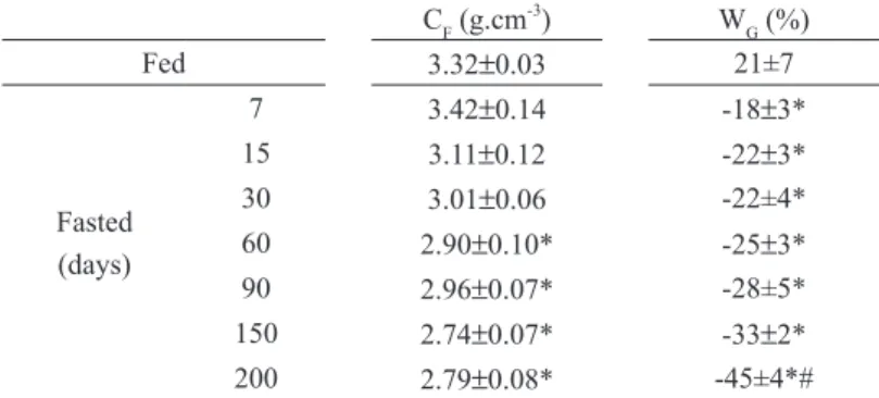
Condition factor and weight gain are presented on
Table I. A significant fall in the condition factor
was observed after 60 days of fasting (Table I). Moreover, during all the experimental period, the fed control animals gained weight (21%) and the
loss of body weight in fasted fish varied from 18%
(7 days) to 45% (200 days of fasting).
Fasting for 15 days or more reduced the liver
weight, but 7 days-fasting induced a significant
decrease in hepatosomatic index (Table II). These index values continued to fall until 60 days of fasting, and then remained constant until 200 days. Gonad weight and gonadosomatic index
were not significantly different during the whole
fasting induced a decrease of 40 - 50% in stomach and gut weights and in their somatic index in comparison to fed animals (Table II). The MAT
mass and its somatic index decreased only after 60 days of fasting (Table II), reaching values 60%
lower than fed fish after 200 days of fasting.
TaBLe ii
Tissue weight and their somatic index of fed and fasted red tilapias (Oreochromis sp).
Tissue Weight (g) Somatic Index (%)
L Go SG MAT HSI GoSI SGSI MATSI
Fed 8.9±0.3 2.2±0.2 13.6±1.7 15.1±1.1 2.27±0.07 0.50±0.04 2.90±0.39 3.29±0.19
Fasted (days)
7 7.1±0.5 1.6±0.3 8.2±0.4* 15.5±2.1 1.55±0.11* 0.35±0.04 1.78±0.12* 3.19±0.30 15 4.6±0.4* 2.0±0.2 7.7±0.4* 11.4±1.7 1.22±0.09* 0.53±0.07 1.79±0.06* 2.80±0.29 30 4.6±0.3∗ 2.2±0.2 7.4±0.5∗ 13.3±1.0 1.08±0.05∗ 0.52±0.06 1.62±0.10∗ 3.11±0.21
60 4.6±0.4∗ 1.9±0.3 7.1±0.4∗ 9.0±0.7∗ 1.04±0.12∗ 0.41±0.06 1.54±0.08∗ 2.07±0.16∗
90 5.4±0.3* 1.5±0.2 7.3±0.5* 9.2±0.9* 1.05±0.04* 0.29±0.03 1.46±0.08* 1.95±0.18* 150 4.5±0.5* 1.8±0.4 6.9±0.8* 6.9±1.6* 1.06±0.07* 0.27±0.05 1.56±0.17* 1.45±0.29* 200 5.1±0.6* 1.4±0.3 7.4±0.8* 5.9±1.1* 1.05±0.08* 0.27±0.06 1.53±0.15* 1.25±0.23*
L: Liver; Go: Gonad; SG: Stomach and Gut; MAT: Mesenteric Adipose Tissue; HSI: Hepatosomatic Index; GoSI: Gonadosomatic Index; SGSI: Stomach and Gut Somatic Index; MATSI: Mesenteric Adipose Tissue Somatic Index. Values represent mean ± SEM (n=10 fish for fed and for each fasting period). *(P<0.05) vs fed.
TaBLe i
Body parameters of fed and fasted red tilapias (Oreochromis sp).
CF (g.cm-3
) WG (%)
Fed 3.32±0.03 21±7
Fasted (days)
7 3.42±0.14 -18±3*
15 3.11±0.12 -22±3*
30 3.01±0.06 -22±4*
60 2.90±0.10* -25±3*
90 2.96±0.07* -28±5*
150 2.74±0.07* -33±2*
200 2.79±0.08* -45±4*#
CF: Condition Factor; WG: Weight Gain. Values represent mean ± SEM (n=10 fish for fed and for each fasting period). *(P<0.05) vs Fed mean; #(P<0.05) 200 vs other fasted periods.
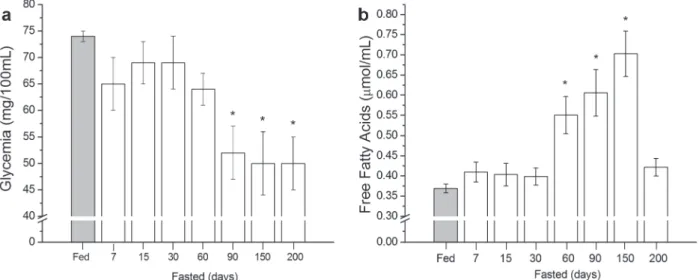
effectof faStingon metabolite levelS
Plasma glucose levels of fed fish (74±1 mg.dL-1)
did not change until 60 days of fasting (Fig. 1a).
After 90 days, glycemia decreased significantly (34%) in relation to fed fish, remaining low and
constant thereafter (~ 50±5 mg.dL-1
). Fasting for 60, 90 and 150 days resulted in 49, 64 and 90% increase of FFA plasma levels, respectively (Fig. 1b). After 200 days of fasting, FFA levels returned to control values (Fig. 1b). Lactate plasma level of
fasted fish did not change significantly during the
different periods of fasting in comparison to fed
fish, ranging from 0.530 ± 0.036 (fed) to 0.716 ±
0.059 mmol.mL-1 (200 days of fasting) (data not shown).
glycogenand total liPidS contentin liverand
muScle
The liver glycogen content in fed tilapia (15.0 ±
up to 60 days of fasting. After 90 to 200 days of fasting, the liver glycogen content fell to values 55% lower than fed state. Muscle glycogen content fell around 42.3% from 60 to 90 days of fasting,
but did not change significantly in the other fasting
periods tested in this work (Table III).
The total lipids content in fed tilapia liver
(10.5 ± 0.4%) increased twice in 90 days of fasting
(Table III), remaining constant until 200 days of fasting. No change was observed in total lipids
con-tent in tilapia muscle until 60 days of fasting (3.1 ±
0.3%); the values remained constant and similar to
fed condition (4.1 ± 0.3%) (Table III). A significant
decrease of muscle lipid content was only observed
after 90 days (2.7% ± 0.2%), which continues to fall until 200 days of fasting (1.5% ± 0.1%).
effectof faStingon meSenteric adiPoSe tiSSue
liPolySiSin vitro
Fatty acids mobilization from fragments of tilapia adipose tissue, incubated in control condition (BASAL medium), fasted for 60 to 200 days, was higher than FFA released from adipose tissue of
fed fish and from adipose tissue of 7, 15 or 30 days fasted fish (Fig. 2). Similar results were also found when adipose tissue from all groups of fish was
figure 1 - Plasma metabolic parameters of fed and fasted red tilapia (Oreochromis sp). Glycemia (a) and free fatty acids (b) were evaluated. Values represent mean ± SEM (n=10 fishes for fed and for each fasting period). *(P<0.05) vs fed.
TaBLe iii
Liver and muscle glycogen and total lipids of fed and fasted red tilapias.
Glycogen (%) Total Lipid (%)
Liver Muscle Liver Muscle
Fed 15.0 ± 0.4 0.52 ± 0.03 10.5 ± 0.4 4.1 ± 0.3
Fasted (days)
incubated in the presence of IBMX, an inhibitor of a cAMP-phosphodiesterase (Fig. 2a), or in presence of ISO, a non-selective beta-adrenoceptor agonist (Fig. 2b). MAT fragments of fasted tilapia, but not those collected from fed tilapias, incubated in the presence of IBMX or ISO showed a significant decrease in FFA release in comparison to MAT fragments incubated in control condition (Fig. 2).
We also found that fed tilapia MAT incubated with alpha 1 and alpha 2 adrenergic receptor agonists and antagonists, and some of their combinations, promoted a MAT lipolysis decrease (Fig. 3). Fatty acids mobilization from fragments of red tilapia MAT, fed and fasted up to 30 days, incubated in the presence of phenylephrine (PHE, 10-5M), showed
that the lipolysis inhibition of this non-selective
alpha-adrenoceptor agonist was 55% for fed fishes and 62% for 15 days fasted fishes (data not shown).
DisCussion
Ectotherms, which have relatively low basal meta-bolic rates, store substantial amounts of lipid in liver and MAT (Albalat et al. 2005). Few studies have an-alyzed the FFA release and the endocrine control of
lipolysis in fish (Murat et al. 1981, Van den Thillart
et al. 2001, Vianen et al. 2002, Albalat et al. 2005). In this study, the FFA release and its control were investigated using red tilapia as a model.
figure 3 - Fatty acids (FA) mobilization (mmol.g-1 MAT.2h) from mesenteric adipose tissue (MAT) of fed red tilapia (Oreochromis sp) to the incubation medium in presence of several lipolytic agents: basal condition (BASAL); dibutyryl-cAMP (AMPc 10-3
M); isoproterenol (ISO 10-5
M); 3-isobutyl-1-methylxanthine (IBMX 10-3
M); Forskolin (FSK 10-5 M); Yohimbine (YHB 10-5
M); Adenosine deaminase (ADA) (10µg. mL-1
); Prazosin (PZS 10-5
M); Prazosin (10-5
M) + Yohimbine (10-5
M); Theophylline (THEO 10-2
M); Phenylephrine (PHE 10-5M); 3-isobutyl-1-methylxanthine (IBMX 10-3M) + NaF(10mM); Arterenol (NOR 10-5
M). * (P<0.05) vs basal. Values are mean ± SEM (n=10 fish).
figure 2 - Fatty acids (FA) mobilization (mmol.g-1
MAT.2h) from mesenteric adipose tissue (MAT) of fed and fasted red tilapia (Oreochromis sp) in basal condition (■) or in presence of 10-3M IBMX (●)
(a) or 10-5M ISO (▲)
(b). * (P<0.05) vs basal; #
(P<0.05) vs fed. Values are mean ± SEM (n=10 fish for fed and for each fasting period). IBMX: 3-isobutyl-1-methylxanthine; ISO:
The condition factor parameter was used to verify the uniformity of the fish used in this work during the different periods of fasting. The condition factor reflects the recent feeding condition or the waste of energy reserves. Changes in these values indicate alterations in the nutritional conditions of animals (Bruton and Allanson 1974, Vazzoler 1996). Only after 60 days of fasting, we observed a reduction in the condition factor. These data are in agreement with other studies carried out with tilapia (Oreochromis mossambicus) and
channel catfish (Ictalurus punctatus) that showed a
low condition factor after 45 days of reduced food availability (Uchida et al. 2003, Peterson and Small 2004).
Hepatosomatic index reflects alterations in the
metabolic activity of the liver, acting as an appro-priate biomarker of the effect of an altered envi-ronment (Rossi et al. 2015). The decrease in liver weight and hepatosomatic index after 15 and 7 days of fasting, respectively, observed in this work corroborate the data obtained previously (Rossi et al. 2015, Bayir et al. 2011, Costas et al. 2011, Bar-cellos et al. 2010, Pérez-Jiménez et al. 2007, Al-balat et al. 2005), indicating that food deprivation
significantly affects the metabolic status of fish. In addition, 7 days of fasting seems to be sufficient to
completely empty the tilapia gut, since reduction of stomach and gut (SG) weight stabilized after 7 days of fasting. These values agree with the data of Figueiredo-Garutti et al. (2002) using Brycon cephalus, a carnivorous fish. The stomach somatic
index of this fish species decreased 11% after 24 h
and the gut somatic index decreased 40% after 72 h of fasting, remaining constant during 15 days of fasting. Gastric emptying in B. cephalus occurred faster than in red tilapia, as a result of shorter gut
of the carnivorous fish (Figueiredo-Garutti et al.
2002). The stomach and gut weight loss observed in this work with red tilapias submitted to pro-longed periods of fasting (60 to 200 days) also sug-gests that these tissues can be mobilized to supply substrates to energy metabolism.
The adaptation of several fish species to long
periods of fasting is explained by use of glucose from hepatic glycogenolysis and gluconeogenesis (Machado et al. 1989). The metabolic changes
during fish fasting are characterized by a sequential
utilization of glycogen, lipid and protein reserves (Collins and Anderson 1995). It has been reported that 3 days without food is enough to induce a decrease in B. cephalus liver glycogen (Figueiredo-Garutti et al. 2002). In the present study, tilapia hepatic glycogen content start reducing during 7 days of fasting, as well as the hepatosomatic
index. This pattern may reflect the mobilization of
glycogen stores to replace the absence of dietary carbohydrate intake. The plasma glucose levels were
kept constant and similar to fed fish until 60 days fasting, similar to what was observed in wolf fish
(Hoplias malabaricus), a Brazilian carnivorous fish
(Machado et al. 1989). Gluconeogenic substrates, as amino acids mobilized from peripheral tissues (white skeletal muscle, for example) and glycerol released, as a result of the rise on lipolysis rate during fasting, could also contribute for the glycemia maintenance in tilapias (Jørgensen et al. 2002). In conclusion, the glycemia maintenance during fasting could be related to (i) the capacity of hepatic glycogen mobilization, mainly during the initial period of fasting, (ii) the activation of hepatic gluconeogenesis and (iii) the reduction of glucose use (Moon and Foster 1995).
A significant glycemia reduction in red tilapias
only occurred after 60 days of fasting, remaining constant until 200 days of fasting. From 60 days until 150 days of fasting, it was observed a FFA plasma levels increase and a MAT weight reduction, indicating a mobilization of tilapia lipid reserves in these periods of fasting. Fatty acid mobilization from MAT in 60 days fasted fish was almost 2
times higher than values in fed fish. These values
prolonged starvation, the resting metabolism of tilapia could decrease markedly (Beamish 1964, Inui and Ohshima 1966, Mehner and Wieser 1994), downregulating the lipolysis pathway. Rossi et al.
(2015) observed a marked and significant decrease
in Hoplosternum littorale plasma triglyceride and hepatic lipid content, indicating that the lipid-reserve could also be accessed during starvation. In contrast, Lewis and Epple (1984), studying eels (Anguilla rostrata) fasted for 6 months, did not find
significant alterations in abdominal fat stores, serum
glucose, liver and muscle glycogen and fatty acids when compared with fed eels. These data show the wide different metabolic responses to prolonged
periods without food among fish species.
No difference was observed between fed and
fasted fish in plasma lactate levels. These results
corroborate a low difference in muscle glycogen
content between fed and fasted fish and suggest a
reduced capacity for gluconeogenesis metabolism from lactate in these animals during food deprived periods, since the plasma lactate allow that the gluconeogenesis occur in the liver via the Cori cycle. Similar results were found in other species
of fish, such as Artic charr (Salvelinus alpinus),
after 140 days of fasting (Jørgensen et al. 2002). In contrast, high plasma FFA levels are a characteristic
marker of food restriction in fish (Farbridge and
Leatherland 1992a, b, Pottinger et al. 2003), which could be used as the main energetic substrates by peripheral tissues.
Comparing the effect of known lipolytic agents in mammals, such as IBMX and ISO, in fatty acids release from tilapias MAT, it was observed that
both drugs promoted a significant decrease in fatty
acids release to the medium after 30 to 200 days of fasting, when compared to lipid mobilization observed in absence of these substances. Moreover, IBMX, ISO and cAMP do not present any effect on MAT lipolysis of fed tilapia. On the other hand, FSK, YHB, ADA, PZS, PZS plus YHB, THEO, PHE, IBMX plus NaF and NOR showed a clear
reduction in FFA mobilization from MAT of fed tilapia. In contradiction, it was demonstrated that
ISO do not alter the FFA mobilization rate in fish,
amphibians and reptiles adipose tissue (Migliorini et al. 1992, Farkas 1967). On the other hand, the lipolytic activity of these tissues is higher in presence of cAMP or xanthine derivatives, cAMP-phosphodiesterase inhibitors, since cAMP activates a triglyceride lipase in mammals (Migliorini et al. 1992). Our results corroborate with previous studies that demonstrated that noradrenaline and ISO inhibit lipolysis trough beta-adrenoceptors (Vianen et al. 2002, Van den Thillart et al. 2001). The reason to the contradictory effect of these drugs observed on tilapias remains elusive. Further experiments, using alpha and beta-adrenoceptors agonists and antagonists and measurements of nucleotide cyclic concentration in adipose tissue, should be done to clarify the physiological importance of these
findings. The physiological signal that promotes
fatty acid mobilization from adipose tissue deposits
in fish remains elusive.
In summary, the results obtained in this work suggest that in tilapia the metabolic adjustment to fasting is characterized by a sequential utilization of glycogen and lipid reserves. In agreement with the increase in plasma free fatty acids, there was a clear increase in the lipolytic activity of fasted tilapia MAT incubated in vitro, indicating a significant
contribution of this tissue to lipid mobilization. We also observed that the increase of fasting period lead to decrease in mesenteric adipose tissue, increase in MAT lipolysis rate and increase in lipid storage in liver. This could be a result of the high levels of FFA released during MAT lipolysis that
are esterified with glycerophosphate in the liver,
2002, Van den Thillart et al. 2001). Taking together, these data suggest that the reduction of lipolysis by adrenergic agonists during stressful situations, such as long periods of fasting, may represent a mechanism to prolong the life span of this specie, by preserving the adipose tissue energy reserve. These data also reinforce previous evidence that catecholamines, different from mammals, are not the lipolytic signal to enhance FFA mobilization during food deprivation in fish (Farkas 1967, Migliorini et al. 1992).
aCKnoWLeDGmenTs
The authors thank Elza Aparecida Filippin and Maria Antonieta R. Garófalo for their technical as-sistance. This work was supported by grants from Fundação de Amparo à Pesquisa do Estado de São Paulo (FAPESP) and from Conselho
Nacio-nal de Desenvolvimento Científico e Tecnológico
(CNPq). During this study, Walter Dias Jr received a fellowship from CNPq.
resumo
Mudanças adaptativas no metabolismo de carboidratos e lipídios induzidas por 7, 15, 30, 60, 90, 150 e 200 dias de jejum foram investigadas em tilápia vermelha (Oreochromis sp.). Níveis de glicose plasmática, lactato e ácidos graxos livres (FFA), glicogênio hepático e muscular, conteúdo de lipídio total e taxas de liberação de FFA de tecido adiposo mesentérico (MAT) foram mensurados. Níveis de glicose plasmática apresentaram diferenças significativas apenas após 90 dias de jejum, quando a glicemia estava 34% menor (50±5mg.dL-1) do que a encontrada em peixes alimentados (74±1mg.dL-1
), permanecendo relativamente constante em até 200 dias de jejum. O conteúdo de glicogênio hepático (»15%) em tilápias alimentadas diminuiu 40% em 7 dias de jejum. Após 60, 90 e 150 dias de jejum, níveis plasmáticos de FFA aumentaram 49%, 64% e 90%, respectivamente, quando comparados com os valores obtidos para peixes alimentados. Em concordância com o aumento de FFA plasmático, o jejum induziu um aumento claro da atividade lipolítica em MAT incubada in vitro. A adição de
isobutilmetilxantina (inibidor da cAMP-fosfodiesterase) e isoproterenol (agonista beta adrenérgico não-seletivo) ao meio de incubação induziu uma redução da lipólise em peixes em jejum, diferentemente do que já foi observado no tecido adiposo de mamíferos. Este estudo permitiu uma avaliação fisiológica da resposta de tilápia vermelha ao jejum.
Palavras-chave: tecido adiposo, adrenoreceptor, lipólise, glicogênio hepático.
referenCes
albalat a, gómeS-requeni P, roJaS P, médale f, kauSHik S, vianen gJ, van den tHillart g, gutiérrez J, Pérez-SáncHez J and navarro i. 2005. Nutritional and hormonal control of lipolysis in isolated gilthead seabream (Sparus aurata) adipocytes. Am J Physiol Regul Integr Comp Physiol 289: 259-265.
barcelloS l, marqueze a, traPP m, quevedo rm
and ferreira d. 2010. The effects of fasting on cortisol, blood glucose and liver and muscle glycogen in adult jundiá Rhamdia quelen. Aquaculture 300: 231-236.
bayir a, Sirkecioglu an, bayir m, Haliloglu Hi,
kocaman em and araS nm. 2011. Metabolic
responses to prolonged starvation, food restriction, and refeeding in the brown trout, Salmo trutta: oxidative stress and antioxidant defenses. Comp Biochem Physiol 159(4): 191-196.
beamiSHFWH. 1964. Influence of starvation on standard and routine oxygen consumption. Trans Am Fish Soc 93: 103-107.
bergmeyer Hu, gaweHn k and graSSl m. 1974. Enzymes as biochemical reagents. In: Bergmeyer HU (Ed), Methods of Enzymatic Analysis, 2nd ed., New York: Academic Press, New York, USA, p. 457-458.
bligH eg and dyer wJ. 1959. A rapid method of total lipid extraction and purification. Can J Biochem Phys 27(8): 911-917.
boyd ce. 2004. Farm-Level Issues in Aquaculture Certification: Tilapia. Report Commissioned by WWF. http://fisheries.tamu.edu/files/2013/09/Farm-Level-Issues-in-Aquaculture-Certification-Tilapia.pdf.
bruton mn and allanSon br. 1974. The growth of Tila-pia mossambica Peters (Pisces, Ciclidae) in Lake Sibaya, South Africa. J Fish Biol 6: 701-715.
carrol nv, longlay rw and roe JH. 1956. The determination of glycogen in liver and muscle by use of anthrone reagent. J Biol Chem 220: 583-593.
caruSo g, mariccHiolo g, micale v, genoveSe
non-specific immune parameters and skin structure. Fish Physiol Biochem 36: 71-83.
clark aS, kelley ra and mitcH we. 1984. Systemic response to thermal injury in rats. Accelerated protein degradation and altered glucose utilization in muscle. J Clin Invest 74: 888-897.
clementS kd and raubenHeimer. 2006. 2 – Feeding and nutrition. In: Evans DH and Claiborne JB (Eds), The physiology of fishes, Boca Raton: CRC Press, Taylor and Francis Group, Florida, USA, p. 47-82.
collinS al and anderSon ta. 1995. The regulation of endogenous energy stores during starvation and refeeding in the somatic tissues of the golden perch. J Fish Biol 47: 1004-1015.
coStaS b, aragão c, ruiz-Jarabo i, vargaS-cHacoff
l, arJona f, diniS mt, mancera Jm and conceição
l. 2011. Feed deprivation in Senegalese sole (Solea sen-egalensis Kaup, 1858) juveniles: effects on blood plasma metabolites and free amino acid levels. Fish Physiol Bio-chem 37: 495-504.
dole vP and meinertz H. 1960. Microdetermination of long-chain fatty acids in plasma and tissues. J Biol Chem 235: 2595-2599.
farbridge kJ and leatHerland Jf. 1992a. Plasma growth hormone levels in fed and fasted rainbow trout (Oncorhynchus mykiss) are decreased following handling stress. Fish Physiol Biochem 10: 67-73.
farbridge kJ and leatHerland Jf. 1992b. Temporal changes in plasma thyroid hormone, growth hormone and free fatty acid concentrations, and hepatic 5’-monodeio-dinase activity, lipid and protein content during chronic fasting and re-feeding in rainbow trout (Oncorhynchus mykiss). Fish Physiol Biochem 10: 245-257.
farkaS t. 1967. The effect of catecholamines and adreno-corticotropic hormone on blood and adipocyte tissue FFA levels in the fish Cyprinus carpio L. Prog Biochem Phar-macol 3: 314-319.
figueiredo-garutti ml, navarro i, caPilla e, Souza
RHS, moraeS g, gutiérrez J and viventi-Paulino
MLM. 2002. Metabolic changes in Brycon cephalus
(Teleostei, Characidae) during post-feeding and fasting. Comp Biochem Phys A 132: 467-476.
gaylord tg, mackenzie dS and gatlinIII dm. 2001. Growth performance, body composition and plasma thyroid hormone status of channel catfish (Ictalurus punctatus) in response to short-term feed deprivation and refeeding. Fish Physiol Biochem 24: 73-79.
Hung SSO and Storebakken t. 1994. Carbohydrate
utilization by rainbow trout is affected by feeding strategy. J Nutr 124: 223-229.
inui y and oHSHima y. 1966. Effect of starvation on me-tabolism and chemical composition of eels. Bull Jpn Soc Sci Fish 33: 181-189.
JørgenSen eH, viJayan mm, aluru n and maule ag. 2002. Fasting modifies Aroclor 1254 impact on plasma cortisol, glucose and lactate responses to a handling disturbance in Artic charr. Comp Biochem Phys C 132: 235-245.
kittilSon Jd, reindl km and SHeridan ma. 2011. Rainbow trout (Oncorhynchus mykiss) possess two hormone-sensitive lipase-encoding mRNAs that are differentially expressed and independently regulated by nutritional state. Comp Biochem Physiol A 158: 52-60.
krogdaHl Å and bakke-mckelleP a. 2005. Fasting and refeeding cause rapid changes in intestinal tissue mass and digestive enzyme capacities of Atlantic salmon (Salmo salar L.). Comp Biochem Physiol A 141: 450-460.
lewiS tl and ePPle a. 1984. Effects of fasting, pancre-atectomy, and hypophsectomy in the yellow eel, Anguila rostrata. Gen Comp Endocr 55(2): 182-194.
li mH and robinSon eH. 2015. 4 - Complete feeds— intensive systems. In: Davis DA (Ed), Feed and Feeding Practices in Aquaculture, Sawston: Oxford, Woodhead Publishing, Cambridge, UK, p. 111-126.
macHado cr, garofalo MAR, roSelino JES,
kettelHut ic and migliorini rH. 1989. Effect of fasting on glucose turnover in a carnivorous fish (Hoplias
sp.). Am J Physiol 256: R612-R615.
magnoni l, vaillancourt e and weber Jm. 2008. In vivo regulation of rainbow trout lipolysis by catechol-amines. J Exp Biol 211: 2460-2466.
meHner t and wieSer w. 1994. Energetics and metabolic correlates of starvation in juvenile perch (Perca fluviatilis). J Fish Biol 45: 325-333.
migliorini rH, lima-verde JS, macHado cr, cardona
GMP, garófaloMARand kettelHut ic. 1992. Con-trol of adipose tissue lipolysis in ectotherm vertebrates. Am J Physiol Regul Integr Comp Physiol 263: R857-R862.
moon tw and foSter gd. 1995. Tissue carbohydrate me-tabolism, gluconeogenesis, and hormonal and environ-mental influences. In: Hochachka PW and Mommsen TP (Eds), Biochemistry and Molecular Biology of Fishes, vol. 4., Amsterdan: Elsevier Science, Netherlands, p. 393-434.
murat Jc, PliSetSkaya em and wooNYS. 1981. Endo-crine control of nutrition in cyclostomes and fish. Comp Biochem Phys A 68: 149-158.
nagay m and ikeda S. 1972. Carbohydrate metabolism in fish. III. Effect of dietary composition on metabolism of glucose U-14
C in carp. Bull Jpn Soc Sci Fish 38: 137-143.
navarro i and gutiérrez J. 1995. Fasting and starvation. In: Hochachka PW and Mommsen TP (Eds), Biochemistry and Molecular Biology of Fishes, Amsterdam: Elsevier, Netherlands, p. 393-434.
Pérez-Jiménez a, cardenete g, Hidalgo mc, garcía -alcázar a, abellán e and moraleS ae. 2012. Metabolic adjustments of Dentex dentex to prolonged starvation and refeeding. Fish Physiol Biochem 38: 1145-1157.
Pérez-Jiménez a, guedeS mJ, moraleS ae and oliva -teleS a. 2007. Metabolic responses to short starvation and refeeding in Dicentrarchus labrax. Effect of dietary composition. Aquaculture 265: 325-335.
PeterSon bc and Small bc. 2004. Effects of fasting on circulating IGF-binding proteins, glucose, and cortisol in channel catfish (Ictatlurus punctatus). Domest Anim Endocrin 26: 231-240.
Pottinger tg, rand-weaver m and SumPter JP. 2003. Overwinter and re-feeding in rainbow trout: plasma growth hormone and cortisol levels in relation to energy mobilization. Comp Biochem Phys B 136: 403-417.
roSSi a, cazenave J, baccHetta c, camPana m and
Parma mJ. 2015. Physiological and metabolic adjustments of Hoplosternum littorale (Teleostei, Callichthyidae) during starvation. Ecol Indic 56: 161-170.
Scorvo filHo Jd, fraScá-Scorvo CMD, alveSJMC
and SouzaFRA. 2010. A tilapicultura e seus insumos, relações econômicas. Rev Bras Zootec 39: 112-118.
ucHida k, kaJimura S, riley lg, Hirano t, aida k and
grau eg. 2003. Effects of fasting on growth hormone/
insulin-like growth factor I axis in the tilapia, Oreochromis mossambicus. Comp Biochem Phys A 134: 429-439.
van den tHillart g, vianen g, Ponce mc, lelieveld
H, nieveen m, van raail m, SteffenS a and
zaagSma J. 2001. Differential role of adrenoceptors in control of plasma glucose and fatty acids in carp, Cyprinus carpio (L.). Am J Physiol Reg Integr Comp Physiol 281: R615-R624.
van raaiJMTM, van den tHillart g, HallemeeScH
m, balm PHM and SteffenS ab. 1995. Effect of arterially infused catecholamines and insulin on plasma glucose and free fatty acids in carp. Am J Physiol Regul Integr Comp Physiol 268: R1163-R1170.
vazzolerAEAM. 1996. Biologia da reprodução de peixes
teleósteos: teoria e prática. Maringá: EDUEM, 169 p.
vianen gJ, obelS PP, van den tHillart ge and
zaagSma J. 2002. Beta-Adrenoceptors mediate inhibi-tion of lipolysis in adipocyte of tilapia (Oreochromis mos-sambicus). Am J Physiol – Endocrinology and Metabolism 282: E318-325.
vigliano f, quiroga m and nieto J. 2002. Metabolic adaptation to food deprivation and refeeding in fish. Rev Ictiol 10: 79-108.